Downloaded 293 times


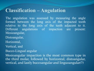


The document discusses the definition, causes, and types of impacted teeth, emphasizing the importance of timely surgical interventions to prevent potential complications. It also outlines various treatment methods, including removal, coronectomy, and surgical exposure for impacted canines. Additionally, the document addresses the anatomical considerations and potential risks associated with the surgical management of impacted teeth, particularly the third molars.


