IRJET - Automated Segmentation and Detection of Melanoma Skin Cancer using Dermoscopy Images
•
0 likes•6 views

https://irjet.net/archives/V7/i3/IRJET-V7I3303.pdf

Recommended
Recommended
More Related Content
What's hot
What's hot (20)
Similar to IRJET - Automated Segmentation and Detection of Melanoma Skin Cancer using Dermoscopy Images
Similar to IRJET - Automated Segmentation and Detection of Melanoma Skin Cancer using Dermoscopy Images (20)
More from IRJET Journal
More from IRJET Journal (20)
Recently uploaded
Recently uploaded (20)
IRJET - Automated Segmentation and Detection of Melanoma Skin Cancer using Dermoscopy Images
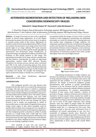
- 1. International Research Journal of Engineering and Technology (IRJET) e-ISSN: 2395-0056 Volume: 07 Issue: 03 | Mar 2020 www.irjet.net p-ISSN: 2395-0072 © 2020, IRJET | Impact Factor value: 7.34 | ISO 9001:2008 Certified Journal | Page 1630 AUTOMATEDSEGMENTATIONAND DETECTION OF MELANOMA SKIN CANCERUSING DERMOSCOPY IMAGES Rakesh N1, Sanjai Kumar N2, Yuvaraj S3, John Berkmans T4 1,2,3 Final Year Student, Dept of Information Technology, Jeppiaar SRR Engineering College, Chennai 4John Berkmans T: Asst Professor, Dept. of Information Technology, Jeppiaar SRR Engineering College, Chennai ---------------------------------------------------------------------***---------------------------------------------------------------------- Abstract - In Image Processing we have such a significant number of clinical image applications. As of late Digital dermoscopy has gotten one of the serious issues to recognize or examine various kinds of skin lesions. Beforehand we needed to utilize Particle SwarmOptimization(PSO)tochoose and compute the parameters of an image and to restrict the specific edges in an image utilizing SIFT (scale invariant element change). There we didn't get the specific aftereffectof improvement and furthermore fringe identification. In proposed strategy, we applied the hereditary calculation(GA) technique to find specific features in skin image. The segmentation procedure of an image will give valuable data and best features. Consequently, we utilize an edge-based segmentation method called DWT (Discrete Wavelet Transform). GA will give precise outcome when contrasted with PSO. In the wake of extracting features from dermo images, it is legitimately feed as input to NN(NeuralNetwork) process. As indicated by the input image, the NN classifier will consolidate and classify. The classifier will distinguish the region of abnormal image or classify it as a normal image. Keywords – skin lesion classification, edge-based segmentation, dermo images. 1.INTRODUCTION Skin malignant growths area unit diseases that emerge from the skin. they're as a result of the advancement of strange cells that may attack or unfold to completely different items of the body. There are3 basic varietiesofskin tumors, basal-cell skin malignant growth (BCC), squamous- cell skin problem (SCC) and skin cancer. The initial 2,aboard varied less traditional skin diseases, area unit called nonmelanoma skinmalignancy(NMSC).Basal-cell malignant growth develops bit by bit and may damage the tissue around it but is maybe not progressing to unfold to inaccessible zones or end in death. It regularly showsupasa simple raised region of skin which {may|that may} be sparkly with very little veins running over it or may gift as a raised territory with Associate in Nursing lesion.Squamous- cell skin malignant growth is absolute to unfold. It usually presents as a tough bump with a flaky high but could likewise frame Associate in Nursing lesion. Melanomas area unit the foremost forceful. Signs incorporate a mole that has modified in size, shape, shading, has noncontinuous edges, has over one shading, is galling or drains. Over ninetieth of cases area unit caused by presentation to bright radiation from the Sun. This presentation builds the danger of every of the 3 primary varieties of skin malignancy. Introduction has distended, principally as a result of a slender layer. Tanning beds area unit another basic wellspring of bright radiation. For melanomas and basal-cell tumors, introduction throughout youth is very harmful. For squamous-cell skintumors,all out presentation, freelance of once it happens, is moreandmore important. Somewhere within the vary of two hundredth and half-hour of melanomas produce from moles. people with lighter skin area unit at higher hazard Fig 1 Types of skin cancer Diminishing introduction to bright radiation and therefore the utilization of emollient provides off a bearing of being eminent techniques for obviation skin cancer and squamous-cell skin problem. it is not clear if emollient influences the danger of basal-cell malignant growth. Nonmelanoma skin problem is usually maintainable. Treatment is for the foremost half by careful expulsion however could, less commonly, embody radiationtreatment or topical prescriptions, for instance, antimetabolite. Treatment of skin cancer could embody a mixture of procedure, therapy, radiation treatment and scentered on treatment. In those people whose malady has unfold to completely different regions of the body, palliative thought can be utilised to enhance personal satisfaction. skin cancer has one in every of the upper endurance rates among diseases, with over eighty of people within the United Kingdom of Great Britain and Northern Ireland and over ninetieth within the u. s. enduring over five years. Skin malignant growth is that the most well-known form of unwellness, all around representing at any rate four- hundredth of malignant growth cases. Basal-cell and squamous-cell skin malignant growths occasionally cause death. The 3 basic varieties of skin malignant growth have gotten more and more regular within the lasttwentytoforty years, significantly in those regions with usually White people.
- 2. International Research Journal of Engineering and Technology (IRJET) e-ISSN: 2395-0056 Volume: 07 Issue: 03 | Mar 2020 www.irjet.net p-ISSN: 2395-0072 © 2020, IRJET | Impact Factor value: 7.34 | ISO 9001:2008 Certified Journal | Page 1631 2. RELATED WORKS 2.1 CNN Model The name “convolutional neural network”indicates that the network employs a mathematical process referred to as convolution. Convolution could be a specialised quite linear operation. Convolutional networks area unit merely neural networks that use convolution in situ of general matrix operation in a minimum of one in every of their layers. CNNs are multilayer perceptrons. Multilayer perceptrons typically mean absolutely connected networks, that is, every vegetative cell in one layer is connected to all or any neurons within the next layer. The "fully- connectedness" of those networks makes them liable to overfitting Therefore, on the dimensions of connectedness and quality, CNNs area unit on the lower extreme. Neurons answer stimuli solely in an exceedingly restricted region of the field of regard called the receptive field. The receptive fields of various neurons partly overlap specified they cowl the complete field of regard. CNNs use comparatively pre- processing compared to alternative image classification algorithms. This suggests that the network learns the filters that in ancient algorithms were hand- designed. A convolutional neural network consists of input layer associated with multiple hidden layers and an output layer. The hidden layers of a CNN generally accommodate a series of convolutional layers thatbend witha multiplication or alternative real. The activation operate is usually a RELU layer, and is later followed by extra convolutionslikepooling layers, absolutelyconnected layersandnormalizationlayers, stated as hidden layers as a result of theirinputsandoutputs area unit disguised by the activation operate and final convolution. Mathematically, it's technically real or cross- correlation. This has significance for the indices in the matrix. 2.2. Edge-Based Segmentation Image segmentation is that the method of partitioning a digital image into multiple segments. Thegoal of segmentation is to alter and/or modification the illustration of a picture into one thing that's additional meaningful and easier to investigate. Image segmentation is usually wont to find objects and limits in pictures.additional exactly, image segmentation is that the method of assignment a label to each element in a picture- specified pixels with an equivalent label share bound characteristics and the results of image segmentation could be a set of segments that put together cowl the complete image, or a group of contours extracted from the image (see edge detection). every of the pixels in an exceedingly region area unit similar with relevancy some characteristic orcomputed property, like color, intensity, or texture. This thresholding technique is predicated on a clip-level (or a threshold value) to show a gray-scale image into a binary image. The key of this technique is to pick out the brink worth (or values once multiple-levels area unit selected). The key plan is that, in contrast to Otsu's technique. New strategies steered the usage of multi-dimensional fuzzyrule- based non-linear thresholds. During this work call over every pixel's membership to a phase is predicated on multi- dimensional rules derived from formal logic and organic process algorithms. Motion primarily based segmentation could be a technique that depends on motion within the image to perform segmentation. The thought is simple: cross-check the variations between a combine of pictures. 2.3 Skin Lesion Classification One of the wide used strategiesbydermatologiststo classify the cancerous skin— skin cancer from traditional skin is that the ABCD rule. it's proven that it is simply learned and quickly calculated and has been tested to be a reliable technique providing an additional objective and duplicatable identification of skin cancer. To calculate the ABCD score, the “asymmetry, border, colors, and diameter” criteria area unit about calculable (semi-quantitatively). every of the standards is then increased by a given weight factor to yield a complete dermoscopy score (TDS). TDS values but four.75 indicates a benign melanocytic lesion, values between four.8 and 5.45 indicate a suspicious lesion, and values of five.45 or bigger area unit extremely connotative skin cancer. Economical technique that classifies skin lesion pictures into 3 categories. Namely, the primary category contains the mar, that could be a benign tumor. The second labels cancerous skin cancer cases comparable to malignant skin tumors derived from melanocytes. The lesions within the third category area unit labelled as keratosis, that is additionally a benign tumor derived from keratinocytes. 3. PROPOSED ARCHITECTURE
- 3. International Research Journal of Engineering and Technology (IRJET) e-ISSN: 2395-0056 Volume: 07 Issue: 03 | Mar 2020 www.irjet.net p-ISSN: 2395-0072 © 2020, IRJET | Impact Factor value: 7.34 | ISO 9001:2008 Certified Journal | Page 1632 4. PROPOSED SYSTEM Dermo dataset is collected. at first dataset is to be pre-processed. Pre-Processing includes Gaussian Blurring, Bi-Lateral Filtered image, Gray-scale image. Next up is that the segmentation of grayscale image by means that of Thresholding. GLCM feature extraction from the divided image. There by the edge-based transformation techniques area unit applied. Then to classify thedermo images,it'ssplit into train information and check information. Train information area unit to be trained underneath the bound model of Machine Learning algorithms. The check accuracy and epochs area unit to be calculated by the check information. 5. MODULES AND DESCRIPTION 5.1. Pre-Processing Gaussian Blurring is employed scale back |to scale back| to cut back} image noise and reduce detail. A bilateral filter could be a non-linear, edge- conserving, replaces the intensity of every element with a weighted average of intensity values from close pixels. Grayscale image mapsthe keep numeric sample values to the achromatic channel of a typical colorspace, that itself is predicated on measured properties of human vision. 5.2 Thresholding for Segmentation Thresholding strategies replace every element in a picture with a black element. One approach is to designate a separate threshold for every of the RGB elements of the image and so mix them with Associate in Nursing AND operation. Hue could be a circular amount it needs circular thresholding. Thresholding strategies work on bimodal distributions, however algorithms have conjointly been developed for unimodal distributions, multimodal distributions, and circular distributions. Automatic thresholding can work best once an honest background to foreground distinction quantitative relation exists. 5.3 GLCM Feature Extraction A GLCM could be a bar chart of co-occurring greyscale values at a given offset over an image. options of the GLCM matrices area unit computed like difference and correlation. Texture could be a feature to partition pictures into regions of interest and to classify those regions. in an exceedingly classification downside these area unit planned for instance that the categories type clusters in feature house. 5.4 Training a Neural Network Train a classifier to label image patches from new images. Fitting a neural network involves using a training dataset to update the model weights to create a good mapping of inputs to outputs. Training process is iterative, solves an optimization problem that finds for parameters (model weights) that result in a minimum error or loss. It is advisable to initialize weights with small random values to break the symmetry between different neurons. 5.5 Classification The main purpose of the artificial neural network is to combine features into more attributes. Classification specifies the class to which data elements belong to and predicts a class for an input variable as well. Cross entropy often goeshand in hand withsoftmax function. The accuracy dependsonthenumberofepochs in order to detect potential overfitting problem 6. CONCLUSION The application of CNNs for skin lesion detection is promising; but, the dearth of huge annotated datasets to coach their models continues to be a barrier to develop strategies unquestionably appropriate for clinical application. During this paper, we tend to propose the CNN model for skin lesion classification in dermo images, that collectively uses the residual learning and a completely unique attention learning mechanism to enhance the discriminative illustration ability of DCNNs. The novel attention learning mechanism is meant to use the feature maps learned by high layers to come up with the eye maps for low layers. Our results show that the projected CNN model will adaptively concentrate on the discriminative components of skin lesions, and so come through the progressive performance in skin lesion classification. Our future work in skin cancer detection, we tend to advocate any analysis on exploitation varied soft computing and computing algorithms with completely different classifiers like Random Forest(RF)classifierandNaïv eBayesanalysing the results for an equivalent. REFERENCES [1] Ferris, L.K., Harkes, J.A., Gilbert, B., et al.: Computer-aided classification of melanocytic lesions exploitation dermoscopic images. J. Am. Acad. Dermatol. 73(5), 769–776 (2015). [2] S.V Shinde, U.V Kulkarni, “Modified Fuzzy Hyperline- Segment Neural Network for Classification with Mixed Attribues”, Fifth International Conference on Computing, Communications and Networking Technologies (ICCCNT), Hefei,China , July 2014. [3] M. Oquab, L. Bottou, I. Laptev, and J. Sivic,” Learning and Transferring Mid-level Image Representations Using Convolutional Neural Networks,”inProc.IEEEConf.Comput. Vis. Pattern Recognit., 2014, pp. 1717-1724. [4] W. AhmadMelanoma recognition framework based on expert definition of ABCD for dermoscopic images Skin Res. Technol., 19 (1) (2013), pp. 93-102.
- 4. International Research Journal of Engineering and Technology (IRJET) e-ISSN: 2395-0056 Volume: 07 Issue: 03 | Mar 2020 www.irjet.net p-ISSN: 2395-0072 © 2020, IRJET | Impact Factor value: 7.34 | ISO 9001:2008 Certified Journal | Page 1633 [5] Germán Capdehourat, Andrés Corez, Anabella Bazzano, Rodrigo Alonso,Pablo Musé, “Toward a Combined Tool to Assist Dermatologists in Melanoma Detection from Dermoscopic Images of Pigmented Skin Lesions”,ELSEVIER, 2011 [6] Krizhevsky, A., Sutskever, I., Hinton, G.E.: Image net classification with deep convolutional neural networks. In: NIPS (2012) [8] N. Otsu, “A threshold selection method from gray- level histograms'', Automatica, vol. 11, nos. 285-296, pp. 23-27, 1975. [9] F. Nachbar, W. Stolz, T. Merkle, A.B. Cognetta, T. Vogt, M. Landthaler, G. Plewig The ABCD rule of dermatoscopy: high prospective value in the diagnosis of doubtful melanocytic skin lesions J. Am. Acad. Dermatol., 30 (4) (1994), pp.