1) The document presents a method for detecting skin lesions using support vector machines (SVM). It involves preprocessing images, segmenting the skin lesion region, extracting features related to shape, color, and texture, and classifying lesions as melanoma or non-melanoma using an SVM classifier.
2) Features extracted include asymmetry, border irregularity, compactness, color ratios in HSV, RGB and LAB color spaces, and texture features from the gray-level co-occurrence matrix.
3) An SVM classifier is used for classification as it can accurately classify data by finding the optimal separating hyperplane that maximizes the margin between the classes. The method achieved efficient classification of lesions.
![Advances in Engineering: an International Journal (ADEIJ), Vol.3, No.2
2
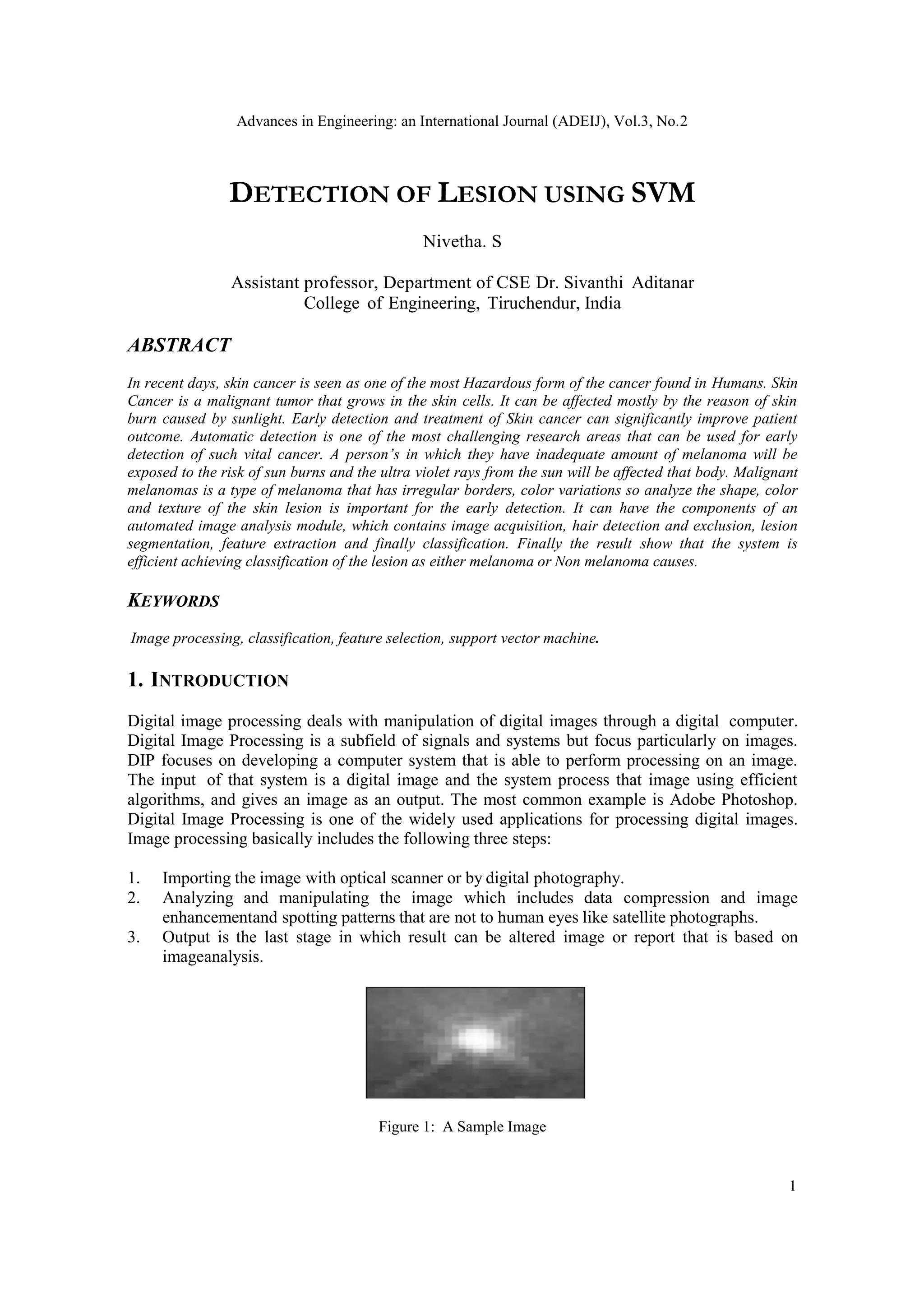
An array or a matrix of pixels arranged in columns and rows. In the sample image figure1, there
may be thousands of pixels that together make up this image. We will zoom that image to the
extent that we are able to see some pixels division. It is shown in the image below.
Figure 2: Pixelized Image
Each pixel figure 2 has a value from 0 (black) to 255 (white). The possible range of the pixel
values depend on the color depth of the image, here 8 bit = 256 tones or gray scales.
The main objective of the proposed work is to automatically analysis the skin lesions with the
help of image processing techniques. The manual segmentation as well as manual diagnosis of
skin cancer lesions are very tedious process. The manual error will be unavoidable in those
manual work. In order to reduce the manual error as well as manual diagnosis, the project is
motivated to provide automatic solution for the skin lesion segmentation & classification.
2. RELATED WORK
Teck Yan Tan, Dermoscopic images are detected using an intelligent agent-based or robotic
system toconduct long-term automatic health monitoring and robust efficient disease diagnosis as
autonomous e- Careers in real-world applications [1]. In this research, we aim to deal with such
challenges by presenting an intelligent decision support system for skin lesion recognition as the
initial step, which could be embedded into an intelligent service robot for health monitoring in
home environments to promote early diagnosis. The system is developed to identify benign and
malignant skin lesions using multiple steps, including pre-processing such as noise removal,
segmentation, and feature extraction from lesion regions, feature selection and classification.
After extracting thousands of raw shape, colour and texture features from the lesion areas, a
Genetic Algorithm (GA) is used to identify the most discriminating significant feature subsets for
healthy and cancerous cases. A Support Vector Machine classifier has been employed to perform
benign and malignant lesion recognition. Evaluated with 1300 images from the Dermo fit
dermoscopy image database, the empirical results indicate that our approach achieves superior
performance in comparison to other related research.
Eliezer Flore set al., Pre-screening systems for the diagnosis of melanocytic skin lesions depend
of the proper segmentation of the image region affected by the lesion. This paper proposes a
feature learning scheme that finds relevant features for skin lesion image segmentation. This work
introduces a new unsupervised dictionary learning method, namely Unsupervised Information-
Theoretic Dictionary Learning (UITDL), and discusses how it can be applied in the segmentation
of skin lesions in macroscopic images [2]. The UITDL approach is adaptive and tends to be
robust to outliers in the trainingdata, and consists of two main stages. In the first stage, a textural
variation image is used to construct an initial feature dictionary and an initial sparse
representation via Non-Negative Matrix Factorization (NMF). In the second stage, the feature
dictionary is optimized by selecting adaptively the number of dictionary atoms. The greedy
approach used for dictionary optimization is quite efficient and flexible enough to be applied to](https://image.slidesharecdn.com/3221adeij01-210416062635/75/DETECTION-OF-LESION-USING-SVM-2-2048.jpg)
![Advances in Engineering: an International Journal (ADEIJ), Vol.3, No.2
3
other dictionary learning problems. Furthermore, the proposed method can be easily extended for
other image segmentation problems. The experimental results suggest that the proposed approach
potentially can provide more accurate skin lesion segmentation results than comparable state-
of-the-art methods [3]. The proposed segmentation method could help to improve the
performance of pre-screening systems for melanocytic skin lesions, which can affect positively
the qualityof the early diagnosis provided to skin lesion patients.
3. THE PROPOSED SYSTEM
Proposed System is an intelligent decision support system for benign and malignant skin lesions
classification. The system includes the following key stages, i.e. pre-processing, skin lesion
segmentation, feature extraction and classification. Figure 3 shows system architecture, which
shows the principal processes of the proposed system. The test image is preprocessed for the
removal of noise as well as contrast adjustment. The skin RGB image is initially converted into
grayscale image for dimension reduction. The grayscale images undergoes for the hair removal
process. The image is now subjected with the ROI segmentation based on active contour
segmentation algorithm. The Segmentation result will be the separated skin lesion. The skin
lesion area is subjected for the feature extraction technique. The texture, shape and color features
are carried out from the skin lesion region. The Support vector machine classifier is already
trained with the two set of features such as benign and malignant images. The test image features
are given into the trained SVM model and the class will be recognized from the SVM. The
following section describes the modules involved in the proposed work.
Figure 3: Block diagram for the proposed work](https://image.slidesharecdn.com/3221adeij01-210416062635/75/DETECTION-OF-LESION-USING-SVM-3-2048.jpg)
![Advances in Engineering: an International Journal (ADEIJ), Vol.3, No.2
4
4. METHODS
PRE-PROCESSING
A pre-processing method is essential for benign and malignant skin lesions classification. This
involves transforming raw data into an understandable format for further processing. In the real
world, data are often inconsistent and incomplete and may contain many errors. Removing any
noise and unnecessary features which cause confusion to classifiers is required. The following
pre-processing is conducted in this research.
Hair Remover: The Enhanced Dull Razor algorithm figure 4 was used to remove hairs from
images where morphological closing image processing was generalized to grey-level images,
followed by identification of the narrow, elongated hair outline.
Bilinear interpolation was implemented to substitute the identified pixels of the hairs. This step
resulted ina smooth fill inward from the borders of the region of interest.
Contrast Enhancement: Subsequently, the image clarity was enhanced by improving the shape
andedges of the image. Image borders were sharpened using contrast enhancement. This process
may also optimize subsequent segmentation accuracy.
Figure 4: Hair removal result;
Left (original image) Right (After hair removal)
Grayscale Conversion: RGB images of lesions, with M× N pixels in size, were transformed to
greyscale by removing hue and saturation using a process which computes the weighted sum of
the colour components.
SEGMENTATION
Image segmentation figure 5 is a technique to determine the shape and size of the border, and to
separate the object from its background based on different features extracted from the image.
After removing the noise from the lesion area, the lesion needs to be separated from the skin, and
therefore the analysis for diagnosis is conducted purely using the necessary area. Previous studies
have proposed several different types of segmentation methods with high accuracies, such as
clustering based, threshold- based and edge-based methods. In this research, the Adaptive Snake
(AS) approach is chosen because of its efficiency indicated by previous research. AS inefficient
for establishing a discriminating analysis that divides the image into two classes of pixels. In the
first instance, the chosen color image is rendered in monochrome. Then corresponding threshold
limits are set within the grey spectrum, and the pixels that occur within the range set by the limits
are selected. Following this, non-lesion pixels are assigned with a value of zero. To extract
multiple features [4], such as colour and area, the segmentation results of this threshold-based
method are plotted into multiple images.](https://image.slidesharecdn.com/3221adeij01-210416062635/75/DETECTION-OF-LESION-USING-SVM-4-2048.jpg)
![Advances in Engineering: an International Journal (ADEIJ), Vol.3, No.2
5
Figure 5: Segmentation result: a) original image b) Segmented lesion region
FEATURE EXTRACTION
After segmentation, image features are extracted for the subsequent classification. Several
methods have been identified for feature extraction. Overall, the majority of related work
employed the ABCDE rules of dermatology for feature extraction. In this research, measurements
such as compactness index, fractal index, and edge abruptness are used in order to indicate border
irregularity.
SHAPE
Asymmetry: A melanocyte lesion may be diagnosed by a number of identifiers, of which one of
the most significant is a lack of symmetrical morphology. In dermatology terms, the ABCDE rule
model rates this aspect as the most crucial factor. In consideration of the symmetry feature, a
number of factors are concurrently relevant, including color, texture and morphology. A three-
fold classification system can be derived from measuring symmetry, with three-class outputs
representing total symmetry, a lack of symmetry along a single axis and a lack of symmetry along
dual axe, respectively. The lesion asymmetry was evaluated by calculating the area with inner and
outer of the lesion, using the formula shown as follows.
where AI represents asymmetry Index. ΔAK represents the area between the two halves of the
lesion andAL denotes the lesion area.
Border Irregularity: Irregularities occurring in the edge of a malignant lesion offer useful
information concerning that lesion’s nature[5]. Typically, the edge of a malignant lesion usually
exhibits four factors of interest, i.e. density, fractal dimension, radial variability and the extent to
which its contourexhibits small irregularities. To identify the lesion border irregularity,
where I represent irregularity with a and b representing the lengths of major and minor axes of the
lesions.P represents the perimeter of the lesion and ΔA indicates the area of corresponding.
Compactness: Another relevant feature is the degree to which the lesion can be described as
compact. In order to determine this aspect, a comparative analysis is performed between the
lesion’s boundary and a circle with a circumference of the same length. It is the former of these
two numerical values that presents a challenge in its assessment. One solution to this issue is to
use the proportions of the most easily measured values of maximum and equivalent lesion
diameter as defined in Equation below](https://image.slidesharecdn.com/3221adeij01-210416062635/75/DETECTION-OF-LESION-USING-SVM-5-2048.jpg)
![Advances in Engineering: an International Journal (ADEIJ), Vol.3, No.2
6
Color: The range of colour types utilized in diagnosing a melanocyte lesion can be broadly
categorized into the following types: black, grey-blue, brown (dark), brown (light),red and white,
which are indicators for a malignant skin lesion. The dermatological analysis allows for the
determination of whether a colour category exists in a particular image and if so, where it exists
[4]. This positional information is noted via a binary mask application, with image segmentation
performed by the dermatological professional (see Figure 5 as an example with separate colour
categories being present). Inthis paper, threetype of colour space including HSV, RGB and LAB
are used.
Ratio of red, green and blue: In the case of red, the ratio represents the average of the red
constituent present in a lesion divided by the mean colour of the surrounding non-lesion skin.
The ratiofor red is expressed as follows:
Texture: The texture of a lesion can be estimated by a number of objective measures derived
fromGeneralized Co-Occurrence Matrix (GCM). Through a body of existing research [7], Grey-
Level Co- Occurrence Matrix (GLCM) has been intensively used as a widely-adopted and
popular methodology. GLCM provides a number of numerical assessment measures, which are
employed in this research with each being grey-level shift-invariant in nature. These enable
sensitive linear shift recognition in terms of the intensity of illumination, such that texture can be
categorized in these terms. Research [15] has demonstrated that a particular point exists beyond
which an elevated G value leads to reduced ability to differentiate in disparity and contrast,
despite maintaining an even level of the other measures[9]. So as to populate a matrix with a
sufficient level of data, an equal quantization to 64 grey levels was carried out, with this number
being above a lower bound of24 and selected based on the findings of existing studies [7].Such
low values do, in addition, minimize the impact of image noise.
It is recommended that the GCM after normalization presents a strong level of density, so as to
provide confident statistical estimation within the distribution of joint probability [10]. In this
research, the measures consist of three color space (RGB, HSV, LAB) to reduce the impact of
deference lighting before the color extraction, plus six color pair (RR, RG, RB, GG,GB, BB).
Three grey level quantization, i.e. 64, 128, 256, are used for every color space. The 12 texture
features represent autocorrelation, correlation, cluster prominence, dissimilarity, entropy, energy,
maximum probability, contrast, homogeneity, cluster shade, inverse difference moment and
variance as mentioned in Haralick [14] and six inter-pixel distances.
SUPPORT VECTOR MACHINE CLASSIFIER
Support vector machines (SVMs) are a set of supervised learning methods used for classification,
regression and outliersdetection. More formally, a support vector machine constructs a
hyperplane or set of hyper planes in a high- or infinite-dimensional space, which can be used for
classification, regression, or other tasks[6]. Intuitively, a good separation is achieved by the
hyperplane[13] that has the largest distance to the nearest training-data point of any class (so-
called functional margin), since in general the larger the margin the lower the generalization error
of the classifier.
SVM ALGORITHM
Classifying data is a common task in machine learning. Suppose some given data points each
belong to one of two classes, and the goal is to decide which class a new data point will be in. In](https://image.slidesharecdn.com/3221adeij01-210416062635/75/DETECTION-OF-LESION-USING-SVM-6-2048.jpg)
![Advances in Engineering: an International Journal (ADEIJ), Vol.3, No.2
7
the case of support vector machines, a data point is viewed as a p-dimensional vector, and
we want to know whether we can separate such points with a (p-1)-dimensional hyperplane.
This is called a linearclassifier [6].
Figure 6: Linearly Separable patterns
There are many hyper planes that might classify the data. One reasonable choice as the best hyper
plane is the one that represents the largest separation, or margin, between the two classes. So we
choose the hyper plane so that the distance from it to the nearest data point on each side is
maximized. If such a hyper plane exists, it is known as the maximum-margin hyper plane and the
linear classifier it defines is known as a maximum margin classifier; [12]or equivalently, the
perception of optimal stability (figure 6).
5. EXPERIMENTAL RESULTS
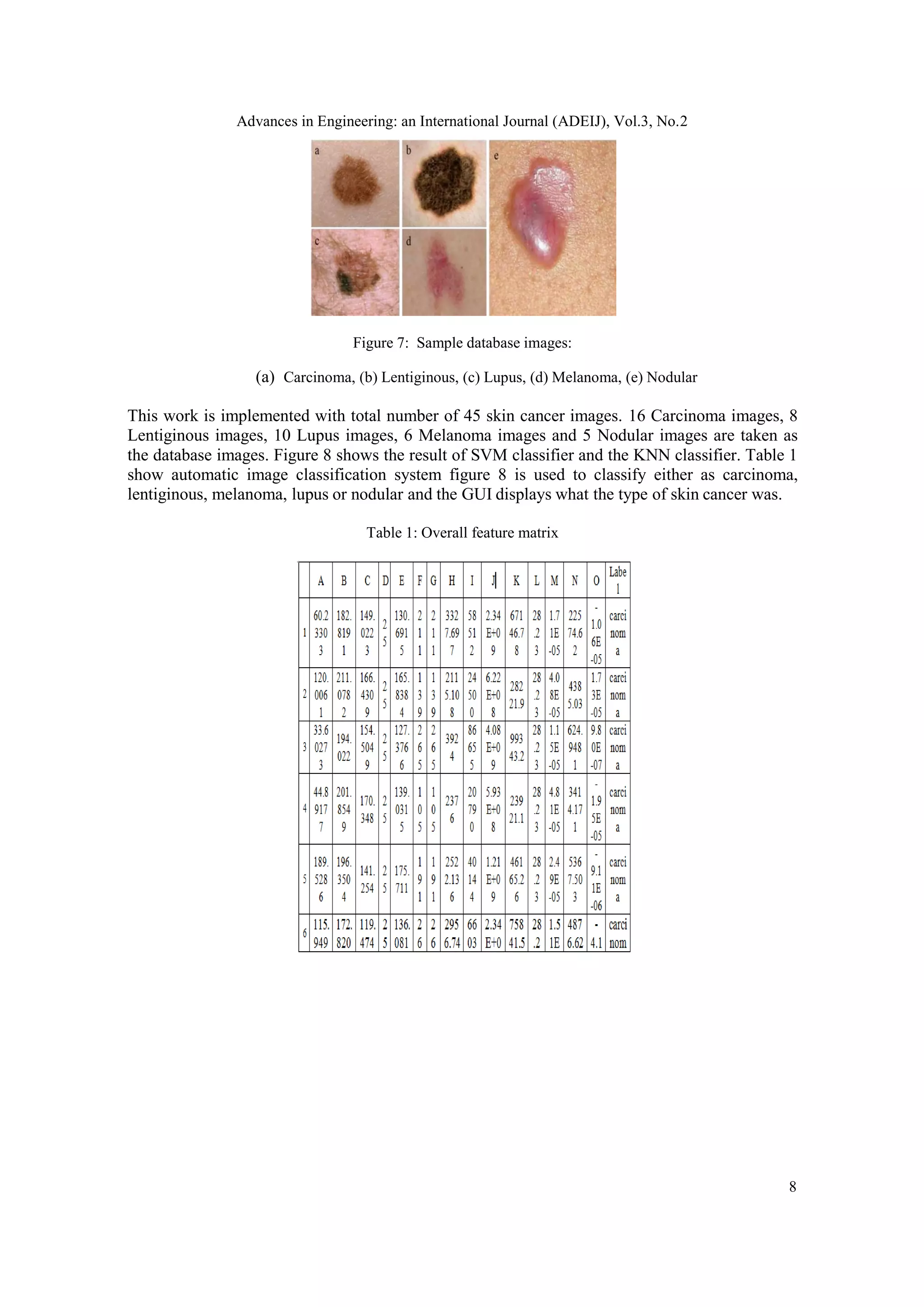
The test database images figure 7 are collected form the online database called university of
Lowa health care. This work constitute the recognition of five different skin cancer disease
automatically. The five disease names are listed over here.
1. Carcinoma
2. Lentiginous
3. Lupus
4. Melanoma
5. Nodular
The five disease images are differing from each other on the way of texture, color and shape. The
following figure depicts the skin diseases[11] which are considered in this work.](https://image.slidesharecdn.com/3221adeij01-210416062635/75/DETECTION-OF-LESION-USING-SVM-7-2048.jpg)
![Advances in Engineering: an International Journal (ADEIJ), Vol.3, No.2
9
Figure 8: Classification results on prepared GUI
6. CONCLUSION
The image processing technique is used to detect and exclude the hair from the dermoscopy
images, preparing it for further segmentation and analysis, resulting in satisfactory classification
results. The proposed automated image analysis process includes image acquisition, hair detect
ion and exclusion, lesion segmentation, feature extraction, and classification. The incidence of
skin cancers has reached a large number of individuals within a given population , especially
among whites, and the trend is still rising. Early detection is vital, especially concerning
melanoma, because surgical excision currently is the only life saving method for skin cancer.
The state of the art is used in the proposed systemfor the dermoscopy image acquisition, which
ensures capturing sharp dermoscopy images with a fixed distance to the skin and consistent
picture quality.
REFERENCES
[1] Rogers HW, Weinstock MA, Feldman SR, Coldiron BM.: “Incidence estimate of nonmelanoma skin
cancer (keratinocyte carcinomas) in the US population”, 2012. JAMA Dermatol 2015; 151(10):1081-
1086.
[2] “Cancer Facts & Figures 2014”. American Cancer Society, 2014.
[3] Siegel, R.L., Miller, K.D., and Jemal, A.: “Cancer statistics, 2016,” CA: A Cancer Journal for
Clinicians, vol. 66, no. 1, pp. 7-30, 2016.
[4] Brady, M.S., Oliveria, S.A., Christos, P.J., Berwick, M., Coit, D.G., Katz, J., Halpern, A.C.: “Patterns
of detection in patients with cutaneous melanoma.” Cancer. 2000 Jul 15;89(2):342-7.
[5] Kittler, H., Pehamberger, H., Wolff, K., Binder, M.: “Diagnostic accuracy of dermoscopy”. In: The
Lancet Oncology. vol. 3, no. 3, pp. 159.165. 2002.
[6] Vestergaard, M.E., Macaskill, P., Holt, P.E., et al.: “Dermoscopy compared with naked eye
examination for the diagnosis of primary melanoma: a meta-analysis of studies performed in a clinical
setting.” Br J Dermatol. Sep 2008;159:669-676.
[7] Bafounta, M.L., Beauchet, A., Aegerter, P., et al.: “Is dermoscopy (epiluminescence microscopy)](https://image.slidesharecdn.com/3221adeij01-210416062635/75/DETECTION-OF-LESION-USING-SVM-9-2048.jpg)
![Advances in Engineering: an International Journal (ADEIJ), Vol.3, No.2
10
useful for the diagnosis of melanoma? Results of a meta-analysis using techniques adapted to the
evaluation of diagnostic tests.” Arch Dermatol. Oct 2001;137:1343-1350.
[8] Abder-Rahman, A.A., Deserno, T.M.,: “A systematic review of automated melanoma detection in
dermatoscopic images and its ground truth data”. In: Proc. SPIE 8318, Medical Imaging 2012: Image
Perception, Observer Performance, and Technology Assessment.
[9] Carli, P., Quercioli, E., Sestini, S., Stante, M., Ricci, L., Brunasso, G., De Giorgi, V.: “Pattern
analysis, not simplified algorithms, is the most reliable method for teaching dermoscopy for
melanoma diagnosis to residents in dermatology”. In: Br J Dermatol. vol. 148, no. 5, pp. 981-4. 2003.
[10] Ganster, H., Pinz, A., Rhrer, R., Wildling, E., Binder, M., Kittler, H.: “Automated Melanoma
Recognition”. In: IEEE Transactions on Medical Imaging, vol. 20, no. 3, 2001.
[11] Al-Masni MA, Al-Antari MA, Choi MT, Han SM, Kim TS: Skin lesion segmentation in dermoscopy
images via deep full resolution convolutional networks. Comput Methods Programs Biomed 162:221-
231,2018
[12] Siegel RL, Miller KD, Jemal A: Cancer statistics, 2019. CA Cancer J Clin 69(1):7-34,2019
[13] Li Y, Shen L: Skin lesion analysis towards melanoma detection using deep learning network. Sensors
18:2,2018
[14] Unver HM, Ayan E: Skin lesion segmentation in dermoscopic images with combination of YOLO
and GrabCut algorithm Diagnostics (Basel) 9(3),2019
[15] Bi L, Feng D, Fulham M, Kim J: Improving skin lesion segmentation via stacked adversarial learning.
In: 2019 IEEE 16th International Symposium on Biomedical Imaging (ISBI 2019), 2019, pp 1100–
1103
[16] Jiang F, Zhou F, Qin J, Wang T, Lei B: Decision-augmented generative adversarial network for skin
lesion segmentation. In: 2019 IEEE 16th International Symposium on Biomedical Imaging (ISBI
2019), 2019, pp 447–450
[17] Khan MA, Javed MY, Sharif M, Saba T, Rehman A: Multi-model deep neural network based features
extraction and optimal selection approach for skin lesion classification. In: 2019 International
Conference on Computer and Information Sciences (ICCIS), 2019, pp 1–7
[18] Stefan Jianu SR, Ichim L, Popescu D: Automatic Diagnosis of Skin Cancer Using Neural Networks.
In: 2019 11th International Symposium on Advanced Topics in Electrical Engineering (ATEE), 2019,
pp 1–4](https://image.slidesharecdn.com/3221adeij01-210416062635/75/DETECTION-OF-LESION-USING-SVM-10-2048.jpg)


![11.[37 46]segmentation and feature extraction of tumors from digital mammograms](https://cdn.slidesharecdn.com/ss_thumbnails/11-37-46segmentationandfeatureextractionoftumorsfromdigitalmammograms-120512235750-phpapp02-thumbnail.jpg?width=640&height=640&fit=bounds)