principles of cardiopulmonary bypass
•Download as PPT, PDF•
140 likes•46,701 views

about heart lung machine

Recommended
More Related Content
What's hot
What's hot (20)
Viewers also liked
Viewers also liked (12)
Similar to principles of cardiopulmonary bypass
Similar to principles of cardiopulmonary bypass (20)
Recently uploaded
Recently uploaded (20)
principles of cardiopulmonary bypass
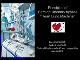
- 1. Principles of Cardiopulmonary bypass “Heart Lung Machine” Ida Simanjuntak Perfusionist Staff National Cardiovascular Center Harapan Kita Agustus 2012
- 2. How it’s work ?
- 4. Definition Cardiopulmonary bypass(CPB) is a form of extracorporeal circulation It temporarily takes over the function of the heart and lungs during surgery, maintaining the circulation of blood and the oxygen content of the body
- 5. Tujuan Umum Cardio Pulmonary By Pass 1. Mempertahankan sirkulasi dan respirasi yang adekuat dengan mengalirkan darah ke suatu sirkuit extracorporal yang berfungsi sebagai jantung dan paru. 2. Menciptakan lapangan operasi yang bersih dari darah. Dengan cara mengalirkan darah keluar jantung dan menghisap darah yang masuk ke jantung, sehingga dokter bedah dapat melakukan koreksi pembedahan/ operasi dengan bebas. ( Jon W. Austin, 1986).
- 6. Componen of Heart Lung Machine Pumps Cannula Reservoir Oxygenator Heat Exchanger Arterial Filter Accessory pump & devices
- 7. PUMP Roller Pump Centrifugal pump
- 8. Roller versus centrifugal pump Roller pump Centrifugal pump Description Nearly occlusive Non occlusive After load independent After load sensitive Advantages Low prime volume Portable, position insensitive Low cost Safe positive and negative pressure No potential for backflow Adapts to venous return Shallow sine-wave pulse Superior for right or left heart bypass Preferred for long-term bypass Protects against massive air embolism Disadvantages Excessive positive and negative pressure Large priming volume Spallation Requires flow meter Tubing rupture, hemolysis Potential passive backward flow Potential for massive air embolism Higher cost Necessary occlusion adjustments
- 10. Vena Cannulas VENOUS CANNULAS AND CANNULATION Three basic approaches for central venous cannulation are used: bicaval, single atrial, or cavoatrial ("two stage") At times, venous cannulation is accomplished via the femoral or iliac vein. This either open or percutaneous cannulation is used for emergency closed cardiopulmonary assist, for support of particularly ill patients,reoperations. Single Canul /Two Stage Double Canul ( SVC, IVC Canul )
- 13. Femoral Cannula Aortic: single outlet hole Venous: multiple inlet holes
- 15. Different ports and Manifold
- 16. BUBBLE DETECTOR can efficiently detect air embolisms between 10 and 250 μm in diameter, as well as simultaneously monitoring for micro bubbles
- 17. FILTERS
- 18. PRINCIPLES OF VENOUS DRAINAGE Venous blood usually enters the circuit by gravity or siphonage into a venous reservoir placed 40 to 70 cm below the level of the heart. “AUGMENTED OR ASSISTED VENOUS RETURN”
- 19. 19
- 20. CARDIOPLEGIA Antegrade cardioplegia is delivered through a small cannula in the aortic root or via handheld cannulas directly into the coronary ostia when the aortic valve is exposed. Pressure Antegrade 150-200 mmHg (Perfusion) 50-100 mmHg (Monitor) Antegrade Cannula
- 21. Retrograde cardioplegia is delivered through a cuffed catheter inserted blindly into the coronary sinus. Proper placement of the retrograde catheter is critical, but not difficult, and is verified by palpation, TEE, color of the aspirated blood, or pressure waveform of a catheter pressure sensor. Complications of retrograde cardioplegia include rupture or perforation of the sinus, hematoma, and rupture of the catheter cuff Retrograde Pressure Retrograde Cannula 100-150 mmHg (Perfusion) 30-50 mmHg (Monitor)
- 23. (A) Aortic root vent, which can also be Vent Cannula used to administer cardioplegic solution after the ascending aorta is clamped. (B) A catheter placed in the right superior pulmonary vein/left atrial junction can be passed through the mitral valve into the left ventricle. (C) Direct venting of the left ventricle at the apex. (D) Venting the main pulmonary artery, which decompresses the left atrium because pulmonary veins lack valves.
- 24. Oxygenator Oxygenation Two types of oxygenators are in current use: the bubble oxygenator and the more widely used mem- brane oxygenator. Both types usually have an integral heat exchanger to control the temperature of the blood Membrane oxygenator with integral venous reservoir
- 25. Oxygenator Studies have shown that membrane oxygenators are less traumatic to blood components (e.g., platelets) and cause less blood loss and protein denaturization than bubble oxygenators (van Oeveren et al. 1985; Hill et al. 1985). Membrane oxygenators also provide sepa- rate control of oxygen and carbon dioxide, which is more difficult to obtain with bubble oxygenators. The indirect blood/gas interface also reduces the occurrence of microemboli (Toner et al. 1997). Furthermore, mem- brane oxygenators require lower priming volumes and eliminate the need for defoaming devices or antifoam agents. However, despite the current preference for membrane oxygenators over bubble oxygenators, the effect of oxygenator type on clinical outcome is not completely certain. Although there is evidence that membrane oxygenators can reduce cerebral injury dur- ing cardiopulmonary bypass (Toner et al. 1997
- 26. HEMOFILTRATION Untukmengurangi Hemodelusi Filtrasi Cairan, faktor inflamasi, hiperkalemia atau azotemia Diintegrasikan dengan sirkuit secara hati2 dan bebas Bubble
- 29. Pre-Bypass 1.Begins with the posting of the operating schedule Perfusionist must assemble specific information about the scheduled procedure Specific information about the scheduled procedure : Surgeon, patient’s data, diagnoses, procedure, time of operation 2. .Review of the patient’s hospital chart Information is recorded on the perfusion record
- 30. 3.Selection of the disposable equipment and perfusion circuit using existing protocols 4.Assembly of the cardiopulmonary bypass circuit 5. Calculation of BSA, BV, cardiac indeks and blood flow 6. Size of cannulae 7. Drug dose l and laboratories 8. Predicted hemoglobin and hematocrits 9. Setting up the HLM & oxygenator 10. Priming the oxygenator 11. Initiating CPB 12. Saffety device on 13. Ice
- 32. Dr Gibbon’s early heart/lung machine Gibbon JH et al. Arch Surg 1937; 34:
- 33. Priming Filling the CPB circuit with blood or blood substitutes after CO2 Flushing Result in hemodilution
- 34. Hemodilution Pt’s Blood Volume Predicted Hct Pre-CPB IV + CPB prime volume Target: < 30% at BT below 30℃ < 25% when BT below 25℃ not below 20%
- 35. Hindari Hct intra CPB < 18 % Hct Untuk memastikan Hct ketika inisiasi CPB: Hctint = initial Hct on CPB EBV = estimated patient blood volume Hct = preoperative Hct Jika diperlukan penambahan RBC maka bisa dikalkulasi dengan : PBV = patient’s blood volume ECCV = extracorporeal circuit volume CPBHct = desired Hct on CPB PtHct = patient’s pre CPB Hct
- 36. Initiating CPB “Lines down” connects between table lines & pump lines (in a sterile manner) Debubble Surgeon : “Heparin in” Anesthesiologist give heparin ACT check. “Speed up (speedy)” fast circulating the priming solutions, make sure no bubble exist. “Stop” debubbling stopped, venous lines clamped. Surgeons prepare to do cannulation ACT > 300 sec Pump suckers on
- 38. Insert drugs and manitol Resirculated of the priming solution Oksigen on Before cannulation of the aortic cannula, surgeon will ask the perfusionist to roll forward, to fill in the tubing with priming solution and to make sure no bubble exist. Reply : “Forward”.. After the aortic cannula is unclamp, surgeon : “Open to you”. Reply : “Open/Ok”, check the pressure fluctuation on the pressure module of the pump. Inform surgeon. Feel for pulsation the arterial line tubing ACT > 480 ready to on bypass
- 40. Continous Monitoring During CPB Reservoir level Blood flow at proper rate/flow rate Pressure line/arterial line pressure Blood pressure/patient’s arterial pressure 50-90 mmHg Oxigen saturation Temperature appropriate ECG Venous oksigen saturation 65%-75%
- 41. Monitoring Blood pressure MAP: in mild to moderate hypothermia normal adult: 60-70 mmHg adult with CAD, DM, and old age: > 60mmHg infants: > 60mmHg CVP: approximate 0 mmHg
- 42. Pump Flow Rate In the normal body temperature adult: 2.2~2.8 L /m2 . min infant: 2.6~3.2 L /m2 . min In hypothermia adult: 1.6~2.2 L /m2 . min Infant: 2.0~2.4 L /m2 . min Adjust according MAP and SvO2
- 43. Monitoring pressure Causes of aortic cannula high line pressure 1. Kink in arterial cannula or line 2. Cannula improperly positioned 3. Clamp too near cannula 4. Cannula to small 5. Arterial systemic blood pressure very high 6. Aortic disection 7. Blockage in arterial filter
- 44. Monitoring Venous Drain Causes of Poor Venous return
- 45. Intermittent Monitoring During CPB blood gas Urine output minimal 0,5-1 ml/kgBB/jam electrolit ACT > 480 sec
- 46. Monitoring Devices Monitoring secara kontinue : SVO2, Suhu vena, Hct ACT > 480 sec Cek ACT dan AGD setiap 30 – 60 menit jika stabil
- 47. Monitoring Blood Gas Coagulation Status and Laboratory Data Menggunakan ACT untuk evaluasi status koagulasi Hb 7,0 – 9,0 gr% Ht 20 – 30 % pO2 arterial AGD : 140-180 mmHg pCO2 arterial AGD : 31 – 45 mmHg BE (-2,5) – (+ 2,5)
- 48. Monitoring Urine Output Urinary volume and renal function Dipengaruhi waktu bypass dan gagal ginjal sebelumnya Volume urine 0,5-1 mL/kg/jam Oligouria / normal + hiperkalemia, hemoglobinemia, hemodilusi berlebihan = indikasi diuretik
- 49. Causes of Urine Production 1. Kinked or disconnected Foley catheter or tubing 2. Catheter with tip obstructed by gel 3. Decreased blood pressure 4. Low pump flows 5. Fluid moving to interstitial space Corrective Action 1. Straighten or connect tubing 2. Push on bladder 3. Give vasopressor 4. Increase flows 5. Use mannitol or lasix
- 50. Hypothermia Advantages: decrease metabolic rate, oxygen requirement decrease rate of degradative reactions, increase tolerance to ischemia reduces K+ necessary for cardiac arrest inhibits intracellular Ca2+ accumulation
- 51. Hypothermia Monitoring: Core temperature: nasopharyngeal or tympanic membrane probes reflect brain temperature Shell temperature: rectal probe or skeletal muscle needle sensor reflect relatively pooly perfused tissues of most of the body’s mass
- 52. Temperature Cardiac Index FIO2 Gas/Blood Flow Ratio 37 C 2,4 L 0,80 1:1 34 C 2,2 L 0,70 0,8 : 1 30 C 2,0 L 0,65 0,7 : 1 28 C 1,8 L 0,60 0,6 : 1 22 C 1,6 L 0,50 0,5 : 1
- 53. Termination of CPB Preparing for Separation (Rewarming) Hipotermia sedang (25-30°C) digunakan untuk memperlambat rewarming. Hipotermia berat (16- 25°C) + circulatory arrest : operasi defek kongenital atau rekonstruksi arkus aorta Kriteria rewarm : naso 37°C, bladder/rectal 35°C atau jempol kaki 30°C Rewarm yang inadekuat mengakibatkan penurunan suhu pasien 2-3°C pasca CPB sampai tiba di ICU mengigil, ↑VO2, gangguan irama jantung, ↑ PVR
- 54. Termination of CPB LAMPS Laboratory data pH, pCO2 darah arteri Acidosis depressant fungsi myocardial, gangguan obat inotropic SvO2, Ht, ACT, konsentrasi heparin Na, K, Ca, HyperK >6 mEq/L (gangguan konduksi, AV blok) HypoK (gangguan irama ventrikel dan atrial) HypoCa akibat hemodilusi, albumin atau produk darah (+sitrat) CaCl2 3-5 mg/kg (memperbaiki kontraksi miocardial dan PVR) Glucosa darah insulin 10-20 unit iv + glukosa prn
- 55. Termination of CPB Anastesia/Machine Analgesia – supplemental opioid Amnesia – benzodiazepine Muscle relaxant – prn Airway and functional oxygen delivery system Anastesia machine on, Adequate oxygen supply Breathing circuit intact, ETT connected Ventilator functional, Ability to ventilate both lungs confirmed Vaporizers off (10 menit sebelum terminasi CPB) untuk mengurangi efek depresi sirkulasi dan menghindari depresi myocardial saat dilepas bypass
- 56. Termination of CPB Monitors Invasive BP monitors – zeroed & calibrated Arterial catheter – radial, femoral, aortic PAC, CVC (right atrial), left atrial catheter ECG Kecepatan, irama, konduksi, iskemia (review semua lead) Bladder catheter – urine output Pulse oxymeter Capnometer/mass spectrometer Safety monitor – oxygen analyzer, circuit pressure alarm, spirometer TEE Temperature (37°C nasofaringeal, 35°C rectal/bladder)
- 57. Termination of CPB Patient/Pump The Heart Cardiac function – contractility, size Rhythm, ventricular filling, air removed, vent removed The Lungs Inflation/deflation, compliance The Field bleeding Oxygenation – blood color Movement – sign of inadequate anasthesia
- 58. Termination of CPB Support Pharmacologic Inotropes Vasodilators Vasoconstrictiors Antidysrhythmics Electrical Atrial/Ventricular Pacing Mechanical Intraaortic Balloon Counterpulsation Left and/or right ventricular assist device
- 60. Termination of CPB Separation Technique CPB : (v. cavae oxygenator aorta) Partial bypass : (v. cavae oxygenator + RV/lungs/LV common return to aorta) Off CPB : (v. cavae heart/lungs aorta)
- 61. After Termination of CPB Setelah kanul aorta dilepas, sisa perfusate bisa diproses kedalam kantung intravena sterile untuk kebutuhan transfusi nantinya. Atau dengan alat cell salvage sehingga darah dicuci dahulu sebelum ditransfusi Pemberian protamine pada beberapa pasien mengakibatkan penurunan hemodinamik sementara. Perfusionis harus terus mengobservasi hemodinamik pasien dan menjaga sirkuit CPB tetap dapat digunakan
- 62. Daftar Pustaka http://www.cts.usc.edu/zglossary-heartlungmachine.html http://www.surgeryencyclopedia.com/Fi-La/Heart-Lung-Machines.html Lippincott Williams & Wilkins 2007 Cardiopulmonary Bypass : Principles and Practice Cardiopulmonary Bypass: Principles and Management: Edited by Kenneth M. Taylor. 1998, Baltimore On Bypass ,Advanced Perfusion Techniques Series: Current Cardiac Surgery Mongero, Linda B.; Beck, James R. (Eds.) 2008, XII, 576 p. 173 illus.
- 63. THANK YOU...
Editor's Notes
- The cardiopulmonary bypass is a form of extracoporeal circulation. It takes over the cardiac and respiratory function temporarily during cardiac surgery, in order to making a silent heart for cardiac surgeon to perform complicating procedures.
- This is the initial prototype of Gibbons CPB machine.
- Priming means filling the bypass machine with fluid, preparing for the connection with the patient’s circulation. In the beginning period of cardiopulmonary bypass history, whole blood was used for priming. However, surgeons found that blood substitutes like crystalloid or colloid solutions made even better prognosis. The reasons we’ll discuss later. As the use of non-red blood cell solutions, the hematocrit of the patient falls down during the cardiopulmonary bypass. It’s called hemodilution.
- When we use crystalloid and other solution as priming solution, hemodilution will be made. We should calculated the predicted hematocrit after the cardiopulmonary bypass. The formula is on the screen, the predicted hemotocrit is dividing the total amount of the patient’s original hemoglobin, which could be product of pre-bypass hematocrit and blood volume, with the total volume in the circuit The target hematocrit could less than 30% when body temperature is below 30 degree celsius, less than 25% when body temperature is below 25 degree celsius. Always remind that never let hematocirt below 20% because some study shows that severe impairment of the oxygen-carrying ability would appear in this low hematocrit.
- Blood pressure during cardiopulmonary bypass should be controlled in order to maintain good tissue perfusion. The aim in the normal adult is over 50 mm mercury. As the patient with worse cardiovascular preserve, the aim should be elevated to 60. And the infant’s target should be over 30 mm mercury. The central venous pressure should approximate to zero, if not, there may be some problem with the venous cannulae drainage. If the blood pressure is inadequate, searching for the etiology and correct it. If hypotension, increase the flow rate to restore the adequate blood volume. After that, if the blood pressure is still low, try some vasoconstrictive agent to raise systemic vascular resistance. If hypertension, fentanyl may be given to correct the inadequate anesthetic level.
- The pump’s flow rate during total cardiopulmonary bypass is the same word as the “cardiac output” in the ordinary time. In the normal body temperature, adult should have the flow rate between 2.2 to 2.8 liter per square-meter per minute. The infant has higher metabolic rate so the flow rate should be higher with the same body surface area. In hypothemic state, the flow may be lower, due to lower metabolic needs. The perfusionist would adjust the flow rate according to the patient’s blood pressure. Higher flow rate leads to higher blood pressure, but the blood cell damage also increases. And according to venous oxygen saturation, which reflects the tissue oxygen extraction ratio.
- K ink in the venous line or cannula Airlock in the venous line Oxygenator or venous reservoir is not positioned low enough Noncardiac suction being used instead of pump suckers
- Hypothermia would be induced during cardiopulmonary bypass surgery. It has several advantages. First, the cells’ metabolic rate and oxygen requirements would decrease in the hypothermic state. Second, cells’ degradation rate decreases and the tolerance of ischemia increases. Third, hypothermia also reduces potassium need for arresting heart and inhibits intracellular calcium accumulation.
- At least two temperature probes should be set. One at nasopharyngeal or tympanic membrane measures the core temperature, which reflect the central, or brain temperature. The other is set at rectal or in the skeletal muscle, which reflects the temperature of peripheral body mass, called shell temperature.