Downloaded 327 times


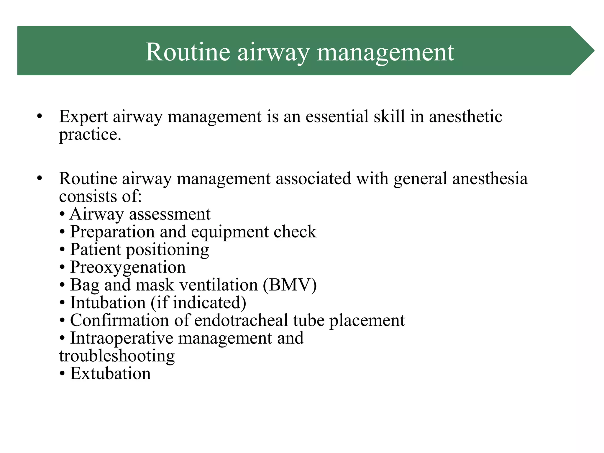


This document provides information on routine airway management techniques for general anesthesia. It discusses airway assessment, equipment, patient positioning, preoxygenation, intubation, tube placement confirmation, and extubation. Difficult airway management techniques are also reviewed, including use of video laryngoscopes, fiberoptic intubation, supraglottic airway devices, surgical airways, and cricothyroidotomy. Factors that increase airway difficulty and algorithms for managing difficult airways are described.


![Anesthesia_for_Airway_management_and_Difficult_airway_Algorithmss[1][1].pptx](https://cdn.slidesharecdn.com/ss_thumbnails/anesthesiaforairwaymanagementanddifficultairwayalgorithmss11-250722203414-f94048f1-thumbnail.jpg?width=640&height=640&fit=bounds)


