1. Cme onCme on cardiaccardiac
arrhythmiaarrhythmia
- Dr. Chintan Parmar → Asst. Prof. → Dept. of Physiology,
- KIMS & RF → Dt. 02/01/2015
2. Sinus (Sinoatrial) Node ActionSinus (Sinoatrial) Node Action
PotentialPotential
- self-excitation - automatic rhythmical
discharge and contraction
- “RMP” of the sinus nodal fiber has a
negativity of about -55 to -60
millivolts.
3. SA Node APSA Node AP
- Because of the high Na ion concentration in the
ECF outside the nodal fiber, as well as a moderate
number of open Na channels,
- +ve Na ions from outside the fibers normally tend to
leak to the inside
4. SA Node APSA Node AP
- the Na-Ca channels become inactivated
within about 100 to 150 milliseconds after opening
- greatly increased numbers of K channels
open
5.
6.
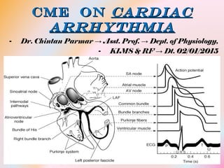
7. Conductive SystemConductive System
- sinus node (sinoatrial or S-A node), in which the
normal rhythmical impulse is generated;
- the internodal pathways that conduct the impulse
from the sinus node to the atrioventricular (A-V) node;
- the A-V node, in which the impulse from the atria is
8.
9. Sinus (Sinoatrial) NodeSinus (Sinoatrial) Node
- It is located in the superior posterolateral
wall of the right atrium immediately below
and slightly lateral to the opening of the superior
vena cava.
- The fibers of this node have almost no
contractile muscle filaments
10. Internodal PathwaysInternodal Pathways
- The ends of the sinus nodal fibers connect
directly with surrounding atrial muscle fibers.
- the action potential spreads through the entire
atrial muscle mass to the A-V node
- anterior interatrial band, passes through the
11. Atrioventricular NodeAtrioventricular Node
- cardiac impulse does not travel from the atria into
the ventricles too rapidly;
- this delay allows time for the atria to empty
their blood into the ventricles before ventricular
contraction begins
12. A–V DelayA–V Delay
- impulse, after traveling through the internodal pathways,
reaches the A-V node about 0.03 second after its
origin in the sinus node.
- Then there is a delay of another 0.09 second in the A-V
node itself before the impulse enters the penetrating
portion of the A-V bundle, where it passes into the
ventricles.
13.
14. Cause of the Slow ConductionCause of the Slow Conduction
- The slow conduction in the nodal and
penetrating A-V bundle fibers is caused mainly
by,
- diminished numbers of gap junctions
between successive cells in the conducting
pathways,
15. One-Way ConductionOne-Way Conduction
- A special characteristic of the A-V bundle
- inability of action potentials to travel backward
from the ventricles to the atria
16. Bundle BranchesBundle Branches
- distal portion of the A-V bundle passes downward in the
ventricular septum toward the apex of the heart
- Then the bundle divides into left and right bundle
branches that lie underneath the endocardium on the 2
respective sides of the ventricular septum
- Each branch spreads downward toward the apex of the
17. Ventricular Purkinje SystemVentricular Purkinje System
- Purkinje fibers lead from the A-V node through the A-
V bundle into the ventricles
- They are very large fibers, even larger than the
normal ventricular muscle fibers,
- they transmit action potentials at a velocity of
19. Ventricular MuscleVentricular Muscle
- the cardiac impulse does not travel directly
outward toward the surface of the heart
- Transmission from the endocardial
surface to the epicardial surface of the
ventricle requires another 0.03 second
20.
21.
22. Why SA Node asWhy SA Node as
Pacemaker ???Pacemaker ???- The Sinus Node as the Pacemaker - 70 to 80
times per minute
- The A-V nodal fibers discharge at an intrinsic
rhythmical rate of 40 to 60 times per minute,
23. ““Ectopic” PacemakerEctopic” Pacemaker
- Sometimes some other part of the heart develops a
rhythmical discharge rate that is more rapid than that of
the sinus node.
- sometimes occurs in the A-V node or in the Purkinje
fibers
24. ““Ectopic” PacemakerEctopic” Pacemaker
- When A-V block occurs — that is, when the
cardiac impulse fails to pass from the atria
into the ventricles through the A-V nodal and
bundle system,
- the atria continue to beat at the normal rate of
rhythm of the sinus node,
25. ““Ectopic” PacemakerEctopic” Pacemaker
- After sudden A-V bundle block, the Purkinje
system does not begin its intrinsic rhythmical
impulses until 5 to 20 seconds later because,
- before the blockage, in a suppressed state
- the ventricles fail to pump blood - person faints
because of lack of blood flow to the brain
26.
27. Parasympathetic (Vagal)Parasympathetic (Vagal)
StimulationStimulation- Acetylcholine ↓ the rate of rhythm of the sinus node
- It ↓ the excitability of the A-V junctional fibers
between the atrial musculature and the A-V node -
slowing transmission
- Weak to moderate vagal stimulation slows the rate of
28. Mechanism of the Vagal EffectsMechanism of the Vagal Effects
- Ach greatly increases the permeability of the
fiber membranes to K ions,
- rapid leakage of K out of the conductive fibers
→ increased negativity inside the fibers →
hyperpolarization → makes this excitable
tissue much less excitable
29. Mechanism of the Vagal EffectsMechanism of the Vagal Effects
- the initial rise of the sinus nodal
membrane potential caused by inward Na
and Ca leakage requires much longer to reach
the threshold potential for excitation → greatly
slows the rate of rhythmicity
- In the A-V node, a state of hyperpolarization
delays conduction of the impulse,
30. Effect of SympatheticEffect of Sympathetic
StimulationStimulation- it increases the rate of sinus nodal
discharge
- it increases the rate of conduction as well as
the level of excitability in all portions of the
heart
- it increases the force of contraction of all
the cardiac musculature
31. Mechanism of the SympatheticMechanism of the Sympathetic
EffectEffect- Norepinephrine increases the permeability of
the fiber membrane to Na and Ca ions.
- In the sinus node, an increase of Na-Ca
permeability causes a more positive resting
potential
32. Mechanism of the SympatheticMechanism of the Sympathetic
EffectEffect- In the A-V node and A-V bundles, increased
Na-Ca permeability makes it easier for the
action potential to excite each succeeding
portion of the conducting fiber bundles,
- decreasing the conduction time from the
atria to the ventricles