
Recommended
More Related Content
Similar to Cardiac impulse conduction and innervation.pptx
Similar to Cardiac impulse conduction and innervation.pptx (20)
Recently uploaded
Recently uploaded (20)
Cardiac impulse conduction and innervation.pptx
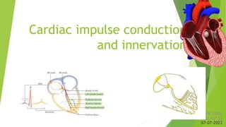
- 1. Cardiac impulse conduction and innervation ATIK GOEL ROLL NO 33 07-07-2023
- 2. SESSION OBJECTIVES Pacemakers of heart Electrical conduction system of heart Time delay in transmission Conduction velocities of various conduction fibres Innervation of heart
- 3. Introduction Cardiac electrophysiology : All processes involved in activation of heart , action potential , conduction system and effects of ANS.
- 4. Heart has 2 special systems for 1. Cardiac impulse generation - pacemakers 2. Impulse conduction This allows atria to contract 1/6th second ahead of ventricle and all ventricle to contract simultaneously
- 5. Pacemaker Generally S A node is pacemaker. It is having highest rate of firing 70 -80 beats /min. If SA node fails : Pacemakers are Av node (40- 60Beats/min) Bundle of his(about 40 beats/min) Purkinje fibres (15-40 beats/min)
- 6. SA node Flattened ellipsoid specialized cardiac muscle 15mm long 3 mm wide 1mm thick. Location : Immediately below and lateral to opening of superior vena cava Normal Pacemaker of heart. It develops from right side of embryo and innervated by right vagus
- 7. Av node Shows AV nodal delay of 0.1 sec. AV nodal delay has following significance : Preventing excess transmission of impulses by SA node . Gives time for atria to contract fully before ventricular depolarisation Located near opening of coronary sinus Conducts Impulses from SA node Defect in it’s conduction causes heart blocks
- 8. Bundle of his and purkinje fibres BUNDLE OF HIS Derived from AV nodal cells They divide into 2 segments and lies on inter ventricular septum They are isolated in a canal till they form purkinje fibres PURKINJE FIBRES They penetrate the ventricular wall Their fast conduction velocity allows ventricle to contract almost altogether
- 10. Pathway of transmission SA NODE INTERNODAL PATHWAY AV NODE BUNDLE OF HIS PURKINJE FIBRES
- 12. SA NODE INTERNODAL TRACTS AV NODE BUNDLE OF HIS PURKINJE FIBRES
- 13. SA NODE INTERNODAL TRACTS AV NODE BUNDLE OF HIS PURKINJE FIBRES Special pathway in arterial wall that transmit impulse from SA to AV node. It has 3 parts Anterior bachman Middle wenckebach Posterior thorel Impulse from sa node can travel directly to atrial muscle fibres
- 14. SA NODE INTERNODAL TRACTS AV NODE BUNDLE OF HIS PURKINJE FIBRES
- 15. SA NODE INTERNODAL TRACTS AV NODE BUNDLE OF HIS PURKINJE FIBRES
- 16. SA NODAL PATHWAY INTERNODAL TRACTS AV NODE BUNDLE OF HIS PURKINJE FIBRES
- 19. CONDUCTION RATE TISSUE CONDUCTION SPEED (m/sec) SAN 0.05 ATRIAL PATHWAY 1 AVN 0.02-0.05 BUNDLE OF HIS 1 PURKINJE SYSTEM 4 VENTRICULAR SYSTEM 1 CONDUCTION VELOCITY DEPENDS ON 1. Diameter of fibre 2. Number of gap junctions
- 20. INNERVATION SYSTEM OF HEART BY VAGUS AND SYMPATHETIC NERVOUS SYSTEM
- 21. VAGUS (Parasympathetic) effects Acetylcholine released at vagal endings increases permeability of fibres to k+ ions. This causes increased negativity inside the fibers called hyperpolarization This makes tissue less excitable Membrane potential of SA nodal fibers decreases to -65 to -75 mv from -55 to -60mv. Therefore initial rise of sinus nodal membrane potential caused by Na and Ca leakage requires more time to reach threshold. They are constantly active, producing a rhythm of 60 – 80 beats per minute. If the vagus nerve was lesioned, the resting heart rate would be around 100 beats per minute. Summary: acetylcholine binds on to M₂ receptors, which act to decrease the slope of the pacemaker potential. This leads to a decrease in heart rate negative chronotropic effect
- 22. SYMPATHETIC EFFECT The sympathetic input into the heart is via the postganglionic fibres from the sympathetic trunk which innervate SA node AV NODE and ventricles mainly. The postganglionic fibres release noradrenaline, which acts on B₁ adrenoreceptors to increase the slope of the pacemaker potential. By increasing permeability to na+ and ca++ ions. This increases the heart rate (a positive chronotropic effect by ca++), as well as the force of contraction (positive inotropic effect).
- 23. Thank you