
Downloaded 2,072 times
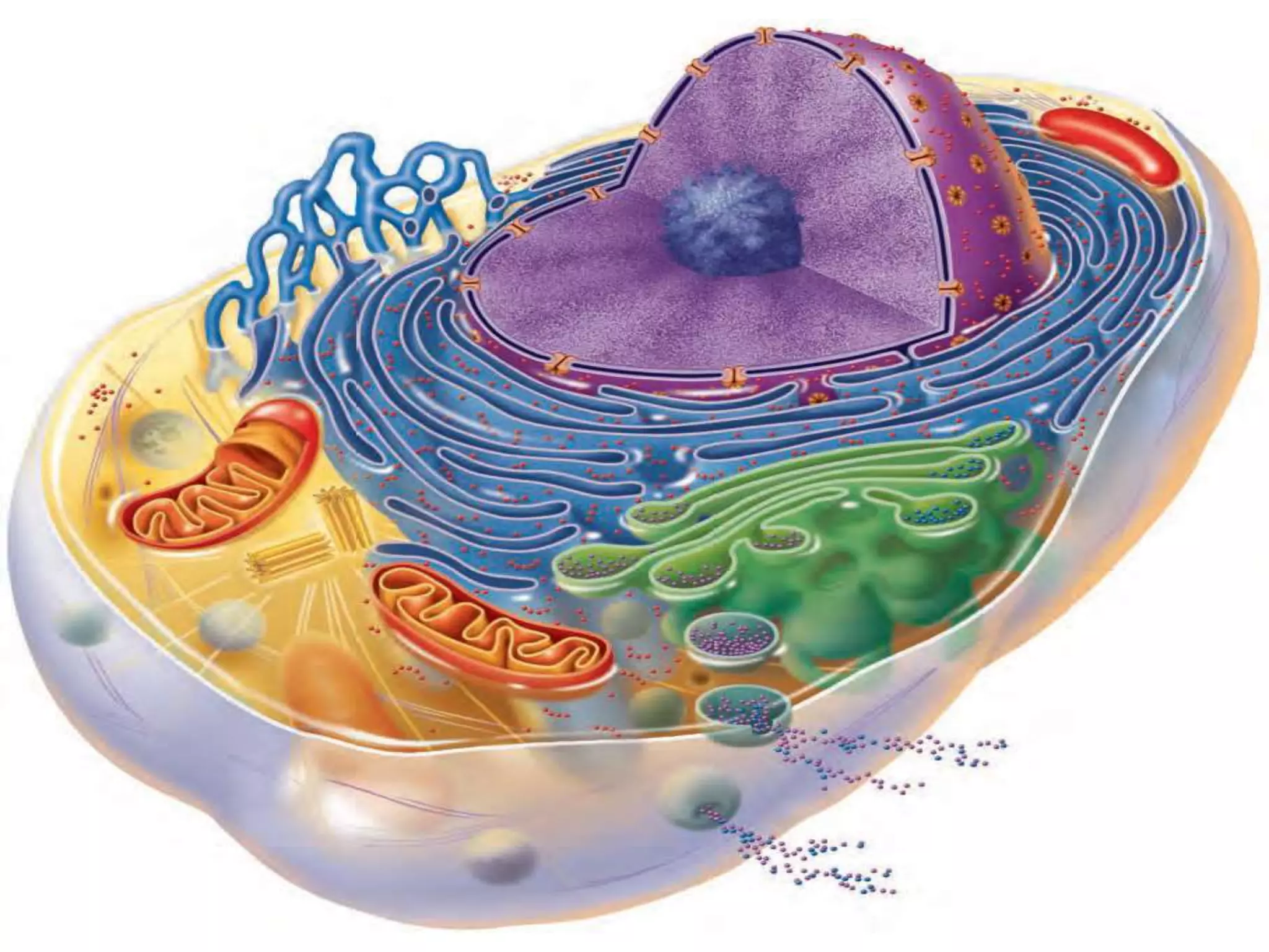


The document summarizes key aspects of cell structure and function. It describes that cells have three main parts - the plasma membrane, cytoplasm, and nucleus. The plasma membrane encloses the cell and regulates what enters and exits. The cytoplasm contains organelles that carry out metabolic functions, and the nucleus houses genetic material and controls cellular activities. Specific organelles like mitochondria, ribosomes, and the endoplasmic reticulum are discussed in detail. The mechanisms of passive transport like diffusion and osmosis, as well as active transport processes, are also summarized.


