
Recommended
More Related Content
What's hot
What's hot (20)
Viewers also liked
Viewers also liked (20)
Similar to What is a Positron Emission Tomography?
Similar to What is a Positron Emission Tomography? (20)
Recently uploaded
Recently uploaded (20)
What is a Positron Emission Tomography?
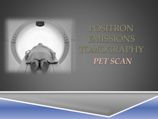
- 1. POSITRON EMISSIONS TOMOGRAPHY PET SCAN
- 2. What is a Pet Scan? • Nuclear 3-D imaging test that uses a radioactive substance called a tracer to look for disease in the body. • Shows how organs and tissues are working at a molecular and cellular level. Scan is non-invasive, but does involve exposure to ionizing radiation. • Best known for its role in detecting cancer imaging.
- 3. How Do Pet Scans work? • A small amount of a radioactive sugar molecule, 18 fluoro-2- deoxyglucose (FDG), is injected into the bloodstream (can also be inhaled as gas or swallowed in pill form). • A PET Scan is used to detect and generate images that indicate areas of high FDG uptake. • Many cancers require more energy than normal cells, and the FDG tracer accumulates in these cells. • This allows cancers to be seen on the Pet images as hot spots.
- 5. Pet scan of a patient showing wide spread of cancer metastasis. A 61-year-old woman with metastasis of breast cancer to the left supraclavicular lymph node
- 6. Scan of a healthy child's brain. Scan of an abused child’s brain
- 7. PET SCAN COMMON USES
- 8. Conditions • Epilepsy • Alzheimer’s Disease • Dementia • Cancer • Heart Disease • Medical Research
- 9. PET SCAN ADVANTAGES • Unlike CT or MRI scans, PET scans can measure cellular-level metabolic changes occurring in an organ or tissue (early stage detection). • CT’s and MRI’s cannot detect changes until the disease has already began to cause changes or damage in the structure of organs or tissues.
- 10. PATIENT COST & TIME $3,000 to $6,000 per scan Takes 2 to 4 hours
- 11. LEARNING OBJECTIVES Select slide transitions Create title slide and text slide with multi-level bulleted lists Insert clips and pictures into a slide with or without content placeholders Bold and italicize text Insert a picture to create background Format slide background Change font and add shadow Apply effects to a shape Print presentation as a handout Align paragraph text Delete and move placeholders Check for spelling errors
- 12. REFERENCES Joseph, U. A. (June 01, 2012). Positron Emission Tomography. Journal of Nuclear Medicine, 53, 6, 1002-1003. Kumar, R., Halanaik, D., & Malhotra, A. (January 01, 2010). Clinical applications of positron emission tomography-computed tomography in oncology. Indian Journal of Cancer, 47, 2.) Politis, M., & Piccini, P. (January 01, 2012). Positron emission tomography imaging in neurological disorders. Journal of Neurology, 259, 9, 1769-80. Society of Nuclear Medicine and Molecular Imaging. (2014). Pet scans: Get the facts. Retrieved from: http://interactive.snm.org/index.cfm?PageID=7988
- 13. POSITRON EMISSIONS TOMOGRAPHY PET SCAN