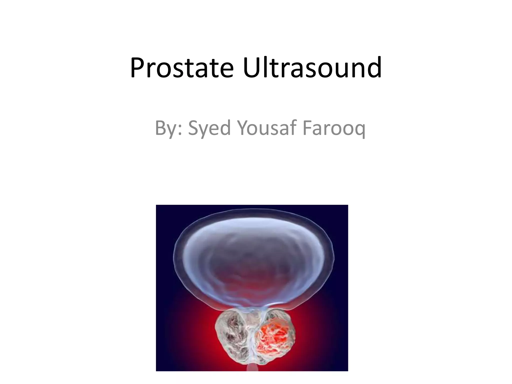
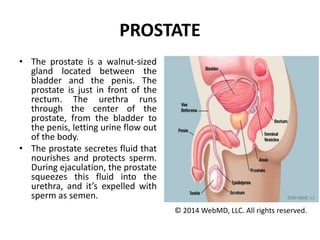
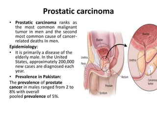
Prostate Ultrasound
By: Syed Yousaf Farooq
PROSTATE
• The prostate is a walnut-sized
gland located between the
bladder and the penis. The
prostate is just in front of the
rectum. The urethra runs
through the center of the
prostate, from the bladder to
the penis, letting urine flow out
of the body.
• The prostate secretes fluid that
nourishes and protects sperm.
During ejaculation, the prostate
squeezes this fluid into the
urethra, and it’s expelled with
sperm as semen.
© 2014 WebMD, LLC. All rights reserved.
Sonographic Features of Prostate
Ultrasound
• Best assessed with transrectal ultrasound.
• Some zonal anatomy distinguishable.
• Outer gland (central and peripheral zones) -
uniform low echogenicity but usually more
echogenic than the inner gland.
• 30 mL is a commonly used upper limit for
normal volume.
Scanning Technique
Transrectal Ultrasound:
• It is ideal to have a small amount of urine in the bladder.
• Ask the patient to try and relax and "bear down" to open the
sphincter as the transducer is inserted slowly. Ensure the
transducer has a latex free dedicated probe cover with plenty of gel.
The highest frequency sector probe 7-12MHz should be used.
• The scanning begins in the axial plane. The seminal vesicles are
examined initially. As the probe is angled caudally the base of the
prostate is seen.
• Once the prostate is examined in its entirety in this plane the probe
is turned 90degrees in a sagittal plane. The probe is angled from
one side across to the other.
• A volume is taken by measuring height x length in the sagittal plane
and x width in the axial plane and multiply by 0.52.
• Look for changes in the contours and echogenicity in each zone.
TRANSABDOMINAL TECHNIQUE:
• The patient lies supine. The patient should
have a half full bladder .500 mls of water 1 hr
before the scan if possible is recommended.
• The probe is angled approximately 30 degrees
caudal using the bladder as a window. Slight
compression to ensure the inferior portion of
the prostate is not obscured by the shadow
artifact from the base of the bladder.
Common Pathology
• Cysts
• Benign Prostatic Hyperplasia (BPH)
• Prostate Carcinoma
• Prostatitis
• Enlarged seminal vesicles
• Stones in the seminal vesicles, Prostate or
ejaculatory ducts.
Prostate cystic disease
• Prostatic cysts are
common, and ~5-8%
men will develop
one. However they
are much more
common in patients
being investigated
for infertility, with
one study showing a
20% prevalence .
Benign prostatic hyperplasia:
• Benign prostatic hyperplasia
(BPH) or benign prostatic
enlargement (BPE) is an extremely
common condition in elderly men
and is a major cause of bladder
outflow obstruction.
Epidemiology:
• By the age of 60, 50% of men have
BPH, and by 90 years of age the
prevalence has increased to 90%.
As such it is often thought of
essentially as a 'normal' part of
aging.
• DISEASE OCCURRENCE IN
PAKISTANI POPULATION:
Pakistan with as many as 50% of the 2
million men older than 65 year are at
risk of bladder outlet obstruction
from BPH.
Sonographic Features
• There is an increase in volume of
the prostate with a calculated
volume exceeding 30 mL (width x
height x length x 0.52).
• The central gland is enlarged, and
is hypoechoic or of mixed
echogenicity.
• Calcification may be seen both
within the enlarged gland as well
as in the pseudocapsule
(representing compressed
peripheral zone).
• Post-micturition residual volume is
typically elevated
• Associated bladder wall
hypertrophy and trabeculation
due to chronically elevated filling
pressures.
Prostatic carcinoma
• Prostatic carcinoma ranks as
the most common malignant
tumor in men and the second
most common cause of cancer-
related deaths In men.
Epidemiology:
• It is primarily a disease of the
elderly male. In the United
States, approximately 200,000
new cases are diagnosed each
year.
• Prevalence in Pakistan:
The prevalence of prostate
cancer in males ranged from 2 to
8% with overall
pooled prevalence of 5%.
Sonographic Features
• On ultrasound,
prostate cancer is
usually seen as a
hypoechoic lesion
(60-70%) in the
peripheral zone of
the gland, but can
be hyperechoic or
isoechoic (30-40%
of lesions).
Prostatitis
• Prostatitis refers to an
infection or inflammation
of the prostate gland that
presents as several
syndromes.
Ultrasound:
• Focal hypoechoic region in
the peripheral zone of the
gland. Discrete fluid
collection suggests abscess
formation. Color Doppler
ultrasound demonstrates
increased flow in the
periphery of the abscess.
Prostate ultrasound (basic)

Prostate ultrasound (basic)

  • 1.
  • 2.
    PROSTATE • The prostateis a walnut-sized gland located between the bladder and the penis. The prostate is just in front of the rectum. The urethra runs through the center of the prostate, from the bladder to the penis, letting urine flow out of the body. • The prostate secretes fluid that nourishes and protects sperm. During ejaculation, the prostate squeezes this fluid into the urethra, and it’s expelled with sperm as semen. © 2014 WebMD, LLC. All rights reserved.
  • 3.
    Sonographic Features ofProstate Ultrasound • Best assessed with transrectal ultrasound. • Some zonal anatomy distinguishable. • Outer gland (central and peripheral zones) - uniform low echogenicity but usually more echogenic than the inner gland. • 30 mL is a commonly used upper limit for normal volume.
  • 4.
    Scanning Technique Transrectal Ultrasound: •It is ideal to have a small amount of urine in the bladder. • Ask the patient to try and relax and "bear down" to open the sphincter as the transducer is inserted slowly. Ensure the transducer has a latex free dedicated probe cover with plenty of gel. The highest frequency sector probe 7-12MHz should be used. • The scanning begins in the axial plane. The seminal vesicles are examined initially. As the probe is angled caudally the base of the prostate is seen. • Once the prostate is examined in its entirety in this plane the probe is turned 90degrees in a sagittal plane. The probe is angled from one side across to the other. • A volume is taken by measuring height x length in the sagittal plane and x width in the axial plane and multiply by 0.52. • Look for changes in the contours and echogenicity in each zone.
  • 5.
    TRANSABDOMINAL TECHNIQUE: • Thepatient lies supine. The patient should have a half full bladder .500 mls of water 1 hr before the scan if possible is recommended. • The probe is angled approximately 30 degrees caudal using the bladder as a window. Slight compression to ensure the inferior portion of the prostate is not obscured by the shadow artifact from the base of the bladder.
  • 6.
    Common Pathology • Cysts •Benign Prostatic Hyperplasia (BPH) • Prostate Carcinoma • Prostatitis • Enlarged seminal vesicles • Stones in the seminal vesicles, Prostate or ejaculatory ducts.
  • 7.
    Prostate cystic disease •Prostatic cysts are common, and ~5-8% men will develop one. However they are much more common in patients being investigated for infertility, with one study showing a 20% prevalence .
  • 8.
    Benign prostatic hyperplasia: •Benign prostatic hyperplasia (BPH) or benign prostatic enlargement (BPE) is an extremely common condition in elderly men and is a major cause of bladder outflow obstruction. Epidemiology: • By the age of 60, 50% of men have BPH, and by 90 years of age the prevalence has increased to 90%. As such it is often thought of essentially as a 'normal' part of aging. • DISEASE OCCURRENCE IN PAKISTANI POPULATION: Pakistan with as many as 50% of the 2 million men older than 65 year are at risk of bladder outlet obstruction from BPH.
  • 9.
    Sonographic Features • Thereis an increase in volume of the prostate with a calculated volume exceeding 30 mL (width x height x length x 0.52). • The central gland is enlarged, and is hypoechoic or of mixed echogenicity. • Calcification may be seen both within the enlarged gland as well as in the pseudocapsule (representing compressed peripheral zone). • Post-micturition residual volume is typically elevated • Associated bladder wall hypertrophy and trabeculation due to chronically elevated filling pressures.
  • 10.
    Prostatic carcinoma • Prostaticcarcinoma ranks as the most common malignant tumor in men and the second most common cause of cancer- related deaths In men. Epidemiology: • It is primarily a disease of the elderly male. In the United States, approximately 200,000 new cases are diagnosed each year. • Prevalence in Pakistan: The prevalence of prostate cancer in males ranged from 2 to 8% with overall pooled prevalence of 5%.
  • 11.
    Sonographic Features • Onultrasound, prostate cancer is usually seen as a hypoechoic lesion (60-70%) in the peripheral zone of the gland, but can be hyperechoic or isoechoic (30-40% of lesions).
  • 12.
    Prostatitis • Prostatitis refersto an infection or inflammation of the prostate gland that presents as several syndromes. Ultrasound: • Focal hypoechoic region in the peripheral zone of the gland. Discrete fluid collection suggests abscess formation. Color Doppler ultrasound demonstrates increased flow in the periphery of the abscess.