SlideShare a Scribd company logo
1 of 57
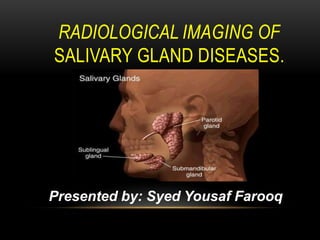
RADIOLOGICAL IMAGING OF
SALIVARY GLAND DISEASES.
Presented by: Syed Yousaf Farooq
Imaging modalities available to assess salivary glands
include:
Plain film radiography.
Sialograph
yCT scan.
MRI.
Diagnostic
ultrasound. Nuclear
scintigraphy.
These imaging modalities play an important role in evaluating a
patient with pain, swelling, or other symptoms related to possible
salivary gland disorders. Imaging helps in differentiating lesions of
salivary glands from those of parapharyngeal space, masticator
space, and mandible, submandibular and submental spaces. In
addition to localizing the lesion, it also aids in determining the extent
of disease, involvement of skull base, mandible, and neural spread in
case of malignant lesions.
Ultrasonography: High resolution Ultrasonography (7.5 – 10 MHz) of
parotid and submandibular salivary glands is actually a quick and
non invasive method for assessing these glands. It helps in the
assessment of vascularity of the gland, whether the lesion is solid or
cystic. It also helps in accurately guiding the FNAC needle to perform
biopsy from the suspected lesion.
It fails to demonstrate the parotid gland in its entirety because of
intervening mandible. It also does not clearly demonstrate the
intraglandular facialnerve branches.
Color Doppler studies of salivary glands indirectly identifies
malignant lesions of salivary glands as these lesions boast high
vascularity in comparison with their benign counterparts.
Radionuclide imaging: Is rarely used to evaluate salivary glands.
Sodium pertechnetate (technetium 99m) is the commonly used
element. This isotope is concentrated and excreted by salivary glands.
Since most salivary gland tumors do not accumulate the radionuclide,
a tumor mass will appear as a filling defecton
this scan. Tumors like warthin’s readily take up pertechnetate resulting
in formation of “Hot spots”.
DEVELOPMENTALANOMALIES.
Aberrant salivary glands.
An aberrant or ectopic is salivary gland tissue that
develop at a site where it is not normally found.
Aplasia and hypoplasia.
Aplasia occurs in combination with congenital
anomalies. Hypoplasia in patient with Melkersen
Rosenthal syndrome. Hyperplasia.
Accessory ducts. Most common developmental
anomalies. Atresia, congenital occlusion or
absence of one or two major salivary gland ducts.
Diverticulae, small pouches of ductal system of
one of the major salivary glands.
Congenital fistula.
Submandibular gland
Sialolithiasis, single (A) and multiple (B) in two different
patients, as detected on CT. Small inset on the right (C)
shows a multilayer structure that is observed in the majority
of calculi (hydroxyapatite)
Acute parotitis. US (longitudinal (A) and transverse (B)) shows an enlarged,
hypoechoic gland with multiple hypoechogenic areas corresponding to acinic
dilatations. Color Doppler register is increased (C).
Acute right-sided parotitis. Transverse, contrast-enhanced, fat-saturated, T1-
weighted SE MR image shows marked enhancement of the right parotid gland
(thick and thin arrows) compared with the left. The superficial subcutaneous
tissue is also inflamed.
Sialolithiasis and sialadenitis, as seen on color Doppler
ultrasound of the submandibular gland. Two small calculi, 2 and
3mm in size, respectively (arrows), are seen as hyperechoic
structures with posterior shadowing. The increased
vascularization of the slightly enlarged gland should be noted.
Recurrent sialadenitis of the parotid gland:
ultrasound and magnetic resonance
sialography findings.
Viral parotitis (mumps) on US. Image A) represents the two pairs
of glands on a longitudinal view, with pathological enlargement
of the right parotid, whereas image B) shows the asymmetry in a
transverse view.
Plunging or cervical Ranula
Left sublingual space extending to the
submandibular space consistent with a
plunging ranula (arrow).
Left parotid hemangioma
Bilateral parotid hemangiomas.
RADIOGRAPHIC FEATURES
CT: Smoothly marginated or Lobulated homogeneous small
spherical mass is the most common appearance. When
larger they can be heterogeneous with foci of necrosis.
Small regions of calcification are common .
When small enhancement tends to be prominent. In larger
tumours enhancement is less marked, but can demonstrate
delayed enhancement. MRI: They are commonly seen as well-
circumscribed and homogeneous when small. Larger tumours
may be heterogeneous.
Ultrasound:Typically hypoechoic. May show a lobulated
distinct border
+/- posterior acoustic enhancement.
Ultrasound is also useful in guiding biopsy (both FNAC and
core biopsies) but needs to be carried out with care to avoid
facial nerve damage .
Angiography (DSA): Typically hypovascular.
A Warthin tumour (or papillary cystadenoma lymphomatosum) is a
benign sharply demarcated tumour of the parotid gland. It is bilateral
in 10-15% of cases.
Epidemiology
They are the 2nd most common (up to 10% of all parotid tumours)
benign parotid tumour (after pleomorphic adenoma) and are the
commonest bilateral or multifocal benign parotid tumour. It typically
occurs in the elderly (6th decade). There is a recognised male
predilection. Patients typically present with painless parotid
swelling.
Morphology
They are often multi-centric (20%) and are usually small (1-4 cm).
They have a typically heterogenous appearance on all modalities,
often with cystic components (30%).
Location
Tends to favour the parotid tail region.
WARTHIN TUMOUR
RADIOGRAPHIC FEATURES
Has a greater tendency to undergo cystic change than any other
salivary gland tumour .
Ultrasound:
Usually seen as a relatively well defined, ovoid, hyperechoic mass. In
some cases
anechoic internal cystic areas may be present. They are often
hypervascular.
CT:
can be often seen bilaterally
classic appearance is a cystic lesion posteriorly within the parotid
with a focal tumour nodule
relatively well defined
cystic changes appear as intra lesion at lower attenuation, no
calcification MRI:
Well defined and can be bilateral.
Computed tomography of Warthin tumor. A: A large well-defined mass involves
the superficial lobe of the left parotid gland. The mass enhances diffusely. B: At
the level of the parotid tail, areas of lower density (arrowheads) are consistent
with cystic components observed in the posterior aspect of the mass.
Bilateral Warthin Tumors of the Parotid Gland on contrast-
enhanced CT
Magnetic resonance imaging of an oncocytoma in the right
parotid tail.
A MUCOEPIDERMOID CARCINOMA IS A TUMOR THAT USUALLY OCCURS IN THE
SALIVARY GLAND
Epidemioloy
Mucoepidermoid carcinomas are seen throughout all adult age groups, but are
most common in middle age (35-65 years of age) . However, it is the most
common malignant salivary gland tumour of childhood . Overall,
mucoepidermoid carcinomas account for :
2.8-15.5% of all salivary gland tumours
1-10% of all major salivary gland
tumours 6.5-41% of minor salivary
gland tumours
In the parotid gland they are the most common malignant primary
neoplasm. Clinical presentation
Mucoepidermoid carcinomas most frequently arise in the parotid gland, and
presents as a painless swelling, with or without facial nerve involvement.
These tumours can however be found anywhere there are salivary glands.
Overall distribution across various glands is as follows :
major salivary glands : 50%.
parotid gland : 40%.
submandibular gland :
7%. sublingual gland :
3%.
minor salivary glands : 50%.
RADIOGRAPHIC FEATURES:
Radiographic appearances largely depend on grade,
making preoperative imaging
important in planning and counseling.
Ultrasound:
Typically a well circumscribed hypoechoic, with an either
part of completely cystic appearance. The lesion stands
out against the relatively hyperechoic normal parotid gland.
MRI:
• Again, imaging is dependent on grade.
• Low grade tumors have similar appearances to
benign mixed tumors.
• T1 - low to intermediate signal ; low signal cystic
spaces
• T2 - intermediate to high signal ; cystic
areas will be high signal T1 C+ (Gd) -
heterogeneous enhancement of solid
components
• High grade tumors on the other hand have lower signal on T2 and
poorly defined margins and infrequent cystic areas.
• T1 - low to
intermediate
signal T2 -
intermediate to
low signal
Computed Tomography:
• Low grade tumors appear as well circumscribed masses, usually
with cystic components. The solid components enhance, and
calcification is sometimes seen. They have appearances similar to
benign mixed tumours.
• High grade tumours on the other hand, have poorly defined
margins, infiltrate locally and appear solid.
Computed tomography of a recurrent adenoid cystic carcinoma in a 49-year-old woman
who had previous right submandibular gland resection. An irregularly infiltrating
enhancing mass is seen in the floor of the mouth and parapharyngeal space. Anterior
fatty infiltration indicates hypoglossal nerve invasion
Adenocarcinoma of the right parotid gland.
Carcinosarcoma of the left parotid gland.
Axial and coronal contrast enhanced CT. The patient had previous surgery for
squamous cell carcinoma involving the parotid gland, these images demonstrate
recurrence within the parotid space. Note the involvement of the skin.
METASTASIS.
Most metastases to the parotid or submandibular
gland are from continuous spread of squamous
cell carcinoma of the pharynx or neck.
Hematogenous metastases have been described
in carcinoma
of the breast, neck, lung, and kidney and in
metastatic melanoma. Despite the vascularity of
the parotid gland, blood borne metastasis is rare.
Lymphatic metastases to the parotid glands also
are possible.
Metastasis to the parotid
glands
THANK YOU

More Related Content

What's hot

Presentation1, mri imaging of the prostate
Presentation1, mri imaging of the prostatePresentation1, mri imaging of the prostate
Presentation1, mri imaging of the prostateAbdellah Nazeer
 
Presentation1.pptx, radiological imaging of beign breast diseases
Presentation1.pptx, radiological imaging of beign breast diseasesPresentation1.pptx, radiological imaging of beign breast diseases
Presentation1.pptx, radiological imaging of beign breast diseasesAbdellah Nazeer
 
Imaging of neurocutaneous syndrome overview
Imaging of neurocutaneous syndrome overviewImaging of neurocutaneous syndrome overview
Imaging of neurocutaneous syndrome overviewcharusmita chaudhary
 
Diagnostic Imaging of Paranasal sinuses and Nose
Diagnostic Imaging of Paranasal sinuses and NoseDiagnostic Imaging of Paranasal sinuses and Nose
Diagnostic Imaging of Paranasal sinuses and NoseMohamed M.A. Zaitoun
 
Imaging findings of metabolic bone diseases
Imaging findings of metabolic bone diseases Imaging findings of metabolic bone diseases
Imaging findings of metabolic bone diseases Pankaj Kaira
 
Intracranial Calcification in Cone Beam CT & Medical CT
Intracranial Calcification in Cone Beam CT & Medical CTIntracranial Calcification in Cone Beam CT & Medical CT
Intracranial Calcification in Cone Beam CT & Medical CTJudy Oh, D.D.S.
 
Radiology Spots PPT- 3 by Dr Chandni Wadhwani
 Radiology Spots PPT- 3 by Dr Chandni Wadhwani Radiology Spots PPT- 3 by Dr Chandni Wadhwani
Radiology Spots PPT- 3 by Dr Chandni WadhwaniChandni Wadhwani
 
Presentation1.pptx, radiological imaging of spinal cord tumour.
Presentation1.pptx, radiological imaging of spinal cord tumour.Presentation1.pptx, radiological imaging of spinal cord tumour.
Presentation1.pptx, radiological imaging of spinal cord tumour.Abdellah Nazeer
 
Salivary gland imaging radiology ppt
Salivary gland imaging radiology pptSalivary gland imaging radiology ppt
Salivary gland imaging radiology pptDr pradeep Kumar
 
radiology Spotters mixed bag
radiology Spotters mixed bagradiology Spotters mixed bag
radiology Spotters mixed bagAnish Choudhary
 
Presentation1, radiological imaging of undescended testis.
Presentation1, radiological imaging of undescended testis.Presentation1, radiological imaging of undescended testis.
Presentation1, radiological imaging of undescended testis.Abdellah Nazeer
 
ANATOMICAL VARIANTS OF CT PNS
ANATOMICAL VARIANTS OF CT PNSANATOMICAL VARIANTS OF CT PNS
ANATOMICAL VARIANTS OF CT PNSsusritha17
 
KEYS OF RADIOLOGY SPOTTERS GIT
KEYS OF RADIOLOGY SPOTTERS GITKEYS OF RADIOLOGY SPOTTERS GIT
KEYS OF RADIOLOGY SPOTTERS GITAnish Choudhary
 
Presentation1.pptx, radiological imaging of extra nodal lymphoma.
Presentation1.pptx, radiological imaging of extra nodal lymphoma.Presentation1.pptx, radiological imaging of extra nodal lymphoma.
Presentation1.pptx, radiological imaging of extra nodal lymphoma.Abdellah Nazeer
 
Diagnostic Imaging of Cerebellopontine Angle Masses
Diagnostic Imaging of Cerebellopontine Angle MassesDiagnostic Imaging of Cerebellopontine Angle Masses
Diagnostic Imaging of Cerebellopontine Angle MassesMohamed M.A. Zaitoun
 
Diagnostic Imaging of Deep Neck Spaces
Diagnostic Imaging of Deep Neck SpacesDiagnostic Imaging of Deep Neck Spaces
Diagnostic Imaging of Deep Neck SpacesMohamed M.A. Zaitoun
 

What's hot (20)

Presentation1, mri imaging of the prostate
Presentation1, mri imaging of the prostatePresentation1, mri imaging of the prostate
Presentation1, mri imaging of the prostate
 
Presentation1.pptx, radiological imaging of beign breast diseases
Presentation1.pptx, radiological imaging of beign breast diseasesPresentation1.pptx, radiological imaging of beign breast diseases
Presentation1.pptx, radiological imaging of beign breast diseases
 
Imaging of neurocutaneous syndrome overview
Imaging of neurocutaneous syndrome overviewImaging of neurocutaneous syndrome overview
Imaging of neurocutaneous syndrome overview
 
Diagnostic Imaging of Paranasal sinuses and Nose
Diagnostic Imaging of Paranasal sinuses and NoseDiagnostic Imaging of Paranasal sinuses and Nose
Diagnostic Imaging of Paranasal sinuses and Nose
 
Imaging findings of metabolic bone diseases
Imaging findings of metabolic bone diseases Imaging findings of metabolic bone diseases
Imaging findings of metabolic bone diseases
 
MRI carcinoma Uterus and Cervix
MRI carcinoma Uterus and  CervixMRI carcinoma Uterus and  Cervix
MRI carcinoma Uterus and Cervix
 
Intracranial Calcification in Cone Beam CT & Medical CT
Intracranial Calcification in Cone Beam CT & Medical CTIntracranial Calcification in Cone Beam CT & Medical CT
Intracranial Calcification in Cone Beam CT & Medical CT
 
Orbit imaging anatomy
Orbit imaging anatomyOrbit imaging anatomy
Orbit imaging anatomy
 
Radiology Spots PPT- 3 by Dr Chandni Wadhwani
 Radiology Spots PPT- 3 by Dr Chandni Wadhwani Radiology Spots PPT- 3 by Dr Chandni Wadhwani
Radiology Spots PPT- 3 by Dr Chandni Wadhwani
 
Presentation1.pptx, radiological imaging of spinal cord tumour.
Presentation1.pptx, radiological imaging of spinal cord tumour.Presentation1.pptx, radiological imaging of spinal cord tumour.
Presentation1.pptx, radiological imaging of spinal cord tumour.
 
Salivary gland imaging radiology ppt
Salivary gland imaging radiology pptSalivary gland imaging radiology ppt
Salivary gland imaging radiology ppt
 
Mri prostate
Mri prostateMri prostate
Mri prostate
 
radiology Spotters mixed bag
radiology Spotters mixed bagradiology Spotters mixed bag
radiology Spotters mixed bag
 
Presentation1, radiological imaging of undescended testis.
Presentation1, radiological imaging of undescended testis.Presentation1, radiological imaging of undescended testis.
Presentation1, radiological imaging of undescended testis.
 
Radiology spotters
Radiology spottersRadiology spotters
Radiology spotters
 
ANATOMICAL VARIANTS OF CT PNS
ANATOMICAL VARIANTS OF CT PNSANATOMICAL VARIANTS OF CT PNS
ANATOMICAL VARIANTS OF CT PNS
 
KEYS OF RADIOLOGY SPOTTERS GIT
KEYS OF RADIOLOGY SPOTTERS GITKEYS OF RADIOLOGY SPOTTERS GIT
KEYS OF RADIOLOGY SPOTTERS GIT
 
Presentation1.pptx, radiological imaging of extra nodal lymphoma.
Presentation1.pptx, radiological imaging of extra nodal lymphoma.Presentation1.pptx, radiological imaging of extra nodal lymphoma.
Presentation1.pptx, radiological imaging of extra nodal lymphoma.
 
Diagnostic Imaging of Cerebellopontine Angle Masses
Diagnostic Imaging of Cerebellopontine Angle MassesDiagnostic Imaging of Cerebellopontine Angle Masses
Diagnostic Imaging of Cerebellopontine Angle Masses
 
Diagnostic Imaging of Deep Neck Spaces
Diagnostic Imaging of Deep Neck SpacesDiagnostic Imaging of Deep Neck Spaces
Diagnostic Imaging of Deep Neck Spaces
 

Similar to Radiological imaging of salivary gland diseases.

Pediatric abdominal tumors
Pediatric abdominal tumorsPediatric abdominal tumors
Pediatric abdominal tumorspassant dorgham
 
Imaging of the neck part ii
Imaging of the neck part iiImaging of the neck part ii
Imaging of the neck part iiWafik Ebrahim
 
Imaging in Pediatric Retroperitoneal Masses
Imaging in Pediatric Retroperitoneal MassesImaging in Pediatric Retroperitoneal Masses
Imaging in Pediatric Retroperitoneal MassesDr.Suhas Basavaiah
 
imaging of Testicular malignancies
imaging of Testicular malignanciesimaging of Testicular malignancies
imaging of Testicular malignanciesvinothmezoss
 
managementofrhabdomyosarcomaver11cdateofpresentation7october2015-copy-2008031...
managementofrhabdomyosarcomaver11cdateofpresentation7october2015-copy-2008031...managementofrhabdomyosarcomaver11cdateofpresentation7october2015-copy-2008031...
managementofrhabdomyosarcomaver11cdateofpresentation7october2015-copy-2008031...RebumaMegersa1
 
Management of rhabdomyosarcoma
Management of rhabdomyosarcomaManagement of rhabdomyosarcoma
Management of rhabdomyosarcomaDr Manas Dubey
 
Amol cardiac tumours
Amol cardiac tumoursAmol cardiac tumours
Amol cardiac tumoursAmol Gulhane
 
Case Report_2.pptx
Case Report_2.pptxCase Report_2.pptx
Case Report_2.pptxZahra1373
 
Retroperitoneal masses radiology
Retroperitoneal masses radiologyRetroperitoneal masses radiology
Retroperitoneal masses radiologyDr. Mohit Goel
 
Pleomorphic adenoma
Pleomorphic adenomaPleomorphic adenoma
Pleomorphic adenomaAhmed Shoeeb
 

Similar to Radiological imaging of salivary gland diseases. (20)

Pediatric abdominal tumors
Pediatric abdominal tumorsPediatric abdominal tumors
Pediatric abdominal tumors
 
Imaging of the neck part ii
Imaging of the neck part iiImaging of the neck part ii
Imaging of the neck part ii
 
Thyroid us reggio-emilia 1-tnt
Thyroid us   reggio-emilia 1-tntThyroid us   reggio-emilia 1-tnt
Thyroid us reggio-emilia 1-tnt
 
MRI and CT of ADRENAL GLAND
MRI and CT of ADRENAL GLANDMRI and CT of ADRENAL GLAND
MRI and CT of ADRENAL GLAND
 
Imaging in Pediatric Retroperitoneal Masses
Imaging in Pediatric Retroperitoneal MassesImaging in Pediatric Retroperitoneal Masses
Imaging in Pediatric Retroperitoneal Masses
 
imaging of Testicular malignancies
imaging of Testicular malignanciesimaging of Testicular malignancies
imaging of Testicular malignancies
 
Testicular tumors
Testicular tumorsTesticular tumors
Testicular tumors
 
retroperitonealmasses-pptx
retroperitonealmasses-pptxretroperitonealmasses-pptx
retroperitonealmasses-pptx
 
Retroperitoneal masses
Retroperitoneal massesRetroperitoneal masses
Retroperitoneal masses
 
managementofrhabdomyosarcomaver11cdateofpresentation7october2015-copy-2008031...
managementofrhabdomyosarcomaver11cdateofpresentation7october2015-copy-2008031...managementofrhabdomyosarcomaver11cdateofpresentation7october2015-copy-2008031...
managementofrhabdomyosarcomaver11cdateofpresentation7october2015-copy-2008031...
 
Management of rhabdomyosarcoma
Management of rhabdomyosarcomaManagement of rhabdomyosarcoma
Management of rhabdomyosarcoma
 
Benign salivary gland tumours
Benign salivary gland tumoursBenign salivary gland tumours
Benign salivary gland tumours
 
retroperitoneal masses.pptx
retroperitoneal masses.pptxretroperitoneal masses.pptx
retroperitoneal masses.pptx
 
The scrotum
The scrotumThe scrotum
The scrotum
 
Amol cardiac tumours
Amol cardiac tumoursAmol cardiac tumours
Amol cardiac tumours
 
Case Report_2.pptx
Case Report_2.pptxCase Report_2.pptx
Case Report_2.pptx
 
Retroperitoneal masses radiology
Retroperitoneal masses radiologyRetroperitoneal masses radiology
Retroperitoneal masses radiology
 
CARCINOMA PENIS
CARCINOMA PENISCARCINOMA PENIS
CARCINOMA PENIS
 
Giant cell tumor
Giant cell tumorGiant cell tumor
Giant cell tumor
 
Pleomorphic adenoma
Pleomorphic adenomaPleomorphic adenoma
Pleomorphic adenoma
 

More from Syed Yousaf Gilani

Ultrasound guided interventional procedure
Ultrasound guided interventional procedureUltrasound guided interventional procedure
Ultrasound guided interventional procedureSyed Yousaf Gilani
 
Gallbladder ultrasound (basic)
Gallbladder ultrasound (basic)Gallbladder ultrasound (basic)
Gallbladder ultrasound (basic)Syed Yousaf Gilani
 
Ultrasound of Fetal anomalies in first trimester
Ultrasound of Fetal anomalies in first trimesterUltrasound of Fetal anomalies in first trimester
Ultrasound of Fetal anomalies in first trimesterSyed Yousaf Gilani
 
Color Doppler Ultrasound - Basic physics
Color Doppler Ultrasound - Basic physicsColor Doppler Ultrasound - Basic physics
Color Doppler Ultrasound - Basic physicsSyed Yousaf Gilani
 
3D and 4D ultrasound in obstetrics
3D and 4D ultrasound in obstetrics3D and 4D ultrasound in obstetrics
3D and 4D ultrasound in obstetricsSyed Yousaf Gilani
 
Ultrasound guided nerves block
Ultrasound guided nerves blockUltrasound guided nerves block
Ultrasound guided nerves blockSyed Yousaf Gilani
 
Deep vein thrombosis Ultrasound
Deep vein thrombosis UltrasoundDeep vein thrombosis Ultrasound
Deep vein thrombosis UltrasoundSyed Yousaf Gilani
 
Musculoskeletal Ultrasound - Basic
Musculoskeletal Ultrasound - BasicMusculoskeletal Ultrasound - Basic
Musculoskeletal Ultrasound - BasicSyed Yousaf Gilani
 
Breast Ultrasound guided procedures
Breast Ultrasound guided proceduresBreast Ultrasound guided procedures
Breast Ultrasound guided proceduresSyed Yousaf Gilani
 

More from Syed Yousaf Gilani (14)

Ultrasound guided interventional procedure
Ultrasound guided interventional procedureUltrasound guided interventional procedure
Ultrasound guided interventional procedure
 
Prostate ultrasound (basic)
Prostate ultrasound (basic)Prostate ultrasound (basic)
Prostate ultrasound (basic)
 
Gallbladder ultrasound (basic)
Gallbladder ultrasound (basic)Gallbladder ultrasound (basic)
Gallbladder ultrasound (basic)
 
Ultrasound of Fetal anomalies in first trimester
Ultrasound of Fetal anomalies in first trimesterUltrasound of Fetal anomalies in first trimester
Ultrasound of Fetal anomalies in first trimester
 
Color Doppler Ultrasound - Basic physics
Color Doppler Ultrasound - Basic physicsColor Doppler Ultrasound - Basic physics
Color Doppler Ultrasound - Basic physics
 
3D and 4D ultrasound in obstetrics
3D and 4D ultrasound in obstetrics3D and 4D ultrasound in obstetrics
3D and 4D ultrasound in obstetrics
 
Ultrasound guided nerves block
Ultrasound guided nerves blockUltrasound guided nerves block
Ultrasound guided nerves block
 
Interventional Ultrasound
Interventional UltrasoundInterventional Ultrasound
Interventional Ultrasound
 
Deep vein thrombosis Ultrasound
Deep vein thrombosis UltrasoundDeep vein thrombosis Ultrasound
Deep vein thrombosis Ultrasound
 
Breast ultrasound techniques
Breast ultrasound techniquesBreast ultrasound techniques
Breast ultrasound techniques
 
Carotid doppler Ultrasound
Carotid doppler UltrasoundCarotid doppler Ultrasound
Carotid doppler Ultrasound
 
Ultrasound of Hernia
Ultrasound of HerniaUltrasound of Hernia
Ultrasound of Hernia
 
Musculoskeletal Ultrasound - Basic
Musculoskeletal Ultrasound - BasicMusculoskeletal Ultrasound - Basic
Musculoskeletal Ultrasound - Basic
 
Breast Ultrasound guided procedures
Breast Ultrasound guided proceduresBreast Ultrasound guided procedures
Breast Ultrasound guided procedures
 

Recently uploaded

Best Rate (Hyderabad) Call Girls Jahanuma ⟟ 8250192130 ⟟ High Class Call Girl...
Best Rate (Hyderabad) Call Girls Jahanuma ⟟ 8250192130 ⟟ High Class Call Girl...Best Rate (Hyderabad) Call Girls Jahanuma ⟟ 8250192130 ⟟ High Class Call Girl...
Best Rate (Hyderabad) Call Girls Jahanuma ⟟ 8250192130 ⟟ High Class Call Girl...astropune
 
VIP Call Girls Pune Vrinda 9907093804 Short 1500 Night 6000 Best call girls S...
VIP Call Girls Pune Vrinda 9907093804 Short 1500 Night 6000 Best call girls S...VIP Call Girls Pune Vrinda 9907093804 Short 1500 Night 6000 Best call girls S...
VIP Call Girls Pune Vrinda 9907093804 Short 1500 Night 6000 Best call girls S...Miss joya
 
Call Girls Colaba Mumbai ❤️ 9920874524 👈 Cash on Delivery
Call Girls Colaba Mumbai ❤️ 9920874524 👈 Cash on DeliveryCall Girls Colaba Mumbai ❤️ 9920874524 👈 Cash on Delivery
Call Girls Colaba Mumbai ❤️ 9920874524 👈 Cash on Deliverynehamumbai
 
Bangalore Call Girls Marathahalli 📞 9907093804 High Profile Service 100% Safe
Bangalore Call Girls Marathahalli 📞 9907093804 High Profile Service 100% SafeBangalore Call Girls Marathahalli 📞 9907093804 High Profile Service 100% Safe
Bangalore Call Girls Marathahalli 📞 9907093804 High Profile Service 100% Safenarwatsonia7
 
Call Girls Service Navi Mumbai Samaira 8617697112 Independent Escort Service ...
Call Girls Service Navi Mumbai Samaira 8617697112 Independent Escort Service ...Call Girls Service Navi Mumbai Samaira 8617697112 Independent Escort Service ...
Call Girls Service Navi Mumbai Samaira 8617697112 Independent Escort Service ...Call girls in Ahmedabad High profile
 
Low Rate Call Girls Pune Esha 9907093804 Short 1500 Night 6000 Best call girl...
Low Rate Call Girls Pune Esha 9907093804 Short 1500 Night 6000 Best call girl...Low Rate Call Girls Pune Esha 9907093804 Short 1500 Night 6000 Best call girl...
Low Rate Call Girls Pune Esha 9907093804 Short 1500 Night 6000 Best call girl...Miss joya
 
Call Girls Darjeeling Just Call 9907093804 Top Class Call Girl Service Available
Call Girls Darjeeling Just Call 9907093804 Top Class Call Girl Service AvailableCall Girls Darjeeling Just Call 9907093804 Top Class Call Girl Service Available
Call Girls Darjeeling Just Call 9907093804 Top Class Call Girl Service AvailableDipal Arora
 
Aspirin presentation slides by Dr. Rewas Ali
Aspirin presentation slides by Dr. Rewas AliAspirin presentation slides by Dr. Rewas Ali
Aspirin presentation slides by Dr. Rewas AliRewAs ALI
 
Bangalore Call Girls Majestic 📞 9907093804 High Profile Service 100% Safe
Bangalore Call Girls Majestic 📞 9907093804 High Profile Service 100% SafeBangalore Call Girls Majestic 📞 9907093804 High Profile Service 100% Safe
Bangalore Call Girls Majestic 📞 9907093804 High Profile Service 100% Safenarwatsonia7
 
Call Girl Number in Vashi Mumbai📲 9833363713 💞 Full Night Enjoy
Call Girl Number in Vashi Mumbai📲 9833363713 💞 Full Night EnjoyCall Girl Number in Vashi Mumbai📲 9833363713 💞 Full Night Enjoy
Call Girl Number in Vashi Mumbai📲 9833363713 💞 Full Night Enjoybabeytanya
 
Premium Call Girls Cottonpet Whatsapp 7001035870 Independent Escort Service
Premium Call Girls Cottonpet Whatsapp 7001035870 Independent Escort ServicePremium Call Girls Cottonpet Whatsapp 7001035870 Independent Escort Service
Premium Call Girls Cottonpet Whatsapp 7001035870 Independent Escort Servicevidya singh
 
Call Girls Service Pune Vaishnavi 9907093804 Short 1500 Night 6000 Best call ...
Call Girls Service Pune Vaishnavi 9907093804 Short 1500 Night 6000 Best call ...Call Girls Service Pune Vaishnavi 9907093804 Short 1500 Night 6000 Best call ...
Call Girls Service Pune Vaishnavi 9907093804 Short 1500 Night 6000 Best call ...Miss joya
 
VIP Service Call Girls Sindhi Colony 📳 7877925207 For 18+ VIP Call Girl At Th...
VIP Service Call Girls Sindhi Colony 📳 7877925207 For 18+ VIP Call Girl At Th...VIP Service Call Girls Sindhi Colony 📳 7877925207 For 18+ VIP Call Girl At Th...
VIP Service Call Girls Sindhi Colony 📳 7877925207 For 18+ VIP Call Girl At Th...jageshsingh5554
 
Call Girl Number in Panvel Mumbai📲 9833363713 💞 Full Night Enjoy
Call Girl Number in Panvel Mumbai📲 9833363713 💞 Full Night EnjoyCall Girl Number in Panvel Mumbai📲 9833363713 💞 Full Night Enjoy
Call Girl Number in Panvel Mumbai📲 9833363713 💞 Full Night Enjoybabeytanya
 
Call Girl Coimbatore Prisha☎️ 8250192130 Independent Escort Service Coimbatore
Call Girl Coimbatore Prisha☎️  8250192130 Independent Escort Service CoimbatoreCall Girl Coimbatore Prisha☎️  8250192130 Independent Escort Service Coimbatore
Call Girl Coimbatore Prisha☎️ 8250192130 Independent Escort Service Coimbatorenarwatsonia7
 
Call Girls Ludhiana Just Call 9907093804 Top Class Call Girl Service Available
Call Girls Ludhiana Just Call 9907093804 Top Class Call Girl Service AvailableCall Girls Ludhiana Just Call 9907093804 Top Class Call Girl Service Available
Call Girls Ludhiana Just Call 9907093804 Top Class Call Girl Service AvailableDipal Arora
 
Call Girls Service Surat Samaira ❤️🍑 8250192130 👄 Independent Escort Service ...
Call Girls Service Surat Samaira ❤️🍑 8250192130 👄 Independent Escort Service ...Call Girls Service Surat Samaira ❤️🍑 8250192130 👄 Independent Escort Service ...
Call Girls Service Surat Samaira ❤️🍑 8250192130 👄 Independent Escort Service ...CALL GIRLS
 
Russian Call Girls in Pune Riya 9907093804 Short 1500 Night 6000 Best call gi...
Russian Call Girls in Pune Riya 9907093804 Short 1500 Night 6000 Best call gi...Russian Call Girls in Pune Riya 9907093804 Short 1500 Night 6000 Best call gi...
Russian Call Girls in Pune Riya 9907093804 Short 1500 Night 6000 Best call gi...Miss joya
 
VIP Russian Call Girls in Varanasi Samaira 8250192130 Independent Escort Serv...
VIP Russian Call Girls in Varanasi Samaira 8250192130 Independent Escort Serv...VIP Russian Call Girls in Varanasi Samaira 8250192130 Independent Escort Serv...
VIP Russian Call Girls in Varanasi Samaira 8250192130 Independent Escort Serv...Neha Kaur
 

Recently uploaded (20)

Best Rate (Hyderabad) Call Girls Jahanuma ⟟ 8250192130 ⟟ High Class Call Girl...
Best Rate (Hyderabad) Call Girls Jahanuma ⟟ 8250192130 ⟟ High Class Call Girl...Best Rate (Hyderabad) Call Girls Jahanuma ⟟ 8250192130 ⟟ High Class Call Girl...
Best Rate (Hyderabad) Call Girls Jahanuma ⟟ 8250192130 ⟟ High Class Call Girl...
 
VIP Call Girls Pune Vrinda 9907093804 Short 1500 Night 6000 Best call girls S...
VIP Call Girls Pune Vrinda 9907093804 Short 1500 Night 6000 Best call girls S...VIP Call Girls Pune Vrinda 9907093804 Short 1500 Night 6000 Best call girls S...
VIP Call Girls Pune Vrinda 9907093804 Short 1500 Night 6000 Best call girls S...
 
Call Girls Colaba Mumbai ❤️ 9920874524 👈 Cash on Delivery
Call Girls Colaba Mumbai ❤️ 9920874524 👈 Cash on DeliveryCall Girls Colaba Mumbai ❤️ 9920874524 👈 Cash on Delivery
Call Girls Colaba Mumbai ❤️ 9920874524 👈 Cash on Delivery
 
Bangalore Call Girls Marathahalli 📞 9907093804 High Profile Service 100% Safe
Bangalore Call Girls Marathahalli 📞 9907093804 High Profile Service 100% SafeBangalore Call Girls Marathahalli 📞 9907093804 High Profile Service 100% Safe
Bangalore Call Girls Marathahalli 📞 9907093804 High Profile Service 100% Safe
 
Call Girls Service Navi Mumbai Samaira 8617697112 Independent Escort Service ...
Call Girls Service Navi Mumbai Samaira 8617697112 Independent Escort Service ...Call Girls Service Navi Mumbai Samaira 8617697112 Independent Escort Service ...
Call Girls Service Navi Mumbai Samaira 8617697112 Independent Escort Service ...
 
Low Rate Call Girls Pune Esha 9907093804 Short 1500 Night 6000 Best call girl...
Low Rate Call Girls Pune Esha 9907093804 Short 1500 Night 6000 Best call girl...Low Rate Call Girls Pune Esha 9907093804 Short 1500 Night 6000 Best call girl...
Low Rate Call Girls Pune Esha 9907093804 Short 1500 Night 6000 Best call girl...
 
Call Girls Darjeeling Just Call 9907093804 Top Class Call Girl Service Available
Call Girls Darjeeling Just Call 9907093804 Top Class Call Girl Service AvailableCall Girls Darjeeling Just Call 9907093804 Top Class Call Girl Service Available
Call Girls Darjeeling Just Call 9907093804 Top Class Call Girl Service Available
 
Aspirin presentation slides by Dr. Rewas Ali
Aspirin presentation slides by Dr. Rewas AliAspirin presentation slides by Dr. Rewas Ali
Aspirin presentation slides by Dr. Rewas Ali
 
Bangalore Call Girls Majestic 📞 9907093804 High Profile Service 100% Safe
Bangalore Call Girls Majestic 📞 9907093804 High Profile Service 100% SafeBangalore Call Girls Majestic 📞 9907093804 High Profile Service 100% Safe
Bangalore Call Girls Majestic 📞 9907093804 High Profile Service 100% Safe
 
Call Girl Number in Vashi Mumbai📲 9833363713 💞 Full Night Enjoy
Call Girl Number in Vashi Mumbai📲 9833363713 💞 Full Night EnjoyCall Girl Number in Vashi Mumbai📲 9833363713 💞 Full Night Enjoy
Call Girl Number in Vashi Mumbai📲 9833363713 💞 Full Night Enjoy
 
Russian Call Girls in Delhi Tanvi ➡️ 9711199012 💋📞 Independent Escort Service...
Russian Call Girls in Delhi Tanvi ➡️ 9711199012 💋📞 Independent Escort Service...Russian Call Girls in Delhi Tanvi ➡️ 9711199012 💋📞 Independent Escort Service...
Russian Call Girls in Delhi Tanvi ➡️ 9711199012 💋📞 Independent Escort Service...
 
Premium Call Girls Cottonpet Whatsapp 7001035870 Independent Escort Service
Premium Call Girls Cottonpet Whatsapp 7001035870 Independent Escort ServicePremium Call Girls Cottonpet Whatsapp 7001035870 Independent Escort Service
Premium Call Girls Cottonpet Whatsapp 7001035870 Independent Escort Service
 
Call Girls Service Pune Vaishnavi 9907093804 Short 1500 Night 6000 Best call ...
Call Girls Service Pune Vaishnavi 9907093804 Short 1500 Night 6000 Best call ...Call Girls Service Pune Vaishnavi 9907093804 Short 1500 Night 6000 Best call ...
Call Girls Service Pune Vaishnavi 9907093804 Short 1500 Night 6000 Best call ...
 
VIP Service Call Girls Sindhi Colony 📳 7877925207 For 18+ VIP Call Girl At Th...
VIP Service Call Girls Sindhi Colony 📳 7877925207 For 18+ VIP Call Girl At Th...VIP Service Call Girls Sindhi Colony 📳 7877925207 For 18+ VIP Call Girl At Th...
VIP Service Call Girls Sindhi Colony 📳 7877925207 For 18+ VIP Call Girl At Th...
 
Call Girl Number in Panvel Mumbai📲 9833363713 💞 Full Night Enjoy
Call Girl Number in Panvel Mumbai📲 9833363713 💞 Full Night EnjoyCall Girl Number in Panvel Mumbai📲 9833363713 💞 Full Night Enjoy
Call Girl Number in Panvel Mumbai📲 9833363713 💞 Full Night Enjoy
 
Call Girl Coimbatore Prisha☎️ 8250192130 Independent Escort Service Coimbatore
Call Girl Coimbatore Prisha☎️  8250192130 Independent Escort Service CoimbatoreCall Girl Coimbatore Prisha☎️  8250192130 Independent Escort Service Coimbatore
Call Girl Coimbatore Prisha☎️ 8250192130 Independent Escort Service Coimbatore
 
Call Girls Ludhiana Just Call 9907093804 Top Class Call Girl Service Available
Call Girls Ludhiana Just Call 9907093804 Top Class Call Girl Service AvailableCall Girls Ludhiana Just Call 9907093804 Top Class Call Girl Service Available
Call Girls Ludhiana Just Call 9907093804 Top Class Call Girl Service Available
 
Call Girls Service Surat Samaira ❤️🍑 8250192130 👄 Independent Escort Service ...
Call Girls Service Surat Samaira ❤️🍑 8250192130 👄 Independent Escort Service ...Call Girls Service Surat Samaira ❤️🍑 8250192130 👄 Independent Escort Service ...
Call Girls Service Surat Samaira ❤️🍑 8250192130 👄 Independent Escort Service ...
 
Russian Call Girls in Pune Riya 9907093804 Short 1500 Night 6000 Best call gi...
Russian Call Girls in Pune Riya 9907093804 Short 1500 Night 6000 Best call gi...Russian Call Girls in Pune Riya 9907093804 Short 1500 Night 6000 Best call gi...
Russian Call Girls in Pune Riya 9907093804 Short 1500 Night 6000 Best call gi...
 
VIP Russian Call Girls in Varanasi Samaira 8250192130 Independent Escort Serv...
VIP Russian Call Girls in Varanasi Samaira 8250192130 Independent Escort Serv...VIP Russian Call Girls in Varanasi Samaira 8250192130 Independent Escort Serv...
VIP Russian Call Girls in Varanasi Samaira 8250192130 Independent Escort Serv...
 

Radiological imaging of salivary gland diseases.

  • 1. RADIOLOGICAL IMAGING OF SALIVARY GLAND DISEASES. Presented by: Syed Yousaf Farooq
  • 2.
  • 3.
  • 4.
  • 5. Imaging modalities available to assess salivary glands include: Plain film radiography. Sialograph yCT scan. MRI. Diagnostic ultrasound. Nuclear scintigraphy. These imaging modalities play an important role in evaluating a patient with pain, swelling, or other symptoms related to possible salivary gland disorders. Imaging helps in differentiating lesions of salivary glands from those of parapharyngeal space, masticator space, and mandible, submandibular and submental spaces. In addition to localizing the lesion, it also aids in determining the extent of disease, involvement of skull base, mandible, and neural spread in case of malignant lesions.
  • 6. Ultrasonography: High resolution Ultrasonography (7.5 – 10 MHz) of parotid and submandibular salivary glands is actually a quick and non invasive method for assessing these glands. It helps in the assessment of vascularity of the gland, whether the lesion is solid or cystic. It also helps in accurately guiding the FNAC needle to perform biopsy from the suspected lesion. It fails to demonstrate the parotid gland in its entirety because of intervening mandible. It also does not clearly demonstrate the intraglandular facialnerve branches. Color Doppler studies of salivary glands indirectly identifies malignant lesions of salivary glands as these lesions boast high vascularity in comparison with their benign counterparts. Radionuclide imaging: Is rarely used to evaluate salivary glands. Sodium pertechnetate (technetium 99m) is the commonly used element. This isotope is concentrated and excreted by salivary glands. Since most salivary gland tumors do not accumulate the radionuclide, a tumor mass will appear as a filling defecton this scan. Tumors like warthin’s readily take up pertechnetate resulting in formation of “Hot spots”.
  • 7.
  • 8.
  • 9.
  • 10.
  • 11.
  • 12.
  • 13. DEVELOPMENTALANOMALIES. Aberrant salivary glands. An aberrant or ectopic is salivary gland tissue that develop at a site where it is not normally found. Aplasia and hypoplasia. Aplasia occurs in combination with congenital anomalies. Hypoplasia in patient with Melkersen Rosenthal syndrome. Hyperplasia. Accessory ducts. Most common developmental anomalies. Atresia, congenital occlusion or absence of one or two major salivary gland ducts. Diverticulae, small pouches of ductal system of one of the major salivary glands. Congenital fistula.
  • 14.
  • 15.
  • 17.
  • 18. Sialolithiasis, single (A) and multiple (B) in two different patients, as detected on CT. Small inset on the right (C) shows a multilayer structure that is observed in the majority of calculi (hydroxyapatite)
  • 19. Acute parotitis. US (longitudinal (A) and transverse (B)) shows an enlarged, hypoechoic gland with multiple hypoechogenic areas corresponding to acinic dilatations. Color Doppler register is increased (C).
  • 20. Acute right-sided parotitis. Transverse, contrast-enhanced, fat-saturated, T1- weighted SE MR image shows marked enhancement of the right parotid gland (thick and thin arrows) compared with the left. The superficial subcutaneous tissue is also inflamed.
  • 21. Sialolithiasis and sialadenitis, as seen on color Doppler ultrasound of the submandibular gland. Two small calculi, 2 and 3mm in size, respectively (arrows), are seen as hyperechoic structures with posterior shadowing. The increased vascularization of the slightly enlarged gland should be noted.
  • 22. Recurrent sialadenitis of the parotid gland: ultrasound and magnetic resonance sialography findings.
  • 23. Viral parotitis (mumps) on US. Image A) represents the two pairs of glands on a longitudinal view, with pathological enlargement of the right parotid, whereas image B) shows the asymmetry in a transverse view.
  • 24.
  • 26. Left sublingual space extending to the submandibular space consistent with a plunging ranula (arrow).
  • 29.
  • 30.
  • 31. RADIOGRAPHIC FEATURES CT: Smoothly marginated or Lobulated homogeneous small spherical mass is the most common appearance. When larger they can be heterogeneous with foci of necrosis. Small regions of calcification are common . When small enhancement tends to be prominent. In larger tumours enhancement is less marked, but can demonstrate delayed enhancement. MRI: They are commonly seen as well- circumscribed and homogeneous when small. Larger tumours may be heterogeneous. Ultrasound:Typically hypoechoic. May show a lobulated distinct border +/- posterior acoustic enhancement. Ultrasound is also useful in guiding biopsy (both FNAC and core biopsies) but needs to be carried out with care to avoid facial nerve damage . Angiography (DSA): Typically hypovascular.
  • 32.
  • 33. A Warthin tumour (or papillary cystadenoma lymphomatosum) is a benign sharply demarcated tumour of the parotid gland. It is bilateral in 10-15% of cases. Epidemiology They are the 2nd most common (up to 10% of all parotid tumours) benign parotid tumour (after pleomorphic adenoma) and are the commonest bilateral or multifocal benign parotid tumour. It typically occurs in the elderly (6th decade). There is a recognised male predilection. Patients typically present with painless parotid swelling. Morphology They are often multi-centric (20%) and are usually small (1-4 cm). They have a typically heterogenous appearance on all modalities, often with cystic components (30%). Location Tends to favour the parotid tail region. WARTHIN TUMOUR
  • 34. RADIOGRAPHIC FEATURES Has a greater tendency to undergo cystic change than any other salivary gland tumour . Ultrasound: Usually seen as a relatively well defined, ovoid, hyperechoic mass. In some cases anechoic internal cystic areas may be present. They are often hypervascular. CT: can be often seen bilaterally classic appearance is a cystic lesion posteriorly within the parotid with a focal tumour nodule relatively well defined cystic changes appear as intra lesion at lower attenuation, no calcification MRI: Well defined and can be bilateral.
  • 35. Computed tomography of Warthin tumor. A: A large well-defined mass involves the superficial lobe of the left parotid gland. The mass enhances diffusely. B: At the level of the parotid tail, areas of lower density (arrowheads) are consistent with cystic components observed in the posterior aspect of the mass.
  • 36. Bilateral Warthin Tumors of the Parotid Gland on contrast- enhanced CT
  • 37.
  • 38.
  • 39. Magnetic resonance imaging of an oncocytoma in the right parotid tail.
  • 40. A MUCOEPIDERMOID CARCINOMA IS A TUMOR THAT USUALLY OCCURS IN THE SALIVARY GLAND Epidemioloy Mucoepidermoid carcinomas are seen throughout all adult age groups, but are most common in middle age (35-65 years of age) . However, it is the most common malignant salivary gland tumour of childhood . Overall, mucoepidermoid carcinomas account for : 2.8-15.5% of all salivary gland tumours 1-10% of all major salivary gland tumours 6.5-41% of minor salivary gland tumours In the parotid gland they are the most common malignant primary neoplasm. Clinical presentation Mucoepidermoid carcinomas most frequently arise in the parotid gland, and presents as a painless swelling, with or without facial nerve involvement. These tumours can however be found anywhere there are salivary glands. Overall distribution across various glands is as follows : major salivary glands : 50%. parotid gland : 40%. submandibular gland : 7%. sublingual gland : 3%. minor salivary glands : 50%.
  • 41. RADIOGRAPHIC FEATURES: Radiographic appearances largely depend on grade, making preoperative imaging important in planning and counseling. Ultrasound: Typically a well circumscribed hypoechoic, with an either part of completely cystic appearance. The lesion stands out against the relatively hyperechoic normal parotid gland.
  • 42. MRI: • Again, imaging is dependent on grade. • Low grade tumors have similar appearances to benign mixed tumors. • T1 - low to intermediate signal ; low signal cystic spaces • T2 - intermediate to high signal ; cystic areas will be high signal T1 C+ (Gd) - heterogeneous enhancement of solid components • High grade tumors on the other hand have lower signal on T2 and poorly defined margins and infrequent cystic areas. • T1 - low to intermediate signal T2 - intermediate to low signal
  • 43. Computed Tomography: • Low grade tumors appear as well circumscribed masses, usually with cystic components. The solid components enhance, and calcification is sometimes seen. They have appearances similar to benign mixed tumours. • High grade tumours on the other hand, have poorly defined margins, infiltrate locally and appear solid.
  • 44.
  • 45.
  • 46.
  • 47. Computed tomography of a recurrent adenoid cystic carcinoma in a 49-year-old woman who had previous right submandibular gland resection. An irregularly infiltrating enhancing mass is seen in the floor of the mouth and parapharyngeal space. Anterior fatty infiltration indicates hypoglossal nerve invasion
  • 48.
  • 49. Adenocarcinoma of the right parotid gland.
  • 50.
  • 51. Carcinosarcoma of the left parotid gland.
  • 52.
  • 53. Axial and coronal contrast enhanced CT. The patient had previous surgery for squamous cell carcinoma involving the parotid gland, these images demonstrate recurrence within the parotid space. Note the involvement of the skin.
  • 54.
  • 55. METASTASIS. Most metastases to the parotid or submandibular gland are from continuous spread of squamous cell carcinoma of the pharynx or neck. Hematogenous metastases have been described in carcinoma of the breast, neck, lung, and kidney and in metastatic melanoma. Despite the vascularity of the parotid gland, blood borne metastasis is rare. Lymphatic metastases to the parotid glands also are possible.
  • 56. Metastasis to the parotid glands