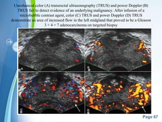
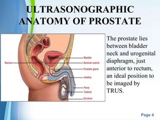
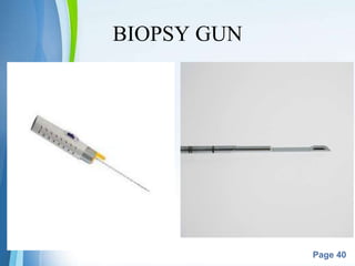
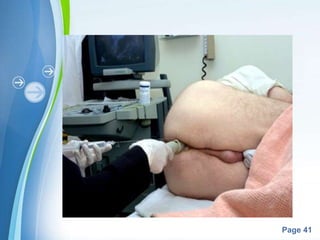
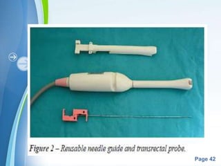
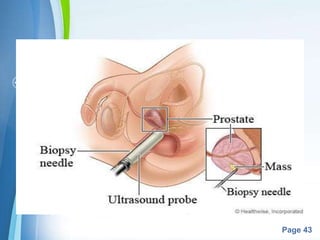
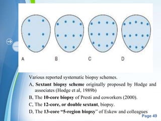
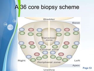
This document describes transrectal ultrasound (TRUS)-guided prostate biopsy techniques. It begins with background on the anatomy of the prostate and ultrasonographic imaging. TRUS-guided biopsy is considered the mainstay for prostate cancer detection and involves using a biopsy gun to obtain core samples under ultrasound guidance. Various biopsy schemes are described, including the original sextant technique and more extensive schemes involving additional cores. Factors such as patient preparation, anesthesia, and antibiotic prophylaxis for biopsies are also outlined. The document provides an overview of TRUS-guided prostate biopsy procedures and technical considerations.
A presentation about Intravenous Urography (Also known as Intravenous Pyeography).
The presentation contains 41 slides, and is divided into 4 parts :
1 - Introduction.
2 - The procedure.
3 - Examples for abnormal findings.
4 - Studies comparing IVU accuracy with KUB & USG with CT Scan.
This presentation was prepared and presented by me in the tutorials of the Radiology Department of Sebha Medical Center.
In this presentation we will discuss about the
Anatomy of Prostate
Technique of Transrectal US
Carcinoma Prostate and
Different modes of prostatic biopsy.
This presentation include biliary anatomy ,indication, contraindication post op care of percutaneus transhepatic biliary drainage with important technique. and advantage and disadvantage of different technique. This is important for radiologist, radiographers, intervention radiologist radiology resident. Thanks
This is about Introduction To renal CT scan Protocol what are the indication and tailoring how to optimize the the right protocol for the patient according to the indication .
Hopping you like it and helping you in daily practice .
Dr Hisham AlKhatib
Consultant Radiologist
uses and indication of radiology in surgeryanimesh kunwar
1.Introduction
2.Diagnostic modalities in radiology
3.Role of radiological imaging in emergency surgical situation
4.Role of radiological imaging in elective surgical situation
5.Conclusion
Five pearls and pitfalls in using head CT for diagnosis of traumatic brain injury. This was presented at the 51st Annual Scientific Meeting of the Royal College of Radiologists of Thailand (6 Aug 2014)
In this presentation we will discuss the basic of axial trauma from head to pelvis. We will discuss the important key points that aids in the diagnosis of axial trauma
Prostate - Excellent Illustrations / Must Watch & Prevent. ery Useful Information. DO NOT MISS to view and read the attached presentation. Please pass it on to your family & friends. Shared via "Sharifah Khatijah Syed Abdul Rahman Al-Attas" <sh_khatijah@yahoo.com
This presentation consist MR procedure of pelvis and hip joint , anatomy and MR planning is shown by picture with positioning block and parameters are included ,it includes basic sequence of both procedures
This slide deck is about Prostate cancer. It is amongst the leading cause of cancer deaths in adult males. This slide deck will provide you with necessary information regarding the symptoms, risk, diagnosis, and possible treatment of prostate cancer. I hope the readers find this slide deck useful & informative
These lecture slides, by Dr Sidra Arshad, offer a quick overview of the physiological basis of a normal electrocardiogram.
Learning objectives:
1. Define an electrocardiogram (ECG) and electrocardiography
2. Describe how dipoles generated by the heart produce the waveforms of the ECG
3. Describe the components of a normal electrocardiogram of a typical bipolar lead (limb II)
4. Differentiate between intervals and segments
5. Enlist some common indications for obtaining an ECG
6. Describe the flow of current around the heart during the cardiac cycle
7. Discuss the placement and polarity of the leads of electrocardiograph
8. Describe the normal electrocardiograms recorded from the limb leads and explain the physiological basis of the different records that are obtained
9. Define mean electrical vector (axis) of the heart and give the normal range
10. Define the mean QRS vector
11. Describe the axes of leads (hexagonal reference system)
12. Comprehend the vectorial analysis of the normal ECG
13. Determine the mean electrical axis of the ventricular QRS and appreciate the mean axis deviation
14. Explain the concepts of current of injury, J point, and their significance
Study Resources:
1. Chapter 11, Guyton and Hall Textbook of Medical Physiology, 14th edition
2. Chapter 9, Human Physiology - From Cells to Systems, Lauralee Sherwood, 9th edition
3. Chapter 29, Ganong’s Review of Medical Physiology, 26th edition
4. Electrocardiogram, StatPearls - https://www.ncbi.nlm.nih.gov/books/NBK549803/
5. ECG in Medical Practice by ABM Abdullah, 4th edition
6. Chapter 3, Cardiology Explained, https://www.ncbi.nlm.nih.gov/books/NBK2214/
7. ECG Basics, http://www.nataliescasebook.com/tag/e-c-g-basics
263778731218 Abortion Clinic /Pills In Harare ,sisternakatoto
263778731218 Abortion Clinic /Pills In Harare ,ABORTION WOMEN’S CLINIC +27730423979 IN women clinic we believe that every woman should be able to make choices in her pregnancy. Our job is to provide compassionate care, safety,affordable and confidential services. That’s why we have won the trust from all generations of women all over the world. we use non surgical method(Abortion pills) to terminate…Dr.LISA +27730423979women Clinic is committed to providing the highest quality of obstetrical and gynecological care to women of all ages. Our dedicated staff aim to treat each patient and her health concerns with compassion and respect.Our dedicated group ABORTION WOMEN’S CLINIC +27730423979 IN women clinic we believe that every woman should be able to make choices in her pregnancy. Our job is to provide compassionate care, safety,affordable and confidential services. That’s why we have won the trust from all generations of women all over the world. we use non surgical method(Abortion pills) to terminate…Dr.LISA +27730423979women Clinic is committed to providing the highest quality of obstetrical and gynecological care to women of all ages. Our dedicated staff aim to treat each patient and her health concerns with compassion and respect.Our dedicated group of receptionists, nurses, and physicians have worked together as a teamof receptionists, nurses, and physicians have worked together as a team wwww.lisywomensclinic.co.za/
Flu Vaccine Alert in Bangalore Karnatakaaddon Scans
As flu season approaches, health officials in Bangalore, Karnataka, are urging residents to get their flu vaccinations. The seasonal flu, while common, can lead to severe health complications, particularly for vulnerable populations such as young children, the elderly, and those with underlying health conditions.
Dr. Vidisha Kumari, a leading epidemiologist in Bangalore, emphasizes the importance of getting vaccinated. "The flu vaccine is our best defense against the influenza virus. It not only protects individuals but also helps prevent the spread of the virus in our communities," he says.
This year, the flu season is expected to coincide with a potential increase in other respiratory illnesses. The Karnataka Health Department has launched an awareness campaign highlighting the significance of flu vaccinations. They have set up multiple vaccination centers across Bangalore, making it convenient for residents to receive their shots.
To encourage widespread vaccination, the government is also collaborating with local schools, workplaces, and community centers to facilitate vaccination drives. Special attention is being given to ensuring that the vaccine is accessible to all, including marginalized communities who may have limited access to healthcare.
Residents are reminded that the flu vaccine is safe and effective. Common side effects are mild and may include soreness at the injection site, mild fever, or muscle aches. These side effects are generally short-lived and far less severe than the flu itself.
Healthcare providers are also stressing the importance of continuing COVID-19 precautions. Wearing masks, practicing good hand hygiene, and maintaining social distancing are still crucial, especially in crowded places.
Protect yourself and your loved ones by getting vaccinated. Together, we can help keep Bangalore healthy and safe this flu season. For more information on vaccination centers and schedules, residents can visit the Karnataka Health Department’s official website or follow their social media pages.
Stay informed, stay safe, and get your flu shot today!
Basavarajeeyam is an important text for ayurvedic physician belonging to andhra pradehs. It is a popular compendium in various parts of our country as well as in andhra pradesh. The content of the text was presented in sanskrit and telugu language (Bilingual). One of the most famous book in ayurvedic pharmaceutics and therapeutics. This book contains 25 chapters called as prakaranas. Many rasaoushadis were explained, pioneer of dhatu druti, nadi pareeksha, mutra pareeksha etc. Belongs to the period of 15-16 century. New diseases like upadamsha, phiranga rogas are explained.
- Video recording of this lecture in English language: https://youtu.be/lK81BzxMqdo
- Video recording of this lecture in Arabic language: https://youtu.be/Ve4P0COk9OI
- Link to download the book free: https://nephrotube.blogspot.com/p/nephrotube-nephrology-books.html
- Link to NephroTube website: www.NephroTube.com
- Link to NephroTube social media accounts: https://nephrotube.blogspot.com/p/join-nephrotube-on-social-media.html
These simplified slides by Dr. Sidra Arshad present an overview of the non-respiratory functions of the respiratory tract.
Learning objectives:
1. Enlist the non-respiratory functions of the respiratory tract
2. Briefly explain how these functions are carried out
3. Discuss the significance of dead space
4. Differentiate between minute ventilation and alveolar ventilation
5. Describe the cough and sneeze reflexes
Study Resources:
1. Chapter 39, Guyton and Hall Textbook of Medical Physiology, 14th edition
2. Chapter 34, Ganong’s Review of Medical Physiology, 26th edition
3. Chapter 17, Human Physiology by Lauralee Sherwood, 9th edition
4. Non-respiratory functions of the lungs https://academic.oup.com/bjaed/article/13/3/98/278874
Muktapishti is a traditional Ayurvedic preparation made from Shoditha Mukta (Purified Pearl), is believed to help regulate thyroid function and reduce symptoms of hyperthyroidism due to its cooling and balancing properties. Clinical evidence on its efficacy remains limited, necessitating further research to validate its therapeutic benefits.
CDSCO and Phamacovigilance {Regulatory body in India}NEHA GUPTA
The Central Drugs Standard Control Organization (CDSCO) is India's national regulatory body for pharmaceuticals and medical devices. Operating under the Directorate General of Health Services, Ministry of Health & Family Welfare, Government of India, the CDSCO is responsible for approving new drugs, conducting clinical trials, setting standards for drugs, controlling the quality of imported drugs, and coordinating the activities of State Drug Control Organizations by providing expert advice.
Pharmacovigilance, on the other hand, is the science and activities related to the detection, assessment, understanding, and prevention of adverse effects or any other drug-related problems. The primary aim of pharmacovigilance is to ensure the safety and efficacy of medicines, thereby protecting public health.
In India, pharmacovigilance activities are monitored by the Pharmacovigilance Programme of India (PvPI), which works closely with CDSCO to collect, analyze, and act upon data regarding adverse drug reactions (ADRs). Together, they play a critical role in ensuring that the benefits of drugs outweigh their risks, maintaining high standards of patient safety, and promoting the rational use of medicines.
2. Powerpoint Templates
Page 2
INTRODUCTION
• There is a significant decline in prostate
cancer related mortality due to early prostate
cancer detection programs.
• Role of PSA screening efforts, introduction
& refinement of systematic transrectal
ultrasonography (TRUS)– guided prostate
biopsy techniques, and increased public
awareness about prostate cancer.
3. Powerpoint Templates
Page 3
• TRUS of the prostate- first described by
Watanabe et al(1968).
• TRUS-guided systematic sextant biopsy
protocol- introduced by Hodge et al(1989).
• TRUS: mainstay of many image-guided
prostate interventions, including prostate
biopsy, brachytherapy, cryotherapy, & high-
intensity focused ultrasonography (HIFU),
evaluation of appropriate patients for treatment
of BPH.
5. Powerpoint Templates
Page 5
• On the basis of pathologic zonal architecture,
prostate is divided into:
Anterior fibromuscular stroma (AFS) that is
devoid of glandular tissue,
transition zone (TZ),
central zone (CZ),
periurethral zone, and
peripheral zone (PZ).
• But these regions are not visible
sonographically as distinct entities.
8. Powerpoint Templates
Page 8
• Normal CZ and PZ(posterior): majority of
adenocarcinomas; homogeneous echogenic
appearance.
• TZ(anterior) is more heterogeneous.
• Calcification along the surgical capsule known
as corpora amylacea highlight the plane between
PZ & TZ
• Small , multiple , diffuse calcifications are
normal
9. Powerpoint Templates
Page 9
• The prostatic urethra traverses the length of the gland
in the midline and thus must be imaged in the sagittal
plane to be simultaneously viewed along the entirety
of its course .
• The distended urethral lumen has a hypoechoic
appearance whereas periurethral calcifications may
produce a thin echogenic outline.
• The smooth muscle of the internal sphincter extends
from the bladder neck, encircling the urethra to the
level of the verumontanum.
10. Powerpoint Templates
Page 10
• These muscle fibers may be visualized
sonographically as a hypoechoic ring around the
upper prostatic urethra, giving it a funneled
appearance proximally as it arises from the bladder
neck.
• On reaching the verumontanum the urethra angles
anteriorly and runs through the remainder of the
gland to exit at the apex of the prostate.
• This angle gives the prostatic urethra an anteriorly
concave appearance when viewed along its entire
course in the sagittal plane.
11. Powerpoint Templates
Page 11
• The paired seminal vesicles (SVs) are
positioned posteriorly at the base of the
prostate.
• They have a smooth, saccular appearance and
should be symmetrical.
• The normal SV measures 4.5 to 5.5 cm in
length and 2 cm in width.
12. Powerpoint Templates
Page 12
A, In the transverse plane with the hypoechoic urethra centrally located
(star) and dotted line representing transverse measurement.
B, Midline sagittal view with the hypoechoic urethra running the length of
the gland, D1 represents longitudinal and D2 anteroposterior measurement.
14. Powerpoint Templates
Page 14
GRAY-SCALE TRUS
• Most common imaging modality for prostate.
• Most commonly used for:
prostate cancer detection
evaluation of other conditions such as
infertility
directing the biopsy of prostate cancer.
Staging of carcinoma prostate (limited role).
15. Powerpoint Templates
Page 15
• Endorectal probes available in both side- and end-
fire models; transmits frequencies of 6 to 10
MHz.
• Newer biplane probes provide simultaneous
sagittal and transverse imaging modes.
• Probes provide a scanning angle approaching 180
degrees to allow simultaneous visualization of the
entire gland in both the transverse and sagittal
planes.
• Increasing frequency yields increased resolution
16. Powerpoint Templates
Page 16
• As the frequency of the probe is increased, the
portion of the image that is in focus (focal range) is
closer to the transducer.
• The commonly used 7-MHz transducer produces a
high resolution image with a focal range from 1 to 4
cm from the transducer (best for PZ where most
cancers arise).
• Lower-frequency transducers (e.g., older 4-MHz
transducers) have a focal range from 2 to 8 cm but at
lower resolution they improve anterior deliniation of
large gland & provides poor internal architecture
visualization
17. Powerpoint Templates
Page 17
TECHNIQUE
• Medium-gray image of normal PZ serves as
the "reference point" for judging lesions as
hypoechoic (darker than the normal PZ),
isoechoic (similar to the normal PZ),
hyperechoic (lighter than the normal PZ), or
anechoic (completely black).
• Patients are scanned in left lateral ducubitus
position
• TRUS should be performed in both transverse
and sagittal planes.
18. Powerpoint Templates
Page 18
VOLUME CALCULATION
• π/6 × transverse diameter × AP diameter ×
longitudinal diameter
• Mature average prostate-20-25 g and remains
constant until age 50.
• For more accurate determination of prostate
volume (Brachytherapy), Planimetry is
required.
• PSA density = serum PSA/gland volume
• Elevated PSAD have sensitivity & specificity
of 75% & 44%, but it is operator dependent.
19. Powerpoint Templates
Page 19
PROSTATE CANCER IMAGING
ON TRUS
• All hypoechoic lesions within the PZ: consider
biopsy.
• A hypoechoic lesion is malignant in 17-57% of cases,
but they are not pathognomonic for cancer.
• Lack of a distinct hypoechoic focus doesn't preclude
biopsy, as 39% of Ca prostate cases are isoechoic, &
1% hyperechoic on conventional gray scale TRUS.
20. Powerpoint Templates
Page 20
• Granulomatous prostatitis, prostatic infarct &
lymphoma- all may produce hypoechoic
lesions.
• Extracapsular extension of prostate cancer,
although not well visualized if present as a
microfocus, is suggested by a focal loss of the
typically bright white periprostatic fat.
21. Powerpoint Templates
Page 21
Classic hypoechoic peripheral zone (PZ) lesion
(dotted line) in the right midgland that transrectal
ultrasonography–guided biopsy proved to be a
Gleason 3 + 3 = 6 adenocarcinoma.
22. Powerpoint Templates
Page 22
CYSTIC LESION OF
PROSTATE
• Simple cysts have the same sonographic appearance
as in any other part of the body: they are thin walled,
are anechoic, and show acoustic enhancement
posterior to the cyst.
• Congenital prostatic cystic lesions may arise from
either müllerian (müllerian duct cysts and prostatic
utricles) or wolffian (ejaculatory duct and seminal
vesicle cysts) structures.
• They appear in the midline as anechoic lesion
23. Powerpoint Templates
Page 23
• Ejaculatory duct cysts are typically small, lie
off of the midline, and may accompany
ejaculatory duct obstruction/obliteration with
azoospermia.
• Seminal vesicle cysts can be caused by
congenital or acquired obstruction of the
ejaculatory duct and are associated with cystic
renal disease; up to two thirds of men with
seminal vesicle cysts may also have renal
agenesis
25. Powerpoint Templates
Page 25
INDICATIONS FOR PROSTATE
BIOPSY
TRUS without Biopsy
• Treatment planning volume measurements: brachytherapy,
cryotherapy, BPH therapy (e.g.TUMT, RFA)
• Volume measurement during hormonal downsizing for EBRT
or brachytherapy
• Placement of fiducial markers for EBRT
• Evaluation of azoospermia: ejaculatory duct cysts, seminal
vesicle cysts, etc.
• Therapeutic aspiration or unroofing of prostatic cysts; drainage
of prostatic abscess
26. Powerpoint Templates
Page 26
TRUS-Directed Biopsy
• Diagnosis of suspected symptomatic prostate cancer (i.e., bone
metastasis, cord compression)
• Screening for prostate cancer in asymptomatic patient > age 50 with
> a 10-year life expectancy (if strong family history or if African-
American, consider screening at age 45)
Prostate nodule or significant prostate asymmetry regardless of PSA
level
PSA > 4.0 ng/dL regardless of age
In men < age 60 to 65 years, consider biopsy if PSA > 2.5 ng/dL
If PSA > 0.6 ng/dL at age 40
Increased PSA velocity (>0.75 ng/dL/year)
Free PSA in considering initial biopsy with PSA < 10 ng/mL: >25%
no biopsy; >10% and <15%, consider biopsy; <10%, biopsy
27. Powerpoint Templates
Page 27
• Prior to intervention in symptomatic BPH (e.g., surgical
therapy or initiation of 5α-reductase inhibitors)
• Prior to cystoprostatectomy or orthotopic urinary diversion
• To diagnose failed radiation therapy before use of second-line
therapy
• Follow-up biopsy (3-6 months) after diagnosis of high-grade
PIN or Atypical small acinar proliferation.
28. Powerpoint Templates
Page 28
• Data from the Prostate Cancer Prevention Trial
have shown that no safe PSA threshold can
rule out prostate cancer in any age range.
• For a serum PSA value between 4.0 and 10.0
ng/mL, using a % free PSA threshold of <25%
allowed detection of 95% of cancers while
eliminating 20% unnecessary biopsies.
29. Powerpoint Templates
Page 29
• Volume-based PSA parameters evaluated to
reduce confounding from BPH.
• These include:
PSA density (PSAD; PSA divided by prostate
volume),
complexed PSA density (complexed PSA
divided by prostate volume), and
PSA transition zone density (PSA divided by
transition zone volume).
30. Powerpoint Templates
Page 30
• PSA levels between 4 and 10 ng/mL and a
normal DRE:
PSAD ≥ 0.15- prostate biopsy recommended.
PSA transition zone volume was the parameter
with highest overall sensitivity & specificity.
31. Powerpoint Templates
Page 31
PSA Dynamics/PSA Velocity(PSAV):
• Rate of change in PSA.
• With PSA levels between 4-10 ng/mL, PSAV ≥ 0.75
ng/mL/year: a specific marker for the presence of
prostate cancer.
• In PSA <4, PSAV > 0.5ng/mL/yr is significant.
• PSAV may play a role in the prediction of life-
threatening prostate cancer .
• A PSAV > 0.35 ng/mL/year 10-15 years prior to
diagnosis- fivefold increased risk of life-threatening
prostate cancer more than a decade later.
33. Powerpoint Templates
Page 33
PATIENT
PREPARATION
• Written, informed consent.
• All anticoagulant therapy (warfarin,
clopidogrel, aspirin/NSAIDs, herbal
supplements) should be stopped 7-10 days
before prostate biopsy.
• For coagulopathic pts., INR should be
below 1.5.
• A small amount of urine can facilitate the
examination .
35. Powerpoint Templates
Page 35
Cleansing enema:
• decreases the amount of faeces in the rectum,
thereby producing a superior acoustic window
for prostate imaging.
• may reduce bacterial seeding of the prostate.
36. Powerpoint Templates
Page 36
Analgesia
• Topical lidocaine jelly.
• Infiltration anesthesia around nerve
bundles.
• Direct infiltration(Intraprostatic
injection).
• Skin & subcut.infiltration for
transperineal biopsy.
37. Powerpoint Templates
Page 37
POSITION
• Left lateral decubitus position with knees and hips
flexed 90 degrees.
• Lithotomy position preferred for transperineal
biopsies, brachytherapy treatment planning, or
placement of fiducial gold markers for external-beam
therapy.
• Lithotomy position is preferred when color Doppler
imaging is used to identify areas of hyperemia for
targeted biopsy of the prostate(distribution of color
Doppler flow within the prostate is dependent on
patient position)
38. Powerpoint Templates
Page 38
TRUS PROSTATE BIOPSY
TECHNIQUES
• Assess prostate volume.
• Prostate imaging in transverse &
sagittal planes(from base to apex).
• Note location and characteristics of any
lesions (i.e., hypoechoic, hyperechoic,
calcifications, contour abnormalities,
cystic structures).
39. Powerpoint Templates
Page 39
• A spring-driven, 18-gauge, needle core biopsy
device or biopsy gun, passed through the
needle guide attached to the ultrasound probe.
• Biopsy gun advances the needle 0.5 cm and
samples subsequent 1.5 cm of tissue with the
tip extending 0.5 cm beyond the area sampled.
45. Powerpoint Templates
Page 45
SEXTANT BIOPSY:
• one core, bilaterally,
each from base, mid,
and apex.
• samples both PZ &
TZ.
• Vast majority of
AdenoCa-
posterolateral PZ.
TRUS BIOPSY SCHEMES
46. Powerpoint Templates
Page 46
EXTENDED CORE BIOPSY
SCHEMES
• Improved cancer detection rates by
incorporating additional laterally
directed cores into the standard
systematic sextant technique.
• At present, 6 cores are considered
inadequate for routine prostate
biopsy for cancer detection.
47. Powerpoint Templates
Page 47
• TZ and SVs are not routinely sampled(low yields for
cancer detection at initial biopsy).
• TZ and anteriorly directed biopsies may
occasionally prove necessary to diagnose prostate
cancer in patients with persistently elevated PSA
levels and prior negative biopsies.
• A role for TZ biopsies in men with gland size > 50
mL, with an additional yield of 15% cancer detection.
48. Powerpoint Templates
Page 48
• Seminal vesicle biopsy is not
routinely performed unless there is a
palpable abnormality, when PSA
value > 30, or if brachytherapy is
being considered.
49. Powerpoint Templates
Page 49
Various reported systematic biopsy schemes.
A, Sextant biopsy scheme originally proposed by Hodge and
associates (Hodge et al, 1989b)
B, The 10-core biopsy of Presti and coworkers (2000).
C, The 12-core, or double sextant, biopsy.
D, The 13-core “5-region biopsy” of Eskew and colleagues
54. Powerpoint Templates
Page 54
• Proper sampling and labelling
• Sample should be sent in 10% formalin filled
bottles
• Preferably each individual sample should be
sent in different bottle, some prefer to send right
& left only separately
• Some pathologists believe strongly that each
site should be specifically identified because
certain locations predisposed to cancer may
look-alike(cooper’s gland at apex , seminal
vesicle at base)
55. Powerpoint Templates
Page 55
REPEAT PROSTATE BIOPSY
• Use of a 2nd prostate biopsy in all cases
of a negative finding on initial biopsy is
justified.
• But 3rd and 4th repeat biopsies should
only be obtained in selected patients
with high suspicion of cancer and/or
poor prognostic factors on the 1st or 2nd
biopsy.
56. Powerpoint Templates
Page 56
• Overall cancer detection rates for repeat prostate
needle biopsy with various biopsy templates ranges
from 10% - 38%.
• Indications for a repeat prostate biopsy include the
following:
1) A highly suspicious DRE (digital rectal examination)
2) A persistently rising serum PSA (> 0.4 – 0.75
ng/ml/yr.)
3) A low free PSA (certainly < 10%, maybe < 22% -
25%)
4) Presence of PIN or atypia on prior biopsy
58. Powerpoint Templates
Page 58
INTERPRETATION
• Benign prostatic hyperplasia
• Acute inflammation
• Chronic granulomatous inflammation
• Atrophy
• High grade prostatic intraepithelial neoplasm
(PIN)
• Suspicious (lesion too small or insufficient
criteria present)
• Adenocarcinoma.
59. Powerpoint Templates
Page 59
ADVANCED USG TECHNIQUES
FOR PROSTATE IMAGING
COLOR & POWER DOPPLER TRUS:
• Color Doppler imaging is based on the
frequency shift in the reflected sound waves
from the frequency of insonation
• Thus it depicts the velocity of blood flow in a
directionally dependent manner.
60. Powerpoint Templates
Page 60
• Color assignment is based on the direction of
blood flow related to the orientation of the
transducer receiving the signal; flow toward
the transducer- red and flow away in shades of
blue; color is not specific for arterial or venous
flow.
61. Powerpoint Templates
Page 61
Transrectal ultrasonography.
• Top image, solid white arrow
depicts hypoechoic lesion
within the peripheral zone
concerning for prostate cancer.
• Lower image depicts
hypervascular area seen with
color Doppler imaging, yellow
and red area corresponds to the
hypoechoic area seen on the
grayscale ultrasonography
above.
62. Powerpoint Templates
Page 62
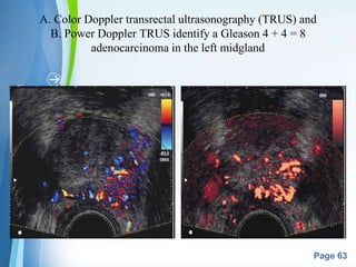
POWER DOPPLER IMAGING (enhanced color
Doppler, color amplitude imaging [CAI], or
color angiography) uses amplitude shift to
detect flow in a velocity and directionally
independent manner.
• Advantages: ability to detect slower flow and
to have less reliance on the Doppler angle,
making it more suitable for detection of
prostate cancer neovascularity.
63. Powerpoint Templates
Page 63
A. Color Doppler transrectal ultrasonography (TRUS) and
B. Power Doppler TRUS identify a Gleason 4 + 4 = 8
adenocarcinoma in the left midgland
64. Powerpoint Templates
Page 64
• Patients with detectable color Doppler flow
within their dominant tumor at the time of
TRUS-guided biopsy are at a 10-fold increased
risk for PSA recurrence after radical retropubic
prostatectomy.
• The presence of increased flow was also
accociated with high gleason grade, increased
incidence of SV invasion & lower incidence of
biochemical disease free survival rate.
65. Powerpoint Templates
Page 65
CONTRAST ENHANCED TRUS(CE-
TRUS)
• Intravenous microbubble ultrasound contrast agents, infused
systemically during gray-scale and TRUS Doppler imaging
amplify flow signals within the microvasculature of prostate
tumors, allowing selective visualization of malignant foci.
• These agents increase the echogenicity of the intravascular
space on grey-scale imaging and provide a dramatic visible
increase in the Doppler signal.
• These are constructed with air or higher-molecular-weight gas
agents encapsulated (albumin or polymer hard shell, lipid- or
surfactant-coated) for longevity.
66. Powerpoint Templates
Page 66
• CE-TRUS + 3D IMAGE
RECONSTRUCTION of enhanced power
doppler.
• GREY-SCALE HARMONIC imaging: better
spatial & temporal resolution.
• FLASH REPLENISHMENT IMAGING:
improved visualisation of vessels.
67. Powerpoint Templates
Page 67
Unenhanced color (A) transrectal ultrasonography (TRUS) and power Doppler (B)
TRUS fail to detect evidence of an underlying malignancy. After infusion of a
microbubble contrast agent, color (C) TRUS and power Doppler (D) TRUS
demonstrate an area of increased flow in the left midgland that proved to be a Gleason
3 + 4 = 7 adenocarcinoma on targeted biopsy
68. Powerpoint Templates
Page 68
OTHER TECHNIQUES
ARTIFICIAL NEURAL NETWORKS
ELASTOGRAPHY:
• New sonography technique.
• employs real-time sonographic imaging of
the prostate at baseline and under varying
degrees of compression.
• Through computerized calculations,
differences in displacement between
ultrasonic images from baseline and during
compression may be visualized, and regions
with decreased tissue elasticity may be
tagged as suggestive of malignancy.
69. Powerpoint Templates
Page 69
Elastography demonstrates an area of decreased compliance in the right base
consistent with an underlying malignancy (blue near arrow). Note color scale
in upper right corner indicating relative tissue “firmness.” Targeted biopsy of
this region revealed a Gleason 4 + 4 = 8 adenocarcinoma
70. Powerpoint Templates
Page 70
ENDORECTAL MRI & MR SPECTROSCOPIC
IMAGING(MRSI):
• MRSI identifies biochemical changes within
the tissue that may predate the appearance of
histological changes.
• MRSI suggestive of malignancy may not have
biopsy detectable PCa at the time of MRSI but
may develop histological cancer at a later date.
71. Powerpoint Templates
Page 71
TAKE HOME MESSAGE
• TRUS ALONE CANNOT DIAGNOSE
PROSTATE CANCER WITHOUT A
TISSUE BIOPSY
• MAINSTAY OF IMAGING FOR
PROSTATE BIOPSY,BRACHYTHERAPY,
CRYOTHERAPY & HIFU
• HYPOECHOIC FOCI SEEN ON GRAY
SCALE TRUS SHOULD BE
CONSIDERED S/O ADENOCARCINOMA
& INCLUDED IN BIOPSY SPECIMEN
72. Powerpoint Templates
Page 72
• 39% OF PROSTATE CANCER ARE NOT VISIBLE
ON ROUTINE GRAY SCALE USG
• SEXTANT BIOPSY IS INADEQUATE, PREFER
10 TO 14 CORE BIOPSY.
• CONTRAST ENHANCED TRUS & BIOPSY ,
COLOUR & POWER DOPPLER IMAGING
MODES MAY IMPROVE CANCER DETECTION
IN FUTURE
• TRUS GRAY SCALE CORE NEEDLE BIOPSY IS
THE GOLD STANDARD FOR DIAGNOSIS OF
PROSATE CANCER AT PRESENT