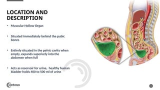
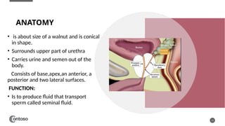
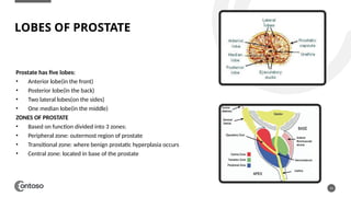
URINARY BLADDER AND PROSTATE
ANATOMY AND USG
TECHNIQUES
BY:
DR. TOOBA BINTE SHAHID
DR. HALA QAMAR CHOUDHRY
• Muscular Hollow Organ
• Situated immediately behind the pubic
bones
• Entirely situated in the pelvic cavity when
empty, expands superiorly into the
abdomen when full
• Acts as reservoir for urine, healthy human
bladder holds 400 to 500 ml of urine
2
LOCATION AND
DESCRIPTION
PARTS OF URINARY BLADDER
3
Dome: Dome or apex is top front part
Base: Bottom back part
Body: Area Between Dome and Base
Neck: Along base of bladder
VASCULATURE
ARTERIES: The main blood supply comes from
internal iliac arteires,which branch into superior
vesical and inferior vesical arteries.
VENUS DRAINAGE:
Most of the bladder’s venous return drains into the
internal iliac vein.
SONOGRAPHIC APPEARANCE OF URINARY BLADDER
5
• Round
• Anechoic
• Fluid filled Structure situated in caudal one third of the
abdomen
SONOGRAPHIC TECHNIQUES FOR URINARY
BLADDER
6
 A curvilinear probe(1-5MHz) is preferred for full visualization of urinary bladder
 Probe is placed midline directly above the pubic symphysis in transverse orientation
 To check for post void residuals, bladder should be full at beginning. Patient should be asked to drink
several glasses of water.
7
TRANSVERSE SCAN PLANE
LONGITUDNAL BLADDER
IMAGE
NORMAL BLADDER WALL THICKNESS
8
The normal thickness of the bladder wall in adults typically
ranges from 2-5 mm when measured using ultrasound
imaging.
Measurement of bladder wall thickness: Calculated as the
distance between the outer and inner surfaces of the bladder
wall.
PRE-VOID AND POST-VOID
ULTRASOUND
MEASUREMNTS.
PRE-VOID:
The amount of urine in the bladder before
urination.
POST-VOID:
Post-void residual describes the amount of urine
left in the bladder after voluntary bladder
emptying.
 To determine post-void residuals the
following formula is used:
length x width x height x 0.53
9
INTERNAL ECHOS WITHIN THE URINARY
BLADDER
10
TRABECULATED URINARY BLADDER
URETEROCELE
11
BLADDER MASS
BLADDER DIVERTICULUM
PROSTATE
Prostate is a gland in male reproductive
system that’s located below the bladder and
in front of the rectum.
ANATOMY
• is about size of a walnut and is conical
in shape.
• Surrounds upper part of urethra
• Carries urine and semen out of the
body.
Consists of base,apex,an anterior, a
posterior and two lateral surfaces.
FUNCTION:
• Is to produce fluid that transport
sperm called seminal fluid.
13
LOBES OF PROSTATE
14
Prostate has five lobes:
• Anterior lobe(in the front)
• Posterior lobe(in the back)
• Two lateral lobes(on the sides)
• One median lobe(in the middle)
ZONES OF PROSTATE
• Based on function divided into 3 zones:
• Peripheral zone: outermost region of prostate
• Transitional zone: where benign prostatic hyperplasia occurs
• Central zone: located in base of the prostate
TRUS IMAGES
15
ZONES:
VASCULATURE
Arteries:
receives arterial supply from prostatic
arteries primarily derived from internal
iliac arteries.
Veins:
The prostatic venous plexus drains blood
into the internal vein.
HOW TO TAKE MEASUREMENTS
SONOGRAPHICALLY
17
Using the formula:
 Prostate gland
volume =AP
diameter x
transverse
diameter x
longitudinal
diameter x 0.52
18
BENIGN PROSTATE
HYPERPLASIA
PROSTATIC CALCIFICATIONS PROSTATIC CALCULI
SONOGRAPHIC APPERANCE
TRUS IMAGES
19
Benign Prostatic Hyperplasia Prostatic Calculi Prostatic Calcification
THANK YOU

Urinary bladder and Prostate anatomy ppt.pptx

  • 1.
    URINARY BLADDER ANDPROSTATE ANATOMY AND USG TECHNIQUES BY: DR. TOOBA BINTE SHAHID DR. HALA QAMAR CHOUDHRY
  • 2.
    • Muscular HollowOrgan • Situated immediately behind the pubic bones • Entirely situated in the pelvic cavity when empty, expands superiorly into the abdomen when full • Acts as reservoir for urine, healthy human bladder holds 400 to 500 ml of urine 2 LOCATION AND DESCRIPTION
  • 3.
    PARTS OF URINARYBLADDER 3 Dome: Dome or apex is top front part Base: Bottom back part Body: Area Between Dome and Base Neck: Along base of bladder
  • 4.
    VASCULATURE ARTERIES: The mainblood supply comes from internal iliac arteires,which branch into superior vesical and inferior vesical arteries. VENUS DRAINAGE: Most of the bladder’s venous return drains into the internal iliac vein.
  • 5.
    SONOGRAPHIC APPEARANCE OFURINARY BLADDER 5 • Round • Anechoic • Fluid filled Structure situated in caudal one third of the abdomen
  • 6.
    SONOGRAPHIC TECHNIQUES FORURINARY BLADDER 6  A curvilinear probe(1-5MHz) is preferred for full visualization of urinary bladder  Probe is placed midline directly above the pubic symphysis in transverse orientation  To check for post void residuals, bladder should be full at beginning. Patient should be asked to drink several glasses of water.
  • 7.
  • 8.
    NORMAL BLADDER WALLTHICKNESS 8 The normal thickness of the bladder wall in adults typically ranges from 2-5 mm when measured using ultrasound imaging. Measurement of bladder wall thickness: Calculated as the distance between the outer and inner surfaces of the bladder wall.
  • 9.
    PRE-VOID AND POST-VOID ULTRASOUND MEASUREMNTS. PRE-VOID: Theamount of urine in the bladder before urination. POST-VOID: Post-void residual describes the amount of urine left in the bladder after voluntary bladder emptying.  To determine post-void residuals the following formula is used: length x width x height x 0.53 9
  • 10.
    INTERNAL ECHOS WITHINTHE URINARY BLADDER 10 TRABECULATED URINARY BLADDER URETEROCELE
  • 11.
  • 12.
    PROSTATE Prostate is agland in male reproductive system that’s located below the bladder and in front of the rectum.
  • 13.
    ANATOMY • is aboutsize of a walnut and is conical in shape. • Surrounds upper part of urethra • Carries urine and semen out of the body. Consists of base,apex,an anterior, a posterior and two lateral surfaces. FUNCTION: • Is to produce fluid that transport sperm called seminal fluid. 13
  • 14.
    LOBES OF PROSTATE 14 Prostatehas five lobes: • Anterior lobe(in the front) • Posterior lobe(in the back) • Two lateral lobes(on the sides) • One median lobe(in the middle) ZONES OF PROSTATE • Based on function divided into 3 zones: • Peripheral zone: outermost region of prostate • Transitional zone: where benign prostatic hyperplasia occurs • Central zone: located in base of the prostate
  • 15.
  • 16.
    VASCULATURE Arteries: receives arterial supplyfrom prostatic arteries primarily derived from internal iliac arteries. Veins: The prostatic venous plexus drains blood into the internal vein.
  • 17.
    HOW TO TAKEMEASUREMENTS SONOGRAPHICALLY 17 Using the formula:  Prostate gland volume =AP diameter x transverse diameter x longitudinal diameter x 0.52
  • 18.
    18 BENIGN PROSTATE HYPERPLASIA PROSTATIC CALCIFICATIONSPROSTATIC CALCULI SONOGRAPHIC APPERANCE
  • 19.
    TRUS IMAGES 19 Benign ProstaticHyperplasia Prostatic Calculi Prostatic Calcification
  • 20.