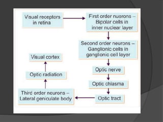
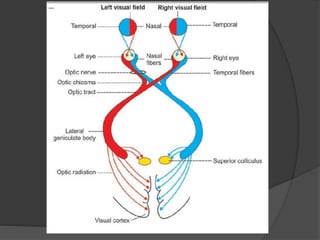
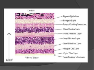
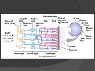
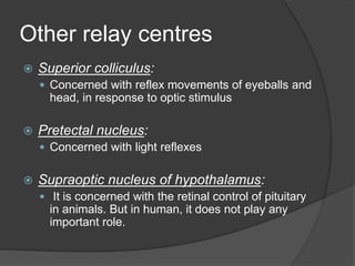
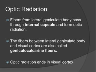
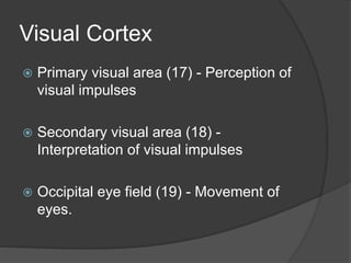
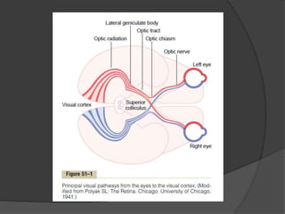
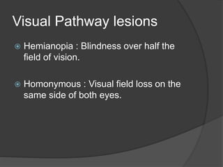
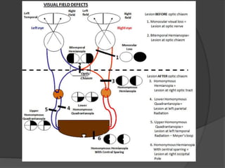
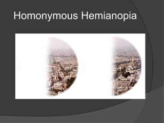
Venkatesh Karthikeyan
II MBBS
Velammal Medical College
Interpret!
 30 year old patient Mr.Siddarth , a
known case of Syphilis, presented to
Department of Ophthalmology, VMCH
with complaints of partial loss of
vision.What type of visual field defect
you suspect ? Why?
Interpret!
 40 year old patient
Mr.Aathreya, a known
diabetic and hypertensive,
presented to Department of
Ophthalmology, VMCH with
complaints of partial loss of
vision in his left eye. He is a
cigarette smoker for past 20
years.
What is Visual Pathway?
 Visual pathway or optic pathway is the
nervous pathway that transmits impulses
from retina to visual centre in cerebral
cortex.
VISUAL RECEPTORS
 Rods and cones
 They synapse with dendrites of bipolar
cells of inner nuclear layer of the retina.
FIRST ORDER NEURONS
 First order neurons (primary neurons)
are bipolar cells in the retina.
 Axons from the bipolar cells synapse
with dendrites of ganglionic cells.
SECOND ORDER
NEURONS
 They are the ganglionic cells in
ganglionic cell layer of retina.
 Axons of the ganglionic cells form optic
nerve.
 Optic nerve leaves the eye and
terminates in lateral geniculate body.
THIRD ORDER NEURONS
 They are in the lateral geniculate body.
 Fibres arising from LGB, reaches the
visual cortex.
OPTIC NERVE
 Formed by the axons of ganglionic cells
 It leaves the eye through optic disc.
 The fibers from temporal part of retina
are in lateral part of the nerve and carry
the impulses from nasal half of visual
field of same eye and vice versa.
„OPTIC CHIASMA
 Medial fibers of each optic nerve cross
the midline and join the uncrossed
lateral fibers of opposite side, to form
the optic tract.
Optic Tract
 Formed by uncrossed fibers of optic
nerve on the same side and crossed
fibers of optic nerve from the opposite
side.
 The fibers turn around the peduncle to
reach the lateral geniculate body in
thalamus.
 Here, many fibers synapse while few
fibers just pass through this and run
towards superior colliculus in midbrain.
 Fibers from fovea do not enter superior
colliculus.
LATERAL GENICULATE
BODY
 Majority of the fibers of optic tract
terminate in lateral geniculate body,
which forms the subcortical center for
visual sensation.
 From here, the geniculocalcarine tract
or optic radiation arises.
Other relay centres
 Superior colliculus:
 Concerned with reflex movements of eyeballs and
head, in response to optic stimulus
 Pretectal nucleus:
 Concerned with light reflexes
 Supraoptic nucleus of hypothalamus:
 It is concerned with the retinal control of pituitary
in animals. But in human, it does not play any
important role.
Optic Radiation
 Fibers from lateral geniculate body pass
through internal capsule and form optic
radiation.
 The fibers between lateral geniculate body
and visual cortex are also called
geniculocalcarine fibers.
 Optic radiation ends in visual cortex
Visual Cortex
 Primary visual area (17) - Perception of
visual impulses
 Secondary visual area (18) -
Interpretation of visual impulses
 Occipital eye field (19) - Movement of
eyes.
Visual Pathway lesions
 Hemianopia : Blindness over half the
field of vision.
 Homonymous : Visual field loss on the
same side of both eyes.
Causes of Optic Nerve
Lesion
 Optic atrophy
 Traumatic avulsion of the optic nerve
 Indirect optic neuropathy
 Acute optic neuritis
Lesions of Chiasma
 Suprasellar aneurysms
 Tumours of pituitary gland
 Third ventricular dilatation due to
obstructive hydrocephalus
 Atheroma of the carotids or posterior
communicating arteries
Optic tract lesions
 Syphilitic meningitis
 Tuberculosis and tumours of optic
thalamus
 Aneurysms of superior cerebellar or
posterior cerebral arteries.
Lesion in Optic Radiation
 Vascular occlusions
 Primary and secondary tumours
 Trauma
Homonymous Hemianopia
Upper Homonymous
Quadrantanopia
Lower Homonymous
Quadrantanopia
Interpret!
 30 year old patient Mr.Siddarth , a
known case of Syphilis, presented to
Department of Ophthalmology, VMCH
with complaints of partial loss of
vision.What type of visual field defect
you suspect ? Why?
 Homonymous hemianopia due to Optic
tract lesion
Interpret!
 40 year old patient
Mr.Aathreya, a known
diabetic and hypertensive,
presented to Department of
Ophthalmology, VMCH with
complaints of partial loss of
vision in his left eye. He is a
cigarette smoker for past 20
years.
 Homonymous hemianopia
with macular sparing!
Thank you 

Visual Pathway - Ophthalmology - Eye

  • 1.
  • 2.
    Interpret!  30 yearold patient Mr.Siddarth , a known case of Syphilis, presented to Department of Ophthalmology, VMCH with complaints of partial loss of vision.What type of visual field defect you suspect ? Why?
  • 3.
    Interpret!  40 yearold patient Mr.Aathreya, a known diabetic and hypertensive, presented to Department of Ophthalmology, VMCH with complaints of partial loss of vision in his left eye. He is a cigarette smoker for past 20 years.
  • 4.
    What is VisualPathway?  Visual pathway or optic pathway is the nervous pathway that transmits impulses from retina to visual centre in cerebral cortex.
  • 9.
    VISUAL RECEPTORS  Rodsand cones  They synapse with dendrites of bipolar cells of inner nuclear layer of the retina.
  • 10.
    FIRST ORDER NEURONS First order neurons (primary neurons) are bipolar cells in the retina.  Axons from the bipolar cells synapse with dendrites of ganglionic cells.
  • 11.
    SECOND ORDER NEURONS  Theyare the ganglionic cells in ganglionic cell layer of retina.  Axons of the ganglionic cells form optic nerve.  Optic nerve leaves the eye and terminates in lateral geniculate body.
  • 12.
    THIRD ORDER NEURONS They are in the lateral geniculate body.  Fibres arising from LGB, reaches the visual cortex.
  • 13.
    OPTIC NERVE  Formedby the axons of ganglionic cells  It leaves the eye through optic disc.  The fibers from temporal part of retina are in lateral part of the nerve and carry the impulses from nasal half of visual field of same eye and vice versa.
  • 14.
    „OPTIC CHIASMA  Medialfibers of each optic nerve cross the midline and join the uncrossed lateral fibers of opposite side, to form the optic tract.
  • 15.
    Optic Tract  Formedby uncrossed fibers of optic nerve on the same side and crossed fibers of optic nerve from the opposite side.  The fibers turn around the peduncle to reach the lateral geniculate body in thalamus.
  • 16.
     Here, manyfibers synapse while few fibers just pass through this and run towards superior colliculus in midbrain.  Fibers from fovea do not enter superior colliculus.
  • 17.
    LATERAL GENICULATE BODY  Majorityof the fibers of optic tract terminate in lateral geniculate body, which forms the subcortical center for visual sensation.  From here, the geniculocalcarine tract or optic radiation arises.
  • 18.
    Other relay centres Superior colliculus:  Concerned with reflex movements of eyeballs and head, in response to optic stimulus  Pretectal nucleus:  Concerned with light reflexes  Supraoptic nucleus of hypothalamus:  It is concerned with the retinal control of pituitary in animals. But in human, it does not play any important role.
  • 19.
    Optic Radiation  Fibersfrom lateral geniculate body pass through internal capsule and form optic radiation.  The fibers between lateral geniculate body and visual cortex are also called geniculocalcarine fibers.  Optic radiation ends in visual cortex
  • 20.
    Visual Cortex  Primaryvisual area (17) - Perception of visual impulses  Secondary visual area (18) - Interpretation of visual impulses  Occipital eye field (19) - Movement of eyes.
  • 23.
    Visual Pathway lesions Hemianopia : Blindness over half the field of vision.  Homonymous : Visual field loss on the same side of both eyes.
  • 25.
    Causes of OpticNerve Lesion  Optic atrophy  Traumatic avulsion of the optic nerve  Indirect optic neuropathy  Acute optic neuritis
  • 26.
    Lesions of Chiasma Suprasellar aneurysms  Tumours of pituitary gland  Third ventricular dilatation due to obstructive hydrocephalus  Atheroma of the carotids or posterior communicating arteries
  • 27.
    Optic tract lesions Syphilitic meningitis  Tuberculosis and tumours of optic thalamus  Aneurysms of superior cerebellar or posterior cerebral arteries.
  • 28.
    Lesion in OpticRadiation  Vascular occlusions  Primary and secondary tumours  Trauma
  • 29.
  • 30.
  • 31.
  • 32.
    Interpret!  30 yearold patient Mr.Siddarth , a known case of Syphilis, presented to Department of Ophthalmology, VMCH with complaints of partial loss of vision.What type of visual field defect you suspect ? Why?  Homonymous hemianopia due to Optic tract lesion
  • 33.
    Interpret!  40 yearold patient Mr.Aathreya, a known diabetic and hypertensive, presented to Department of Ophthalmology, VMCH with complaints of partial loss of vision in his left eye. He is a cigarette smoker for past 20 years.  Homonymous hemianopia with macular sparing!
  • 34.