SlideShare a Scribd company logo
1 of 175
Lecture 3Lecture 3
Ivano-Frankivsk National Medical UniversityIvano-Frankivsk National Medical University
The Department of Human anatomyThe Department of Human anatomy
Prepared by PhD TetyanaPrepared by PhD Tetyana
Knyazevych - ChornaKnyazevych - Chorna
Splanchnology.
Digestive system
Respiratory
System
Urinary System
The Reproductive
System
 Splanchnology-branch of anatomy, which
studies the structure of the internal
organs.
 The internal organs (viscera) are
responsible for important functions of the
organism: homeostasis, metabolism,
water and gases turnover, excretion of
metabolic products and reproduction.
 ISCOMS – 2009ISCOMS – 2009
 ISCOMS – InternationalISCOMS – International
Student Congress of MedicalStudent Congress of Medical
Sciences – is one of theSciences – is one of the
world’s leading studentworld’s leading student
conferences in the medicalconferences in the medical
sciences.sciences.
 the University Medical Center ofthe University Medical Center of
GroningenGroningen (the(the NetherlandsNetherlands ))  
WithWith Prof. Robin WarrenProf. Robin Warren
MD PhD, Nobel PrizeMD PhD, Nobel Prize
laureate in 2005laureate in 2005
  hehe is credited with the 1979 re-is credited with the 1979 re-
discovery of the discovery of the bacteriumbacterium
Helicobacter pyloriHelicobacter pylori, together with , together with 
Barry MarshallBarry Marshall..
 Digestive systemDigestive system is the foodis the food
processing system of humanprocessing system of human
body. The whole digestivebody. The whole digestive
system is in the form of a long,system is in the form of a long,
hollow, twisted and turnedhollow, twisted and turned
tube, called the alimentarytube, called the alimentary
canal, which starts from thecanal, which starts from the
oral cavity and ends at theoral cavity and ends at the
anus. The overall process ofanus. The overall process of
digestion and absorption ofdigestion and absorption of
food occurs in this tube. Thefood occurs in this tube. The
tube is divided into differenttube is divided into different
parts on the basis of structureparts on the basis of structure
and function of each part.and function of each part.
Necessary Organs Of the digestive system:Necessary Organs Of the digestive system:
 ORAL CAVITYORAL CAVITY
 ESOPHAGUSESOPHAGUS
 STOMACHSTOMACH
 SMALL INTESTINESMALL INTESTINE
 LARGE INTESTINELARGE INTESTINE
 ANUSANUS
Accessory Organs of the digestive system:Accessory Organs of the digestive system:
 LIVER AND GALL BLADDERLIVER AND GALL BLADDER
 PANCREASPANCREAS
 SALIVARY GLANDSSALIVARY GLANDS
 TEETHTEETH
 TONGUETONGUE
Functions of digestive system:Functions of digestive system:
 IngestionIngestion
 Digestion: break down of large particles of foodDigestion: break down of large particles of food

mechanical digestionmechanical digestion

chemical digestionchemical digestion
 PropulsionPropulsion
 peristalsisperistalsis
 segmentationsegmentation
 Secretion:Secretion:
 digestive enzymesdigestive enzymes
 hormoneshormones
 Absorption:Absorption:
 from external environment into internal environmentfrom external environment into internal environment
 across mucosaacross mucosa
 Elimination of wastes (defecation)Elimination of wastes (defecation)
Oral Cavity (mouth)Oral Cavity (mouth)
Entrance to the GI tract.Entrance to the GI tract.
 Initial site of digestion:Initial site of digestion:
 mechanical digestion (viamechanical digestion (via
mastication)mastication)
 chemical digestion (viachemical digestion (via
enzymes in saliva).enzymes in saliva).
 Bounded anteriorly by theBounded anteriorly by the
teeth and lipsteeth and lips
 Bounded posteriorly by the oropharynx.Bounded posteriorly by the oropharynx.
 Superior boundary is formed by theSuperior boundary is formed by the
hard and soft palates.hard and soft palates.
 Floor, or inferior surface, of the oralFloor, or inferior surface, of the oral
cavitycavity
 the tonguethe tongue
 FaucesFauces represent the opening between the oralrepresent the opening between the oral
cavity and the oropharynx.cavity and the oropharynx.
 Fauces are bounded by paired muscular folds:Fauces are bounded by paired muscular folds:
 glossopalatine arch (anterior fold)glossopalatine arch (anterior fold)
 pharyngopalatine arch (posterior fold)pharyngopalatine arch (posterior fold)
 Palatine tonsils are housed between the arches.Palatine tonsils are housed between the arches.
TeethTeeth
 Collectively known as theCollectively known as the
dentitiondentition..
 Responsible forResponsible for masticationmastication
 first part of the mechanicalfirst part of the mechanical
digestion.digestion.
 A tooth has:A tooth has:
 crowncrown
 neckneck
 one or moreone or more rootsroots
 Collectively, the roots, theCollectively, the roots, the
dental alveoli, and thedental alveoli, and the
periodontal ligament that bindsperiodontal ligament that binds
the roots to the alveolarthe roots to the alveolar
TOOTH TYPES ARE :TOOTH TYPES ARE :
INCISORSINCISORS
CANINECANINE
PREMOLARSPREMOLARS
MOLARSMOLARS
COMPONENTS OF THE TOOTHCOMPONENTS OF THE TOOTH
INCLUDE:INCLUDE:
ENAMEL
DENTIN
PULP
CEMENTUM
 20 deciduous teeth, also called “milk teeth,”
erupt between 6 months and 30 months after
birth.
 These teeth are eventually lost and replaced by
32 permanent teeth.
THE LARGETHE LARGE SALIVARY GLANDSSALIVARY GLANDS ARE :ARE :
 PAROTIDPAROTID
 SUBMANDIBULARSUBMANDIBULAR
 SUBLINGUALSUBLINGUAL
GLANDS PRODUCE SEROUS AND MUCOSALGLANDS PRODUCE SEROUS AND MUCOSAL
SECRETIONSSECRETIONS
Components of salivaComponents of saliva
 Water:Water: makes up 99%makes up 99%
 AmylaseAmylase: first step of chemical digestion: first step of chemical digestion
 LysozymeLysozyme: antimicrobial: antimicrobial
FunctionsFunctions
 Moisten foodMoisten food
 Food molecules into solution: tasteFood molecules into solution: taste
 Form bolus: for swallowingForm bolus: for swallowing
 Cleanse oral cavity.Cleanse oral cavity.
TONSILS INCLUDETONSILS INCLUDE ::
PHARYNGEAL, PALATINE,PHARYNGEAL, PALATINE,
LINGUALLINGUAL
THETHE PHARYNXPHARYNX INCLUDES :INCLUDES :
 NASOPHARYNXNASOPHARYNX
 OROPHARYNXOROPHARYNX
 LARYNGOPHARYNXLARYNGOPHARYNX
SwallowingSwallowing
Swallowing involves coordinatedSwallowing involves coordinated
activity of tongue, soft palateactivity of tongue, soft palate
pharynx and oesophagus.pharynx and oesophagus.
The first (buccal) phase isThe first (buccal) phase is
voluntary, food being forcedvoluntary, food being forced
into the pharynx by the tongue.into the pharynx by the tongue.
After this the process is reflex.After this the process is reflex.
The tongue blocks the mouth,The tongue blocks the mouth,
soft palate closes off the nosesoft palate closes off the nose
and the larynx rises so that theand the larynx rises so that the
epiglottis closes off theepiglottis closes off the
trachea. Food thus moves intotrachea. Food thus moves into
the pharynx and onwards bythe pharynx and onwards by
peristalsis aided by gravity.peristalsis aided by gravity.
If we try to talk whilst swallowingIf we try to talk whilst swallowing
food may enter the respiratoryfood may enter the respiratory
passages and a cough reflexpassages and a cough reflex
expels the bolus.expels the bolus.
EsophagusEsophagus
 A straight muscular tube about 25-30A straight muscular tube about 25-30
cm longcm long
 It begins at the level of the cricoidIt begins at the level of the cricoid
cartilage, inferior to the larnyx behindcartilage, inferior to the larnyx behind
the trachea and extends through thethe trachea and extends through the
chest cavity, pierces the diaphragm atchest cavity, pierces the diaphragm at
the esophageal hiatus , and meets withthe esophageal hiatus , and meets with
the stomach at an opening called thethe stomach at an opening called the
cardiac orifice.cardiac orifice.
 It transports food to the stomach andIt transports food to the stomach and
secretes mucus, which aids transport.secretes mucus, which aids transport.
 The inferior segment is constrictedThe inferior segment is constricted
forming the lower esophageal sphincterforming the lower esophageal sphincter
which, along with the diaphragm,which, along with the diaphragm,
closes to prevent back flow ofcloses to prevent back flow of
stomach contentsstomach contents
StomachStomach
 An enlarged segment of theAn enlarged segment of the
tract that functions mainly intract that functions mainly in
storing food and mixing it withstoring food and mixing it with
gastric juice (creating a pastegastric juice (creating a paste
called chyme).called chyme).
Other functions of stomachOther functions of stomach
include:include:
 Chemical digestion of proteinsChemical digestion of proteins
 Secretion of intrinsic factor –Secretion of intrinsic factor –
a chemical that is necessarya chemical that is necessary
for vitamin B12 absorption.for vitamin B12 absorption.
 Destruction of ingestedDestruction of ingested
bacteria via secretedbacteria via secreted
hydrochloric acid.hydrochloric acid.
Gross anatomyGross anatomy
 CardiaCardia
 Cardiac orificeCardiac orifice
 FundusFundus
 BodyBody
 PylorusPylorus
 Pyloric sphincterPyloric sphincter
 Pyloric orificePyloric orifice
 Greater curvatureGreater curvature
 Greater omentumGreater omentum
 Lesser curvatureLesser curvature
 Lesser omemtumLesser omemtum
 THE WALL OF THE STOMACHTHE WALL OF THE STOMACH
INCLUDESINCLUDES ::
 EXTERNAL SEROSAEXTERNAL SEROSA
 MUSCLE LAYERMUSCLE LAYER
( CIRCULAR,( CIRCULAR,
LONGITUDINAL, OBLIQUE )LONGITUDINAL, OBLIQUE )
 SUBMUCOSASUBMUCOSA
 INTERIOR MUCOSALINTERIOR MUCOSAL
 The mucous membraneThe mucous membrane
lining the stomach islining the stomach is
thick and vascular. It isthick and vascular. It is
thrown into numerousthrown into numerous
folds, known as rugae,folds, known as rugae,
which are predominantlywhich are predominantly
longitudinal in direction.longitudinal in direction.
On distention of theOn distention of the
stomach, these foldsstomach, these folds
flatten out. It contains theflatten out. It contains the
glands and the gastricglands and the gastric
pits.pits.
GASTRIC PITS ARE THEGASTRIC PITS ARE THE
OPENINGS INTO THE GASTRICOPENINGS INTO THE GASTRIC
GLANDSGLANDS
 Mucous neck cellsMucous neck cells – found in the– found in the
upper portion of the gland. Secreteupper portion of the gland. Secrete
acidic mucus and function as stemacidic mucus and function as stem
cells for surface mucous cells.cells for surface mucous cells.
 Chief cellsChief cells – primary function is– primary function is
the secretion of pepsinogen, anthe secretion of pepsinogen, an
inactive form of the protease,inactive form of the protease,
pepsin. Pepsinogen is activated bypepsin. Pepsinogen is activated by
HCl and by pepsin itself.HCl and by pepsin itself.
 Parietal cellsParietal cells – found in the– found in the
midportion of the glands. Secretemidportion of the glands. Secrete
hydrochloric acid (which gives thehydrochloric acid (which gives the
stomach its low pH – usually 1-3)stomach its low pH – usually 1-3)
as well as intrinsic factor.as well as intrinsic factor.
 Enteroendocrine cellsEnteroendocrine cells – secrete– secrete
multiple hormones into the plasma.multiple hormones into the plasma.
An example is gastrin, a hormoneAn example is gastrin, a hormone
that regulates the stomach’sthat regulates the stomach’s
motility and secretory activity.motility and secretory activity.
Stomach has three main functions;Stomach has three main functions;
 Storage of food:Storage of food:  It stores food for a variable amount It stores food for a variable amount
of time (depending on the nature of food) in order toof time (depending on the nature of food) in order to
make it appropriate for digestion and absorption in themake it appropriate for digestion and absorption in the
small intestine. The storing capacity of stomach in ansmall intestine. The storing capacity of stomach in an
average adult is about 1500 ml.average adult is about 1500 ml.
 Mixing of food:Mixing of food:  It mixes the food with its own It mixes the food with its own
secretions (gastric secretions) to form a semifluidsecretions (gastric secretions) to form a semifluid
chyme.chyme.
 Controlling the rate of delivery of chyme:Controlling the rate of delivery of chyme:   TheThe
stomach also controls the rate of delivery of chyme tostomach also controls the rate of delivery of chyme to
the small intestine so that efficient digestion andthe small intestine so that efficient digestion and
absorption can take place.absorption can take place.
  
Small intestineSmall intestine
is the longest part of alimentaryis the longest part of alimentary
canal. It extends from thecanal. It extends from the
pylorus of the stomach to thepylorus of the stomach to the
junction between cecum andjunction between cecum and
ileum. Much of digestion andileum. Much of digestion and
absorption of food takes placeabsorption of food takes place
in the small intestine.in the small intestine. IngestedIngested
nutrients spend at leastnutrients spend at least 1212
hours in the small intestine.hours in the small intestine.
The small intestine is divided into threeThe small intestine is divided into three
parts:parts:
-- DuodenumDuodenum
-- JejunumJejunum
-- Ileum.Ileum.
Duodenum:Duodenum:
It is the first part of smallIt is the first part of small
intestine that joins theintestine that joins the
stomach. It is a C-stomach. It is a C-
shaped tube, about 10shaped tube, about 10
inches longinches long (25,4 sm)(25,4 sm). It. It
lies between thelies between the
stomach and jejunumstomach and jejunum
TThe duodenum is divided into fourhe duodenum is divided into four
partsparts::
 superiorsuperior
 descendingdescending
 inferior/horizontainferior/horizonta ll
 ascendingascending
The organs and structuresThe organs and structures
surrounding the duodenumsurrounding the duodenum
includes :includes :
 Superior (above) – liver andSuperior (above) – liver and
gallbladdergallbladder
 Anterior (in front) – gallbladder andAnterior (in front) – gallbladder and
transverse colontransverse colon
 Posterior (behind) – inferior venaPosterior (behind) – inferior vena
cava, right psoas major muscle andcava, right psoas major muscle and
aortaaorta
 Lateral – pancreasLateral – pancreas
A A liquid mixtureliquid mixture of food and gastric secretions of food and gastric secretions
enters the superior duodenum from the pylorusenters the superior duodenum from the pylorus
of the stomach, triggering the release ofof the stomach, triggering the release of
pancreas-stimulating hormones from glands inpancreas-stimulating hormones from glands in
the duodenal wall. Ducts fromthe duodenal wall. Ducts from
the the pancreaspancreas and  and gallbladdergallbladder enter at enter at
the descending duodenum, bringing bicarbonatethe descending duodenum, bringing bicarbonate
to neutralize the acid in the gastric secretions,to neutralize the acid in the gastric secretions,
pancreatic enzymes to further digestion,pancreatic enzymes to further digestion,
and and bile saltsbile salts to emulsify fat. The mucous to emulsify fat. The mucous
lining of the last two segments of the duodenumlining of the last two segments of the duodenum
begins the absorption of nutrients, in particularbegins the absorption of nutrients, in particular
iron and calcium, before the food contents enteriron and calcium, before the food contents enter
the next part of the small intestine, the the next part of the small intestine, the jejunumjejunum..
The Jejunum and IleumThe Jejunum and Ileum
 TheThe jejunumjejunum
 middle region of the small intestine.middle region of the small intestine.
 approximately 2.5 meters (7.5 feet)approximately 2.5 meters (7.5 feet)
 makes up approximately two-fifths of the smallmakes up approximately two-fifths of the small
intestine’s total length.intestine’s total length.
 primary region for chemical digestion andprimary region for chemical digestion and
nutrient absorptionnutrient absorption
 TheThe ileumileum
 is the last region of the small intestine.is the last region of the small intestine.
 about 3.6 meters (10.8 feet) in lengthabout 3.6 meters (10.8 feet) in length
 forms approximately three-fifths of the smallforms approximately three-fifths of the small
intestine.intestine.
 terminates at the ileocecal valveterminates at the ileocecal valve
 sphincter that controls the entry of materials into thesphincter that controls the entry of materials into the
large intestine.large intestine.
 Ileocecal valve, as the name indicates, is aIleocecal valve, as the name indicates, is a
structure present at the junction of ileum andstructure present at the junction of ileum and
cecum. It is a rudimentary valve and consists ofcecum. It is a rudimentary valve and consists of
two horizontal folds of mucous membrane thattwo horizontal folds of mucous membrane that
project around the opening of the ileum. Ileocecalproject around the opening of the ileum. Ileocecal
valve controlvalve controlss the flow of contents from ileum intothe flow of contents from ileum into
the large intestine.the large intestine.
Large IntestineLarge Intestine
 approximate length of 1.5 meters (5approximate length of 1.5 meters (5
feet)feet)
 diameter of 6.5 centimeters (2.5diameter of 6.5 centimeters (2.5
inches).inches).
 Absorbs most of the waterAbsorbs most of the water andand
electrolytes from the remaining digestedelectrolytes from the remaining digested
material.material.
 Watery material that first enters theWatery material that first enters the
large intestine soonlarge intestine soon solidifiessolidifies andand
becomesbecomes faeces.faeces.
 Stores faecal material until the body isStores faecal material until the body is
ready toready to defaecate.defaecate.
 Absorbs a very small percentage ofAbsorbs a very small percentage of
nutrientsnutrients still remaining in the digestedstill remaining in the digested
material.material.
 Composed of four segments:Composed of four segments:
 thethe caecumcaecum,, coloncolon,, rectumrectum,, analanal
canalcanal
      
Structures Structures 
       a. cecum with vermiform appendix        a. cecum with vermiform appendix 
       b. ascending, transverse, descending colon        b. ascending, transverse, descending colon 
       c. sigmoid colon, rectum        c. sigmoid colon, rectum 
            
Haustra are pouches in wall of large intestine Haustra are pouches in wall of large intestine 
                  
Millions of bacteria colonize the large intestine. TheyMillions of bacteria colonize the large intestine. They
breakdown indigestible carbohydrate residues andbreakdown indigestible carbohydrate residues and
produce many B vitamins as well as most of the body’sproduce many B vitamins as well as most of the body’s
vitamin K supply.vitamin K supply.
Typically 100 g of feces are produced per day consisting ofTypically 100 g of feces are produced per day consisting of
75% water and 25% solids. Solids include dead bacteria,75% water and 25% solids. Solids include dead bacteria,
fat, inorganic matter, protein, undigested plant fibers,fat, inorganic matter, protein, undigested plant fibers,
bile pigments, and shed epithelial cells.  bile pigments, and shed epithelial cells.       
The liverThe liver is an accessory digestive organ thatis an accessory digestive organ that
has multiple functions including:has multiple functions including:
 Carbohydrate metabolism – storage andCarbohydrate metabolism – storage and
release of glucoserelease of glucose
 Removal of drugs, toxins, and foreignRemoval of drugs, toxins, and foreign
chemicals from the plasmachemicals from the plasma
 Storage of vitamins (A, D, E, and K) andStorage of vitamins (A, D, E, and K) and
minerals (iron and copper)minerals (iron and copper)
 Protein metabolismProtein metabolism
 Lipid metabolismLipid metabolism
 Synthesis of plasma proteins (e.g., albumin,Synthesis of plasma proteins (e.g., albumin,
fibrin, etc.)fibrin, etc.)
 Phagocytosis of old RBCs and of pathogens.Phagocytosis of old RBCs and of pathogens.
 Production of bile (0.5-1 L/day).Production of bile (0.5-1 L/day).
 The liverThe liver
 composed of four incompletely separatedcomposed of four incompletely separated
lobeslobes
 supported by two ligamentssupported by two ligaments
 Right lobeRight lobe
 Left lobeLeft lobe
 Falciform ligamentFalciform ligament
 Round ligamentRound ligament
 Caudate lobeCaudate lobe
 Quadrate lobeQuadrate lobe
 The gallbladderThe gallbladder is a thin-walledis a thin-walled
green muscular sac found on thegreen muscular sac found on the
ventral surface of the liver.ventral surface of the liver.
 It functions primarily in the storageIt functions primarily in the storage
and concentration of bile.and concentration of bile.
 The liver continuously produces bile.The liver continuously produces bile.
However the hepatopancreaticHowever the hepatopancreatic
sphincter is normally closed. Thissphincter is normally closed. This
results in bile backing up into theresults in bile backing up into the
common bile duct, cystic duct, andcommon bile duct, cystic duct, and
ultimately into the gallbladder.ultimately into the gallbladder.
 When fatty chyme arrives in the smallWhen fatty chyme arrives in the small
intestine, intestinal glands secrete theintestine, intestinal glands secrete the
hormone cholecystokinin.hormone cholecystokinin.
 CCK causes gallbladder contractionCCK causes gallbladder contraction
and relaxation of the hepatopancreaticand relaxation of the hepatopancreatic
sphincter, letting bile flow into thesphincter, letting bile flow into the
duodenum.duodenum.
The pancreasThe pancreas
 Mostly retroperitoneal and deep to the greater curvature ofMostly retroperitoneal and deep to the greater curvature of
the stomach.the stomach.
 TheThe headhead of the pancreas sits next to the duodenum as itof the pancreas sits next to the duodenum as it
emanates from and loops away from the pylorus. Theemanates from and loops away from the pylorus. The bodybody
extends behind the stomach and itsextends behind the stomach and its tailtail ends at the spleen.ends at the spleen.
 The pancreas primarily consists ofThe pancreas primarily consists of aciniacini – small clusters of– small clusters of
enzyme secreting cells. These acinar cells empty theirenzyme secreting cells. These acinar cells empty their
secretion into small ducts. Small ducts coalesce into largersecretion into small ducts. Small ducts coalesce into larger
ducts that empty into theducts that empty into the main pancreatic ductmain pancreatic duct , which, which
runs centrally along the long axis of the pancreasruns centrally along the long axis of the pancreas
 The major function of the acinar and duct cells is theThe major function of the acinar and duct cells is the
secretion ofsecretion of pancreatic juicepancreatic juice (1.5 L/day).(1.5 L/day).
 Acinar cells contribute digestive enzymes to the pancreaticAcinar cells contribute digestive enzymes to the pancreatic
juice including:juice including: proteases, pancreatic lipase,proteases, pancreatic lipase,
pancreatic amylase.pancreatic amylase.
 Scattered amidst the pancreatic acini are the hormone-Scattered amidst the pancreatic acini are the hormone-
producingproducing islets of Langerhansislets of Langerhans . Their major function is. Their major function is
the regulation of blood glucose levels.the regulation of blood glucose levels.
 PeritoneumPeritoneum
 Parietal peritoneumParietal peritoneum
 Visceral peritoneumVisceral peritoneum
 Peritoneal cavityPeritoneal cavity
 Intraperitoneal organsIntraperitoneal organs
 Mesoperitoneal organsMesoperitoneal organs
 Retroperitoneal organsRetroperitoneal organs
The The respiratory systemrespiratory system is the anatomical system of is the anatomical system of
an organism that introduces respiratory gases to thean organism that introduces respiratory gases to the
interior and performs gas exchange. In humans theinterior and performs gas exchange. In humans the
respiratory system include airways, lungs, and therespiratory system include airways, lungs, and the
respiratory muscles. Molecules of oxygen and carbonrespiratory muscles. Molecules of oxygen and carbon
dioxide are passively exchanged, by diffusion,dioxide are passively exchanged, by diffusion,
between the gaseous external environment andbetween the gaseous external environment and
the blood. This exchange process occurs in thethe blood. This exchange process occurs in the
alveolar region of the lungsalveolar region of the lungs
General Functions of Respiratory
System:
 1. O2 (oxygenoxygen)) and CO2 (carbon dioxidecarbon dioxide)
exchange between blood and air
 2. speech and vocalization
 3. sense of smell
 4. helps control acid base balance of body
 5. breathing movements help promote
blood and lymph flow
Main Organs:
 nose
 pharynx
 larynx
 trachea
 primary bronchi
 lungs:
 bronchioles
 alveoli/respiratory membrane
these organs can also be subdivided
into:
 upper respiratory tract
Nose-pharynx-larynx
 lower respiratory tract
respiratory organs of the thorax
 Functional classifications:Functional classifications:
 ConductingConducting portion: transports air.portion: transports air.
 RespiratoryRespiratory portion: carries out gas exchange.portion: carries out gas exchange.
 respiratory bronchiolesrespiratory bronchioles
 alveolar ductsalveolar ducts
 air sacs called alveoliair sacs called alveoli
 Upper respiratory tract is all conductingUpper respiratory tract is all conducting
 Lower respiratory tract has bothLower respiratory tract has both
conducting and respiratory portionsconducting and respiratory portions
Nose
 separated from mouth by hard and
soft palate
 each nasal cavity is divided into 3
passageways by turbinates
(choncae)
turbulent passageways to insure that
all air makes contact with mucous
membranes
membranes are heavily vascualrized
 remove bacteria, debris and
particles
 warms and moisturizes air entering
lungs
 also contains receptors for smell
nasolacrimal ducts drain into nasal
cavity
Paranasal sinuses:Paranasal sinuses:
 In four skull bonesIn four skull bones
 paired air spacespaired air spaces
 decrease skull bone weightdecrease skull bone weight
 warm and moisten air
 sound resonance
 Named for the bonesNamed for the bones
in which they are housed.in which they are housed.
 frontalfrontal
 ethmoidalethmoidal
 sphenoidalsphenoidal
 maxillarymaxillary
 Communicate with the nasal cavity by ducts.Communicate with the nasal cavity by ducts.
 Covered with the same pseudostratified ciliatedCovered with the same pseudostratified ciliated
columnar epithelium as the nasal cavity.columnar epithelium as the nasal cavity.
Pharynx (throat)
- Is situated from base of skull to junction with
esophagus and trachea
- made of muscle and lined with mucous membrane
- junction between digestive and respiratory systems
divided into three regions:
a. Nasopharynx
 behind nose to level of soft palate
 includes uvula, tonsils (adenoids)
 auditory tube (eustachian tube)
b. Oropharynx
 behind mouth
 from soft palate to level of
hyoid bone
 palatine and lingual tonsils
c. Laryngopharynx
 from hyoid bone to
esophagus/larynx
Larynx (voice box)
 enlarged beginning portion of trachea
 composed of cartilage and muscles
 opening into larynx = glottis (prevent food
from entering lower respiratory system sound)
 9 cartilages:
 three individual piecesthree individual pieces
 Thyroid cartilageThyroid cartilage
 Cricoid cartilageCricoid cartilage
 EpiglottisEpiglottis
 three cartilage pairsthree cartilage pairs
 Arytenoids: on cricoidArytenoids: on cricoid
 Corniculates: attach toCorniculates: attach to
arytenoidsarytenoids
 Cuniforms:in aryepiglottic foldCuniforms:in aryepiglottic fold
Sound ProductionSound Production
 Two pairs of ligamentsTwo pairs of ligaments
 Inferior ligaments, calledInferior ligaments, called vocal ligamentsvocal ligaments
 covered by a mucous membranecovered by a mucous membrane
 vocal folds: ligament and mucosa.vocal folds: ligament and mucosa.
 are “true vocal cords”are “true vocal cords”
 they produce sound when air passes between themthey produce sound when air passes between them
 Superior ligaments, calledSuperior ligaments, called vestibular ligamentsvestibular ligaments
 Covered by mucosaCovered by mucosa
 vestibular folds: ligament and mucosavestibular folds: ligament and mucosa
 Are “false vocal cords”Are “false vocal cords”
 no function in sound productionno function in sound production
 protect the vocal folds.protect the vocal folds.
 The vestibular folds attach to the corniculateThe vestibular folds attach to the corniculate
cartilagescartilages ..
 The tension, length,The tension, length, and position of theand position of the
vocal folds determine the quality of thevocal folds determine the quality of the
sound.sound.
 Longer vocal folds produce lower soundsLonger vocal folds produce lower sounds
 More taunt, higher pitchMore taunt, higher pitch
 Loudness based on force of airLoudness based on force of air
 Rima glottidisRima glottidis: opening between the vocal: opening between the vocal
foldsfolds
 GlottisGlottis: rima glottidis and the vocal folds: rima glottidis and the vocal folds
Trachea
 extends from larynx to bronchi
(immediately anterior to the esophagus,immediately anterior to the esophagus,
inferior to the larynx, superior to theinferior to the larynx, superior to the
primary bronchi of the lungs.primary bronchi of the lungs. )
 surrounded by 15 to 2015 to 20 “C” – shaped
bands of cartilage ends joined by bands
of muscle tissue (holds walls open,
prevents collapse)
 lined by pseudostratified ciliated
columnar epithelium
 At the level of theAt the level of the sternal anglesternal angle, the, the
trachea bifurcates into two smaller tubes,trachea bifurcates into two smaller tubes,
called thecalled the right and left primary bronchiright and left primary bronchi..
 Each primary bronchus projects laterallyEach primary bronchus projects laterally
toward each lung.toward each lung.
 The most inferior tracheal cartilageThe most inferior tracheal cartilage
separates the primary bronchi at theirseparates the primary bronchi at their
origin and forms an internal ridge calledorigin and forms an internal ridge called
thethe carina.carina.
Bronchi
 bronchi resemble trachea in structure
(also supported by C-shaped cartilages)
 also have lots of elastic connective
tissue
 Right primary bronchusRight primary bronchus
 shorter, wider, and more vertically orientedshorter, wider, and more vertically oriented
than the left primary bronchus.than the left primary bronchus.
 Foreign particles are more likely to lodge inForeign particles are more likely to lodge in
the right primary bronchus.the right primary bronchus.
 each bronchus enters lung and
continues to divide into smaller and
smaller branches = bronchi, then into
microscopic bronchioles
 because of the extensive branching it
formes bronchial tree
 Primary bronchiPrimary bronchi
 enter the hilum of each lungenter the hilum of each lung
 Also entering hilum:Also entering hilum:
 pulmonary vesselspulmonary vessels
 lymphatic vesselslymphatic vessels
 nerves.nerves.
 Secondary bronchi (or lobar bronchi)Secondary bronchi (or lobar bronchi)
 Branch of primary bronchusBranch of primary bronchus
 left lung:left lung:
 two lobestwo lobes
 two secondary bronchitwo secondary bronchi
 right lungright lung
 three lobesthree lobes
 three secondary bronchi.three secondary bronchi.
 Tertiary bronchi (or segmental bronchi)Tertiary bronchi (or segmental bronchi)
 Branch of secondary bronchiBranch of secondary bronchi
 left lung is supplied by 8 to 10 tertiary bronchi.left lung is supplied by 8 to 10 tertiary bronchi.
 right lung is supplied by 10 tertiary bronchiright lung is supplied by 10 tertiary bronchi
 supply a part of the lung called a bronchopulmonarysupply a part of the lung called a bronchopulmonary
segment.segment.
Bronchioles
 smallest branches of “respiratory tree”
 <1mm diameter
 no cartilage rings (but larger branches may have small
patches of cartilage)
 lined with ciliated cuboidal epithelium and layer of smooth
muscle
 asthma affects the smallest terminal bronchioles
Respiratory Bronchioles, AlveolarRespiratory Bronchioles, Alveolar
Ducts, and AlveoliDucts, and Alveoli
 Contain small saccular outpocketings calledContain small saccular outpocketings called
alveoli.alveoli.
 An alveolus is about 0.25 to 0.5 millimeter inAn alveolus is about 0.25 to 0.5 millimeter in
diameter.diameter.
 Its thin wall is specialized to promote diffusionIts thin wall is specialized to promote diffusion
of gases between the alveolus and the blood inof gases between the alveolus and the blood in
the pulmonary capillaries.the pulmonary capillaries.
 Gas exchange can take place in the respiratoryGas exchange can take place in the respiratory
bronchioles and alveolar ducts as well as in thebronchioles and alveolar ducts as well as in the
lungs, which contain approximately 300–400lungs, which contain approximately 300–400
million alveoli.million alveoli.
 The spongy natureThe spongy nature of the lung is due to theof the lung is due to the
packing of millions of alveoli together.packing of millions of alveoli together.
Gross Anatomy of the LungsGross Anatomy of the Lungs
 Each lung has a conical shape.Each lung has a conical shape.
 Its wide, concave base rests upon the muscular diaphragm.Its wide, concave base rests upon the muscular diaphragm.
 Its relatively blunt superior region, called the apex orIts relatively blunt superior region, called the apex or
(cupola), projects superiorly to a point that is slightly(cupola), projects superiorly to a point that is slightly
superior and posterior to the clavicle.superior and posterior to the clavicle.
 Both lungs are bordered by the thoracic wall anteriorly,Both lungs are bordered by the thoracic wall anteriorly,
laterally, and posteriorly, and supported by the rib cage.laterally, and posteriorly, and supported by the rib cage.
 Toward the midline, the lungs are separated from eachToward the midline, the lungs are separated from each
other by the mediastinum.other by the mediastinum.
 The relatively broad, rounded surface in contact with theThe relatively broad, rounded surface in contact with the
thoracic wall is called the costal surface of the lung.thoracic wall is called the costal surface of the lung.
Pleura and Pleural CavitiesPleura and Pleural Cavities
 The outer surface of each lung and the adjacentThe outer surface of each lung and the adjacent
internal thoracic wall are lined by a serousinternal thoracic wall are lined by a serous
membrane called pleura, which is formed frommembrane called pleura, which is formed from
simple squamous epithelium.simple squamous epithelium.
 The outer surface of each lung is tightlyThe outer surface of each lung is tightly
covered by the visceral pleura, while thecovered by the visceral pleura, while the
internal thoracic walls, the lateral surfaces ofinternal thoracic walls, the lateral surfaces of
the mediastinum, and the superior surface ofthe mediastinum, and the superior surface of
the diaphragm are lined by the parietal pleura.the diaphragm are lined by the parietal pleura.
 The parietal and visceral pleural layers areThe parietal and visceral pleural layers are
continuous at the hilum of each lung.continuous at the hilum of each lung.
Pleura and Pleural CavitiesPleura and Pleural Cavities
 The potential space between theseThe potential space between these
serous membrane layers is a pleuralserous membrane layers is a pleural
cavity.cavity.
 The pleural membranes produce a thin,The pleural membranes produce a thin,
serous fluid that circulates in the pleuralserous fluid that circulates in the pleural
cavity and acts as a lubricant, ensuringcavity and acts as a lubricant, ensuring
minimal friction during breathing.minimal friction during breathing.
27-83
General Structure and FunctionsGeneral Structure and Functions
of the Urinary Systemof the Urinary System
 Urinary System organsUrinary System organs
 remove waste products from the bloodremove waste products from the blood
(there are toxic and accumulate in blood)(there are toxic and accumulate in blood)
 then from the bodythen from the body
 Major homeostatic systemMajor homeostatic system
27-84
General Structure and FunctionsGeneral Structure and Functions
of the Urinary Systemof the Urinary System
 Organs of the Urinary System:Organs of the Urinary System:
 KidneysKidneys
 UretersUreters
 Urinary BladderUrinary Bladder
 UrethraUrethra
 Primary organs: kidneysPrimary organs: kidneys
 filter waste products from the bloodstreamfilter waste products from the bloodstream
 convert the filtrate into urine.convert the filtrate into urine.
 The Urinary Tract:The Urinary Tract:
 Includes:Includes:
 uretersureters
 urinary bladderurinary bladder
 urethraurethra
 Because they transport the urine out of the body.Because they transport the urine out of the body.
85
86
87
27-88
Functions of the Urinary SystemFunctions of the Urinary System
 Removing waste products from the bloodstream.Removing waste products from the bloodstream.
 Storage of urine.Storage of urine.
 the urinary bladder is an expandable, muscular sac that canthe urinary bladder is an expandable, muscular sac that can
store as much as 1 liter of urinestore as much as 1 liter of urine
 Excretion of urine.Excretion of urine.
 Blood volume regulation.Blood volume regulation.
 the kidneys control the volume of interstitial fluid and bloodthe kidneys control the volume of interstitial fluid and blood
under the direction of certain hormonesunder the direction of certain hormones
 Regulation of erythrocyte production.Regulation of erythrocyte production.
 as the kidneys filter the blood, they are also indirectlyas the kidneys filter the blood, they are also indirectly
measuring the oxygen level in the bloodmeasuring the oxygen level in the blood
 Erythropoietin (EPO): hormone produced by kidneyErythropoietin (EPO): hormone produced by kidney
 Released if blood oxygen levels fallReleased if blood oxygen levels fall
 Stimulates RBC production in red bone marrowStimulates RBC production in red bone marrow
27-89
Kidneys: Gross and SectionalKidneys: Gross and Sectional
AnatomyAnatomy
 RetroperitonealRetroperitoneal
 Anterior surface covered with peritoneumAnterior surface covered with peritoneum
 Posterior surface against posterior abdominalPosterior surface against posterior abdominal
wallwall
 Superior pole: T-12Superior pole: T-12
 Inferior pole: L-3Inferior pole: L-3
 Right kidney ~ 2cm lower than leftRight kidney ~ 2cm lower than left
 Adrenal gland on superior poleAdrenal gland on superior pole
90
27-91
Kidneys: Gross and SectionalKidneys: Gross and Sectional
AnatomyAnatomy
 Hilum: concave medial borderHilum: concave medial border
 Renal sinus: internal spaceRenal sinus: internal space
 Houses blood vessels, lymphatic vessels,Houses blood vessels, lymphatic vessels,
nervesnerves
 Houses renal pelvis, renal calycesHouses renal pelvis, renal calyces
 Also fatAlso fat
27-92
Kidneys: Gross and SectionalKidneys: Gross and Sectional
AnatomyAnatomy
 Surrounding tissues, from deep to superficial:Surrounding tissues, from deep to superficial:
 Fibrous capsule (renal capsule)Fibrous capsule (renal capsule)
 Dense irregular CTDense irregular CT
 Covers outer surfaceCovers outer surface
 Perinephric fat (adipose capsule)Perinephric fat (adipose capsule)
 Also called perirenal fatAlso called perirenal fat
 Completely surrounds kidneyCompletely surrounds kidney
 Cushioning and insulationCushioning and insulation
 Renal fasciaRenal fascia
 Dense irregular CTDense irregular CT
 Anchors kidney to posterior wall and peritoneumAnchors kidney to posterior wall and peritoneum
 Paranephric fatParanephric fat
 Between renal fascia and peritoneumBetween renal fascia and peritoneum
93
27-94
Kidneys: Gross and SectionalKidneys: Gross and Sectional
AnatomyAnatomy
 Sectioned on a coronal plane:Sectioned on a coronal plane:
 Renal CortexRenal Cortex
 Renal archesRenal arches
 Renal columnsRenal columns
 Renal MedullaRenal Medulla
 Divided into renal pyramids (8 to 15 per kidney)Divided into renal pyramids (8 to 15 per kidney)
 Base against cortexBase against cortex
 Apex called renal papillaApex called renal papilla
27-95
Kidneys: Gross and SectionalKidneys: Gross and Sectional
AnatomyAnatomy
 Minor calyx:Minor calyx:
 Funnel shapedFunnel shaped
 Receives renal papillaReceives renal papilla
 8 to 15 per kidney, one per pyramid8 to 15 per kidney, one per pyramid
 Major calyxMajor calyx
 Fusion of minor calycesFusion of minor calyces
 2 to 3 per kidney2 to 3 per kidney
 Major calyces merge to form renal pelvisMajor calyces merge to form renal pelvis
 Renal LobeRenal Lobe
 Pyramid plus some cortical tissuePyramid plus some cortical tissue
 8 to 15 per kidney8 to 15 per kidney
96
97
27-98
Blood Supply to the KidneyBlood Supply to the Kidney
 About 20 to 25% of cardiac output toAbout 20 to 25% of cardiac output to
kidneyskidneys
 Path:Path:
 Renal artery to segmental arteries toRenal artery to segmental arteries to
interlobar arteries to arcuate arteries tointerlobar arteries to arcuate arteries to
interlobular arteries to:interlobular arteries to:
 Afferent arteriole to glomerulus to efferentAfferent arteriole to glomerulus to efferent
arteriole to peritubular capilaries and vasaarteriole to peritubular capilaries and vasa
rectarecta
99
27-100
Blood Supply to the KidneyBlood Supply to the Kidney
 Blood plasma is filtered across the glomerulusBlood plasma is filtered across the glomerulus
into the glomerular space.into the glomerular space.
 Once the blood plasma is filteredOnce the blood plasma is filtered
 blood leaves the glomerulusblood leaves the glomerulus
 enters an efferent arteriole.enters an efferent arteriole.
 efferent arteriole is still carrying oxygenatedefferent arteriole is still carrying oxygenated
bloodblood
 a gas and nutrient exchange with the kidney tissuesa gas and nutrient exchange with the kidney tissues
has not yet occurred.has not yet occurred.
27-101
Blood Supply to the KidneyBlood Supply to the Kidney
 The efferent arterioles branch into one of twoThe efferent arterioles branch into one of two
types of capillary networks:types of capillary networks:
 peritubular capillariesperitubular capillaries
 vasa rectavasa recta
 these capillary networks are responsiblethese capillary networks are responsible
for the actual exchange of gases andfor the actual exchange of gases and
nutrientsnutrients
 Peritubular capillaries:Peritubular capillaries: primarily in cortexprimarily in cortex
 Vasa recta:Vasa recta: surround the thin tubes thatsurround the thin tubes that
project into the medulla.project into the medulla.
27-102
Blood Supply to the KidneyBlood Supply to the Kidney
 Path for veins:Path for veins:
 Interlobar veins to arcuate veins to interlobarInterlobar veins to arcuate veins to interlobar
veins to the renal veinveins to the renal vein
103
27-104
NephronsNephrons
 The functional filtration unit in the kidney.The functional filtration unit in the kidney.
 Consists of the following:Consists of the following:
 Renal corpuscleRenal corpuscle
 GlomerulusGlomerulus
 Glomerular capsule (Bowman’s capsule)Glomerular capsule (Bowman’s capsule)
 Proximal convoluted tubule (PCT)Proximal convoluted tubule (PCT)
 Nephron loop (loop of Henle)Nephron loop (loop of Henle)
 Ascending loop of HenleAscending loop of Henle
 Descending loop of HenleDescending loop of Henle
 Distal convoluted tubule (DCT)Distal convoluted tubule (DCT)
 collectively called thecollectively called the renal tubulerenal tubule
 In both kidneys: approximatelyIn both kidneys: approximately 2.5 million nephrons.2.5 million nephrons.
 Are microscopic: measure aboutAre microscopic: measure about 5 centimeters5 centimeters inin
length.length.
27-105
NephronsNephrons
 Cortical NephronsCortical Nephrons
 Near peripheral edge of cortexNear peripheral edge of cortex
 Short nephron loopsShort nephron loops
 Have peritubular capillariesHave peritubular capillaries
 Juxtamedullary nephronsJuxtamedullary nephrons
 Near corticomedullary borderNear corticomedullary border
 Long nephron loopsLong nephron loops
 Have vasa rectaHave vasa recta
106
27-107
Urine FormationUrine Formation
 Three processesThree processes
 FiltrationFiltration
 Renal corpuscle: forms filtrateRenal corpuscle: forms filtrate
 From blood to tubuleFrom blood to tubule
 ReabsorptionReabsorption
 Mostly PCTMostly PCT
 Water and salt: rest of nephronWater and salt: rest of nephron
 From tubule to bloodFrom tubule to blood
 SecretionSecretion
 From blood to tubuleFrom blood to tubule
108
27-109
Urinary Tract : UretersUrinary Tract : Ureters
 long, fibromuscular tubeslong, fibromuscular tubes
 conduct urine from the kidneys to the urinary bladder.conduct urine from the kidneys to the urinary bladder.
 average 25 centimeters in lengthaverage 25 centimeters in length
 retroperitoneal.retroperitoneal.
 ureters originate at the renal pelvisureters originate at the renal pelvis
 extend inferiorly to enter the posterolateral wall of theextend inferiorly to enter the posterolateral wall of the
base of the urinary bladder.base of the urinary bladder.
 wall is composed of three concentric tunics.wall is composed of three concentric tunics.
 mucosamucosa
 muscularismuscularis
 adventitia.adventitia.
27-110
Urinary Tract – Urinary BladderUrinary Tract – Urinary Bladder
 The urinary bladder:The urinary bladder:
 expandable, muscular containerexpandable, muscular container
 serves as a reservoir for urineserves as a reservoir for urine
 positioned immediately superior and posterior to the pubicpositioned immediately superior and posterior to the pubic
symphysis.symphysis.
 in femalesin females
 the urinary bladder is in contact with the uterus posterosuperiorlythe urinary bladder is in contact with the uterus posterosuperiorly
and with the vagina posteroinferiorly.and with the vagina posteroinferiorly.
 in malesin males
 it is in contact with the rectum posterosuperiorly and is immediatelyit is in contact with the rectum posterosuperiorly and is immediately
superior to the prostate gland.superior to the prostate gland.
 is a retroperitoneal organ.is a retroperitoneal organ.
 when empty exhibits an upside-down pyramidal shape.when empty exhibits an upside-down pyramidal shape.
 Filling with urine distends it superiorly until it assumes an ovalFilling with urine distends it superiorly until it assumes an oval
shape.shape.
27-111
Urinary Tract – Urinary BladderUrinary Tract – Urinary Bladder
 TrigoneTrigone
 posteroinferior triangular area of the urinary bladder wallposteroinferior triangular area of the urinary bladder wall
 formed by imaginary linesformed by imaginary lines
 connect the two posterior ureteral openingsconnect the two posterior ureteral openings
 and the anterior urethral opening.and the anterior urethral opening.
 The trigone remains immovable as the urinaryThe trigone remains immovable as the urinary
bladder fills and evacuates.bladder fills and evacuates.
 It functions as a funnelIt functions as a funnel
 directs urine into the urethra as the bladder wall contractsdirects urine into the urethra as the bladder wall contracts
 four tunicsfour tunics
 mucosamucosa
 submucosasubmucosa
 Muscularis: called the detrusor muscleMuscularis: called the detrusor muscle
 adventitia.adventitia.
 Internal urethral sphincter (smooth muscle)Internal urethral sphincter (smooth muscle)
112
113
114
28-115
Human AnatomyHuman Anatomy
The Reproductive SystemThe Reproductive System
28-116
28-117
Comparison of the Female andComparison of the Female and
Male Reproductive SystemsMale Reproductive Systems
 Female typically produces and releases aFemale typically produces and releases a
singlesingle oocyte monthly.oocyte monthly.
 MaleMale producesproduces 100,000,000’s100,000,000’s of (sperm)of (sperm)
daily.daily.
 male gametes aremale gametes are storedstored for a shortfor a short
timetime
 if they are not expelled from the bodyif they are not expelled from the body
within that period, they arewithin that period, they are resorbedresorbed
28-118
Anatomy of the MaleAnatomy of the Male
Reproductive SystemReproductive System
 Internal organs: testes, spermaticInternal organs: testes, spermatic
cord, ductus deferens, seminalcord, ductus deferens, seminal
glands, prostate.glands, prostate.
 External organs include:External organs include:
 The penisThe penis
 The scrotumThe scrotum
 The male urethraThe male urethra
28-119
TestesTestes
 Small, oval organSmall, oval organ
 Housed in the scrotumHoused in the scrotum
 Produces:Produces:
 SpermSperm
 Androgens (Androgens (testosteronetestosterone).).
 CoveringsCoverings
 Serous membrane called tunica vaginalisSerous membrane called tunica vaginalis
 Parietal layerParietal layer
 Visceral layer.Visceral layer.
 Tunica albugineaTunica albuginea
 Forms internal septaForms internal septa
 250 lobules per testis250 lobules per testis
 Each lobule has up to 4 seminiferousEach lobule has up to 4 seminiferous
tubulestubules
 Two types of cellTwo types of cell
 Sustentacular cellsSustentacular cells
 Germ cellsGerm cells
 Interior is called mediastinum testis.Interior is called mediastinum testis.
28-120
28-121
TestesTestes
 Sperm develop in the semineferous tubulesSperm develop in the semineferous tubules
 Interstitial spaces: surroundInterstitial spaces: surround the seminiferous tubules.the seminiferous tubules.
 Contain interstitial (Leydig) cellsContain interstitial (Leydig) cells
 produce hormones calledproduce hormones called androgensandrogens..
 Several types ofSeveral types of androgensandrogens
 most common one ismost common one is testosteronetestosterone..
 the adrenal cortex secretes a small amount of androgensthe adrenal cortex secretes a small amount of androgens
 the vast majority of androgen release is viathe vast majority of androgen release is via interstitialinterstitial
cellscells in the testisin the testis
 These hormones cause males to develop the classicThese hormones cause males to develop the classic
characteristics:characteristics:
 axillary and pubic hairaxillary and pubic hair
 deeper voicedeeper voice
 sperm production.sperm production.
28-122
28-123
TestesTestes
 Series of tubes:Series of tubes:
 SeminiferousSeminiferous
tubulestubules
 Straight ductsStraight ducts
 Rete testisRete testis
 Efferent ductuleEfferent ductule
 EpididymisEpididymis
 Ductus deferensDuctus deferens
28-124
The testicular tunicsThe testicular tunics
1- testis; (orchis)
2- epididymis
3- ductus deferens
4- tunica vaginalis testis
5- cavum vaginalis testis
6- fascia spermatica interna
7- m. cremaster
8- fascia cremasterica
9- fascia spermatica externa
10-tunica dartos
11-cutis
28-125
Spermatic CordSpermatic Cord
 The blood vesselsThe blood vessels
and nerves to theand nerves to the
testis travel fromtestis travel from
within the abdomenwithin the abdomen
to the scrotum in ato the scrotum in a
multilayeredmultilayered
structure called thestructure called the
spermatic cord.spermatic cord.
28-126
Developmemt of spermDevelopmemt of sperm
 Called spermatogenesisCalled spermatogenesis
 Occurs in the seminiferous tubulesOccurs in the seminiferous tubules
 Process:Process:
 SpermatogoniumSpermatogonium
 Primary spermatocytePrimary spermatocyte
 Secondary spermatocyteSecondary spermatocyte
 SpermatidSpermatid
 SpermiogenesisSpermiogenesis
 Spermatid matures into spermatozoonSpermatid matures into spermatozoon
28-127
28-128
28-129
EpididymisEpididymis
 The epididymis is a comma-shaped structure composed ofThe epididymis is a comma-shaped structure composed of
anan internal ductinternal duct and an external covering of connectiveand an external covering of connective
tissue.tissue.
 ItsIts headhead lies on the superior surface of the testis, while thelies on the superior surface of the testis, while the
bodybody andand tailtail are posterior to the testis.are posterior to the testis.
 Internally, the epididymis contains a long, convolutedInternally, the epididymis contains a long, convoluted ductduct
of the epididymisof the epididymis, which is approximately, which is approximately 4 to 5 meters4 to 5 meters inin
length.length.
 Sperm must reside in the epididymis for a period of time toSperm must reside in the epididymis for a period of time to
becomebecome maturemature andand fully motilefully motile..
 If they are expelledIf they are expelled too soontoo soon, they, they lack the motilitylack the motility
necessary to travel through the female reproductive tractnecessary to travel through the female reproductive tract
and fertilize an oocyte.and fertilize an oocyte.
 If sperm are not ejected from the male reproductive systemIf sperm are not ejected from the male reproductive system
in a timely manner,in a timely manner, the old sperm degeneratethe old sperm degenerate in thein the
epididymis.epididymis.
28-130
Ductus DeferensDuctus Deferens
 When sperm leave the epididymis,When sperm leave the epididymis,
they enter thethey enter the ductus deferensductus deferens,,
also called thealso called the vas deferensvas deferens..
 The ductus deferens is a thick-The ductus deferens is a thick-
walled tube that travels within thewalled tube that travels within the
spermatic cordspermatic cord, through the, through the
inguinal canal, and within theinguinal canal, and within the
pelvic cavity before it reaches thepelvic cavity before it reaches the
prostate gland.prostate gland.
 TheThe ampullaampulla of the ductusof the ductus
deferens unites with the proximaldeferens unites with the proximal
region of the seminal vesicle toregion of the seminal vesicle to
form the terminal portion of theform the terminal portion of the
reproductive duct system, calledreproductive duct system, called
thethe ejaculatory ductejaculatory duct..
28-131
1 -1 - lobula testislobula testis
2 -2 - septula testisseptula testis
3 -3 - tubuli seminiferitubuli seminiferi
contorticontorti
4 -4 - tubuli seminiferi rectitubuli seminiferi recti
5 -5 - rete testisrete testis
6 -6 - ductuli efferentesductuli efferentes
testistestis
7 -7 - ductus epididymidisductus epididymidis
8 -8 - ductus deferensductus deferens
9 -9 - canalis inguinaliscanalis inguinalis
10 -10 - vesicula seminalisvesicula seminalis
11 -11 - ductus excretoriusductus excretorius
12 -12 - ductus ejaculatoriusductus ejaculatorius
28-132
UrethraUrethra
 Transports semen from theTransports semen from the
ejaculatory duct to the outside ofejaculatory duct to the outside of
the body.the body.
 Subdivided into:Subdivided into:
 prostatic urethraprostatic urethra that extendsthat extends
through the prostate glandthrough the prostate gland
 membranous urethramembranous urethra thatthat
travels through the urogenitaltravels through the urogenital
diaphragmdiaphragm
 penile urethrapenile urethra that endsthat ends
through the penisthrough the penis
 Sperm leave the body throughSperm leave the body through
the urethra.the urethra.
28-133
Accessory GlandsAccessory Glands
 The vagina has aThe vagina has a highly acidichighly acidic environment to prevent bacterialenvironment to prevent bacterial
growth.growth.
 SpermSperm cannot survivecannot survive in this type of environment, so anin this type of environment, so an alkalinealkaline
secretion calledsecretion called seminal fluidseminal fluid is needed tois needed to lessen the aciditylessen the acidity of theof the
vagina and bring pH values closer to neutral.vagina and bring pH values closer to neutral.
 As the sperm travel through the reproductive tract (a process that canAs the sperm travel through the reproductive tract (a process that can
take several days), they aretake several days), they are nourishednourished by nutrients within the seminalby nutrients within the seminal
fluid.fluid.
 The components ofThe components of seminal fluidseminal fluid are produced by accessory glands:are produced by accessory glands:
 seminal vesiclesseminal vesicles
 prostate glandprostate gland
 bulbourethral glandsbulbourethral glands
28-134
28-135
Seminal VesiclesSeminal Vesicles
 TheThe paired seminal vesiclespaired seminal vesicles are located on the posterior surface ofare located on the posterior surface of
the urinary bladder adjacent to the ampulla of the ductus deferens.the urinary bladder adjacent to the ampulla of the ductus deferens.
 Each seminal vesicle is an elongated, pouchlike hollow organEach seminal vesicle is an elongated, pouchlike hollow organ
approximately 5–8 centimeters long.approximately 5–8 centimeters long.
 It is the proximal portion of each seminal vesicle that merges with aIt is the proximal portion of each seminal vesicle that merges with a
ductus deferens to form theductus deferens to form the ejaculatory ductejaculatory duct..
 The seminal vesicles secrete aThe seminal vesicles secrete a viscous, whitish-yellow alkaline fluidviscous, whitish-yellow alkaline fluid
containing bothcontaining both fructosefructose and prostaglandins.and prostaglandins.
 The fructose is aThe fructose is a sugarsugar that nourishes the sperm as they travelthat nourishes the sperm as they travel
through the female reproductive tract, while the prostaglandinsthrough the female reproductive tract, while the prostaglandins
promote thepromote the widening and slight dilationwidening and slight dilation of the external os of theof the external os of the
cervix.cervix.
28-136
Prostate GlandProstate Gland
 A compact encapsulated organ that weighs about 20 grams and isA compact encapsulated organ that weighs about 20 grams and is
shaped like a walnut, measuring approximately 2 cm by 3 cm by 4shaped like a walnut, measuring approximately 2 cm by 3 cm by 4
cm.cm.
 Located immediatelyLocated immediately inferiorinferior to the bladder.to the bladder.
 Secretes aSecretes a slightly milky fluidslightly milky fluid that is weakly acidic and rich in citricthat is weakly acidic and rich in citric
acid, seminalplasmin, and prostate-specific antigen (PSA).acid, seminalplasmin, and prostate-specific antigen (PSA).
 citric acid is a nutrient forcitric acid is a nutrient for sperm healthsperm health
 seminalplasmin is an antibiotic thatseminalplasmin is an antibiotic that combats urinary tractcombats urinary tract
infectionsinfections
 PSA acts as an enzyme to help liquefy semen followingPSA acts as an enzyme to help liquefy semen following
ejaculationejaculation
28-137
Bulbourethral GlandsBulbourethral Glands
 Paired, pea-shapedPaired, pea-shaped
 Also called Cowper’s glandsAlso called Cowper’s glands
 Location:Location:
 within the urogenital diaphragmwithin the urogenital diaphragm
 on each side of the membranous urethra.on each side of the membranous urethra.
 Each gland has a short ductEach gland has a short duct
 projects into the base of the penisprojects into the base of the penis
 enters the spongy urethra.enters the spongy urethra.
 secretory productsecretory product
 clear, viscous mucin (forms mucus when mixed with water).clear, viscous mucin (forms mucus when mixed with water).
 mucin protects the urethramucin protects the urethra
 serves as a lubricant during sexual intercourse.serves as a lubricant during sexual intercourse.
28-138
ScrotumScrotum
a skin-covered sac that houses:a skin-covered sac that houses:
 male gonadsmale gonads
 first portion of the duct systemfirst portion of the duct system
 Testes exposed to elevated temperaturesTestes exposed to elevated temperatures
 Skin of the scrotal sac becomesSkin of the scrotal sac becomes thinthin
 TheThe cremaster musclecremaster muscle relaxesrelaxes
 The testes temperature becomesThe testes temperature becomes lessless than normalthan normal
body temperature.body temperature.
 TheThe oppositeopposite occurs if the testes are exposedoccurs if the testes are exposed
toto coldcold..
28-139
28-140
28-141
SemenSemen
 Combination of seminal fluid from theCombination of seminal fluid from the
accessory glands and sperm.accessory glands and sperm.
 Called theCalled the ejaculateejaculate (when released during(when released during
ejaculation)ejaculation)
 normally about 3 to 5 millilitersnormally about 3 to 5 milliliters
 contains approximatelycontains approximately 200 to 500 million200 to 500 million
spermatozoaspermatozoa..
 Average transit time: aboutAverage transit time: about 2 weeks2 weeks
 from release of sperm into the lumen of thefrom release of sperm into the lumen of the
seminiferous tubules, passage through the ductseminiferous tubules, passage through the duct
system, and appearance in the ejaculate.system, and appearance in the ejaculate.
28-142
Anatomy of the FemaleAnatomy of the Female
Reproductive SystemReproductive System
INTERNAL organsINTERNAL organs of the female are:of the female are:
the ovaries, uterine tubes, vaginathe ovaries, uterine tubes, vagina
EXTERNALEXTERNAL organs comprise the strructuresorgans comprise the strructures
associated with the pudendal cleftassociated with the pudendal cleft
28-143
28-144
28-145
28-146
OvaryOvary (ovarium, oophoron)(ovarium, oophoron)
 The principal gland of female genial system,The principal gland of female genial system,
which produces the oocytes and hormoneswhich produces the oocytes and hormones
(estrogen and progesterone)(estrogen and progesterone)
 It is a paired flattend ovoid organ sized 1x2x3It is a paired flattend ovoid organ sized 1x2x3
cmcm
 The principal substance of ovary is divided intoThe principal substance of ovary is divided into
the ovarian cortex and ovarian medullathe ovarian cortex and ovarian medulla
28-147
Ovarian FolliclesOvarian Follicles
 Within the cortex areWithin the cortex are
thousands of ovarianthousands of ovarian
follicles.follicles.
 Several different kinds ofSeveral different kinds of
ovarian follicles, eachovarian follicles, each
representing a differentrepresenting a different
stage of development.stage of development.
 Oogenesis:Oogenesis:
 maturation of a primarymaturation of a primary
oocyte to a secondaryoocyte to a secondary
oocyte.oocyte.
28-148
28-149
28-150
Before BirthBefore Birth
 The process ofThe process of oogenesisoogenesis occurs in a female fetusoccurs in a female fetus beforebefore
birthbirth. At this time, the ovary contains. At this time, the ovary contains primordial germprimordial germ
cellscells calledcalled oogoniaoogonia, which are, which are diploiddiploid cells, meaningcells, meaning
they have 23 pairs of chromosomes.they have 23 pairs of chromosomes.
 During the fetal period, theDuring the fetal period, the oogoniaoogonia start the process ofstart the process of
meiosismeiosis , but they are, but they are stopped at prophase Istopped at prophase I . At this. At this
point, the cells are calledpoint, the cells are called primary oocytesprimary oocytes ..
 At birth, the ovary of a female child is estimated toAt birth, the ovary of a female child is estimated to
contain approximatelycontain approximately 1.5 to 2 million primordial follicles1.5 to 2 million primordial follicles
within its cortex.within its cortex.
 The primary oocytes in the primordial follicles remainThe primary oocytes in the primordial follicles remain
arrested inarrested in prophase Iprophase I until afteruntil after pubertypuberty ..
28-151
From Puberty to MenopauseFrom Puberty to Menopause
 During childhood ovaries areDuring childhood ovaries are inactiveinactive , and, and no folliclesno follicles
developdevelop..
 Atresia occursAtresia occurs , in which some primordial follicles regress, in which some primordial follicles regress
or break down.or break down.
 By the time she reachesBy the time she reaches pubertypuberty only about 400,000only about 400,000
primordial follicles remain.primordial follicles remain.
 At puberty, the hypothalamus releasesAt puberty, the hypothalamus releases GnRHGnRH
(gonadotropin-releasing hormone), which stimulates the(gonadotropin-releasing hormone), which stimulates the
anterior pituitary to releaseanterior pituitary to release FSHFSH (follicle-stimulating(follicle-stimulating
hormone) andhormone) and LHLH (luteinizing hormone).(luteinizing hormone).
 TheThe levelslevels of FSH and LHof FSH and LH varyvary in a cyclical pattern andin a cyclical pattern and
produce aproduce a monthly ovarian cycle.monthly ovarian cycle.
 TheThe threethree phases of the ovarian cycle: arephases of the ovarian cycle: are the follicularthe follicular
phasephase,, ovulationovulation, and the, and the luteal phase.luteal phase.
28-152
The Three Phases of the Ovarian CycleThe Three Phases of the Ovarian Cycle
 Follicular phaseFollicular phase occupies days 1–13 of an approximate 28-dayoccupies days 1–13 of an approximate 28-day
ovarian cycle.ovarian cycle.
 OvulationOvulation occurs onoccurs on day 14day 14 of a 28-day ovarian cycle and isof a 28-day ovarian cycle and is
defined as the release of the secondary oocyte from a vesiculardefined as the release of the secondary oocyte from a vesicular
follicle.follicle.
 only one ovary ovulates each monthonly one ovary ovulates each month
 Luteal phaseLuteal phase occurs duringoccurs during days 15–28days 15–28 when the remainingwhen the remaining
follicle cells in the ruptured vesicular follicle turn into a corpusfollicle cells in the ruptured vesicular follicle turn into a corpus
luteum.luteum.
 secretes progesterone and estrogen that stabilize and buildsecretes progesterone and estrogen that stabilize and build
up theup the uterine lininguterine lining, and prepare for possible implantation, and prepare for possible implantation
of a fertilized oocyteof a fertilized oocyte
 has a life span of about 10–13 days if the secondary oocytehas a life span of about 10–13 days if the secondary oocyte
is not fertilizedis not fertilized
 it regresses and becomes a corpus albicansit regresses and becomes a corpus albicans
 the uterine lining to be shed as menstruationthe uterine lining to be shed as menstruation
 menarchemenarche
28-153
Regions of the UterusRegions of the Uterus
 FundusFundus
 BodyBody
 IsthmusIsthmus
 CervixCervix
 Cervical canalCervical canal
 Internal ostiumInternal ostium
 External ostiumExternal ostium
28-154
Wall of the UterusWall of the Uterus
 Composed ofComposed of three concentric tunics:three concentric tunics:
 PerimetriumPerimetrium
 MyometriumMyometrium
 EndometriumEndometrium
 TheThe outer tunicouter tunic of most of the uterus is aof most of the uterus is a serosaserosa called thecalled the
perimetriumperimetrium..
 continuous with the broad ligamentcontinuous with the broad ligament
 TheThe myometriummyometrium is the thick,is the thick, middlemiddle tunic of the uterine walltunic of the uterine wall
formed fromformed from three intertwining layers of smooth musclethree intertwining layers of smooth muscle..
 in thein the nonpregnantnonpregnant uterus, the muscle cells are less thanuterus, the muscle cells are less than
0.25 millimeters in length0.25 millimeters in length
 during the course of a pregnancy, smooth muscle cellsduring the course of a pregnancy, smooth muscle cells
increase both in size and in numberincrease both in size and in number
28-155
28-156
The Uterus Serves FourThe Uterus Serves Four
FunctionsFunctions
 Site for implantation.Site for implantation.
 pre-embryo implants into the inner uterine wall and becomespre-embryo implants into the inner uterine wall and becomes
connected to the uterine liningconnected to the uterine lining
 Supports, protects, and nourishes the developing embryo/fetusSupports, protects, and nourishes the developing embryo/fetus
 forms a vascular connection with the mother’s uterine wall thatforms a vascular connection with the mother’s uterine wall that
later develops into thelater develops into the placentaplacenta
 Ejects the fetus at birthEjects the fetus at birth after maternal oxytocin levels increase toafter maternal oxytocin levels increase to
initiate theinitiate the uterine contractions of labor.uterine contractions of labor.
 Site for menstruation.Site for menstruation.
 if an oocyte is not fertilized or after a baby is expelled, theif an oocyte is not fertilized or after a baby is expelled, the
muscular wall of the uterus contracts and sheds its inner liningmuscular wall of the uterus contracts and sheds its inner lining
as menstruationas menstruation
28-157
After MenopauseAfter Menopause
 The time when a woman is nearing menopause is calledThe time when a woman is nearing menopause is called
perimenopause.perimenopause.
 estrogen levels begin to dropestrogen levels begin to drop, and, and
 a woman may experience irregular periods,a woman may experience irregular periods, skip some periodsskip some periods, or have, or have
very light periodsvery light periods
 When a woman hasWhen a woman has stopped having monthly menstrualstopped having monthly menstrual
cycles for 1 year and is not pregnantcycles for 1 year and is not pregnant , she is said to be in, she is said to be in
menopause.menopause.
 The age at onset typically is betweenThe age at onset typically is between 45 and 55 years45 and 55 years
 follicles stop maturing, and significant amounts of estrogen andfollicles stop maturing, and significant amounts of estrogen and
progesterone are no longer being secretedprogesterone are no longer being secreted
 a woman’sa woman’s endometrial lining does not growendometrial lining does not grow, and she, and she no longer has ano longer has a
menstrual periodmenstrual period
28-158
Uterine TubesUterine Tubes
 TheThe uterine tubesuterine tubes, also called the, also called the fallopian tubesfallopian tubes oror oviductsoviducts,,
extend laterally from both sides of the uterus toward the ovaries.extend laterally from both sides of the uterus toward the ovaries.
 In these tubes, theIn these tubes, the secondary oocyte is fertilizedsecondary oocyte is fertilized, and the, and the pre-pre-
embryoembryo begins to develop as it travels toward the uterus.begins to develop as it travels toward the uterus.
 Usually it takes the pre-embryo aboutUsually it takes the pre-embryo about 5 to 6 days5 to 6 days to reach theto reach the
lumen of the uterus.lumen of the uterus.
 Parts: lined with mucosa (simple ciliated columnar ep),Parts: lined with mucosa (simple ciliated columnar ep),
muscularis, serosamuscularis, serosa
 InfundibulumInfundibulum
 AmpullaAmpulla
 IsthmusIsthmus
 Interstitial segmentInterstitial segment
28-159
28-160
28-161
Uterine (Menstrual) Cycle andUterine (Menstrual) Cycle and
MenstruationMenstruation
 The menstrual phaseThe menstrual phase occurs approximately duringoccurs approximately during days 1–5days 1–5
of the cycle. This phase is marked byof the cycle. This phase is marked by sloughing of the functionalsloughing of the functional
layerlayer and lasts through the period ofand lasts through the period of menstrual bleedingmenstrual bleeding..
 The proliferative phaseThe proliferative phase follows, spanning approximatelyfollows, spanning approximately
days 6–14days 6–14. The initial development of the functional layer of the. The initial development of the functional layer of the
endometrium overlaps the time ofendometrium overlaps the time of follicle growthfollicle growth andand estrogenestrogen
secretion.secretion.
 The last phase is the secretory phaseThe last phase is the secretory phase , which occurs at, which occurs at
approximatelyapproximately days 15–28days 15–28. During the secretary phase,. During the secretary phase,
increased progesterone secretion from the corpus luteumincreased progesterone secretion from the corpus luteum
results inresults in increased vascularization and development of uterineincreased vascularization and development of uterine
glandsglands..
 If the oocyte isIf the oocyte is notnot fertilized, the corpus luteum degenerates,fertilized, the corpus luteum degenerates,
and the progesterone level drops dramatically.and the progesterone level drops dramatically.
 Without progesterone, the functional layer lining sloughs off, andWithout progesterone, the functional layer lining sloughs off, and
thethe next menstrual phase beginsnext menstrual phase begins..
28-162
Menstrual cyclesMenstrual cycles
28-163
VaginaVagina
 The vagina isThe vagina is
 thick-walled, fibromuscular tubethick-walled, fibromuscular tube
 forms the inferior-most region of the female reproductive tractforms the inferior-most region of the female reproductive tract
 measures about 10 centimeters in length in an adult female.measures about 10 centimeters in length in an adult female.
 The vagina connects the uterus with the outside of the bodyThe vagina connects the uterus with the outside of the body
anteroventrallyanteroventrally
 functions as the birth canal.functions as the birth canal.
 Also the copulatory organ of the femaleAlso the copulatory organ of the female
 Serves as the passageway for menstruation.Serves as the passageway for menstruation.
 The vaginal wall is heavily invested with both blood vessels andThe vaginal wall is heavily invested with both blood vessels and
lymphatic vessels.lymphatic vessels.
 The vagina’s relatively thin, distensible wall consists of threeThe vagina’s relatively thin, distensible wall consists of three
tunics:tunics:
 an inner mucosa, a middle muscularis, and an outer adventitiaan inner mucosa, a middle muscularis, and an outer adventitia
28-164
External GenitaliaExternal Genitalia
 The external sex organs of the female, are collectively called theThe external sex organs of the female, are collectively called the
vulvavulva..
 TheThe mons pubismons pubis is an expanse of skin and subcutaneousis an expanse of skin and subcutaneous
connective tissue immediately anterior to the pubic symphysis.connective tissue immediately anterior to the pubic symphysis.
 covered withcovered with pubic hairpubic hair in postpubescent femalesin postpubescent females
 labia majoralabia majora
 labia minoralabia minora
 Contain the vestibuleContain the vestibule
 Urethral orificeUrethral orifice
 Vaginal orificeVaginal orifice
 Clitoris located at the anterior regions of theClitoris located at the anterior regions of the
labia minoralabia minora
 glansglans
 prepuce−prepuce−an external fold of the labia minora thatan external fold of the labia minora that
forms a hoodlike covering over the clitoris.forms a hoodlike covering over the clitoris.
28-165
DevelopmentDevelopment
 The reproductive organs are developed from the The reproductive organs are developed from the 
intermediate mesodermintermediate mesoderm. The permanent organs. The permanent organs
of the adult are preceded by a set of structuresof the adult are preceded by a set of structures
which are purely embryonic, and which with thewhich are purely embryonic, and which with the
exception of the ducts disappear almost entirelyexception of the ducts disappear almost entirely
before the end of fetal life. These embryonicbefore the end of fetal life. These embryonic
structures are the structures are the WolffianWolffian and  and Müllerian ductsMüllerian ducts,,
also known as mesonephric andalso known as mesonephric and
paramesonephric ducts, respectively. Theparamesonephric ducts, respectively. The
Wolffian duct remains as the duct in males, andWolffian duct remains as the duct in males, and
the Müllerian as that of the female.the Müllerian as that of the female.
28-166
Development of male gonadsDevelopment of male gonads
From indifferent primordia developsFrom indifferent primordia develops
seminiferous tubules, which connects to theseminiferous tubules, which connects to the
mesonephros and its duct to form semenmesonephros and its duct to form semen
transporting pathways. The mesonephrictransporting pathways. The mesonephric
duct eventually transforms into the ductusduct eventually transforms into the ductus
deferens while the paramesonephric ductdeferens while the paramesonephric duct
disappear.disappear.
28-167
Development of female gonadsDevelopment of female gonads
 The primordia tissue differentiates into theThe primordia tissue differentiates into the
cortex and medulla. Each ovary containscortex and medulla. Each ovary contains
approximately 1 million of primordial cells.approximately 1 million of primordial cells.
Each oogonium surrounded by a singleEach oogonium surrounded by a single
layer of epithelial cells forms a primarylayer of epithelial cells forms a primary
follicle.follicle.
28-168
Descent of the gonadsDescent of the gonads
 The gonadal primordia appear at the levelThe gonadal primordia appear at the level
of L4-L5. However, neither ovaries norof L4-L5. However, neither ovaries nor
testes remain at the place of origin buttestes remain at the place of origin but
descend caudally.descend caudally.
 By the 3By the 3rdrd
month of development, the testismonth of development, the testis
reaches the pelvic cavity and by the 7reaches the pelvic cavity and by the 7thth
month – the deep inguinal ring.month – the deep inguinal ring.
28-169
28-170
28-171
28-172
Homologous structuresHomologous structures
28-173
28-174
Congenital anomalies:Congenital anomalies:
 EpispadiasEpispadias: Urethral opening on: Urethral opening on
the dorsum of the penis.the dorsum of the penis.
 HypospadiasHypospadias: Urethra opens on: Urethra opens on
the ventral aspect of the penis orthe ventral aspect of the penis or
scrotum.scrotum.
 Exstrophy of bladder withExstrophy of bladder with
epispadias.epispadias.
 MicropenisMicropenis..
 Bifid penis & double penisBifid penis & double penis ::
Occurs if the genital tubercle splits.Occurs if the genital tubercle splits.
EpispadiassHypospadias
Hypospadias Exstrophy & Epispadias
28-175
Thank you for attention!Thank you for attention!

More Related Content

What's hot

Development of pharyngeal apparatus
Development of pharyngeal apparatusDevelopment of pharyngeal apparatus
Development of pharyngeal apparatusMohamed Autifi
 
Middle ear anatomy raju 1
Middle ear anatomy raju 1Middle ear anatomy raju 1
Middle ear anatomy raju 1Raju Mandal
 
Pterygopalatine fossa
Pterygopalatine fossaPterygopalatine fossa
Pterygopalatine fossaChitransha03
 
The abdomen. Regions, muscles, weak points of walls of abdomen
The abdomen. Regions, muscles, weak points of walls of abdomen The abdomen. Regions, muscles, weak points of walls of abdomen
The abdomen. Regions, muscles, weak points of walls of abdomen Eneutron
 
Pituitary gland- Anatomy, histology and embryology
Pituitary gland- Anatomy, histology and embryologyPituitary gland- Anatomy, histology and embryology
Pituitary gland- Anatomy, histology and embryologyDr Laxman Khanal
 
Embrology of the respiratory system
Embrology of the respiratory systemEmbrology of the respiratory system
Embrology of the respiratory systemOriba Dan Langoya
 
Cavity of larynx
Cavity of larynxCavity of larynx
Cavity of larynxJinijazz93
 
Intra-Embryonic Mesoderm (General Embryology)
Intra-Embryonic Mesoderm (General Embryology)Intra-Embryonic Mesoderm (General Embryology)
Intra-Embryonic Mesoderm (General Embryology)Dr. Sherif Fahmy
 
03.18.09(d): Ear Histology
03.18.09(d): Ear Histology03.18.09(d): Ear Histology
03.18.09(d): Ear HistologyOpen.Michigan
 
Neck triangles anatomy
Neck triangles anatomyNeck triangles anatomy
Neck triangles anatomyJamil Anwar
 
Submandibular salivary gland
Submandibular salivary glandSubmandibular salivary gland
Submandibular salivary glandChitransha03
 

What's hot (20)

Cranial cavity
Cranial cavityCranial cavity
Cranial cavity
 
Nasal cavity
Nasal cavityNasal cavity
Nasal cavity
 
Development of pharyngeal apparatus
Development of pharyngeal apparatusDevelopment of pharyngeal apparatus
Development of pharyngeal apparatus
 
Middle ear anatomy raju 1
Middle ear anatomy raju 1Middle ear anatomy raju 1
Middle ear anatomy raju 1
 
Pterygopalatine fossa
Pterygopalatine fossaPterygopalatine fossa
Pterygopalatine fossa
 
The abdomen. Regions, muscles, weak points of walls of abdomen
The abdomen. Regions, muscles, weak points of walls of abdomen The abdomen. Regions, muscles, weak points of walls of abdomen
The abdomen. Regions, muscles, weak points of walls of abdomen
 
Pituitary gland- Anatomy, histology and embryology
Pituitary gland- Anatomy, histology and embryologyPituitary gland- Anatomy, histology and embryology
Pituitary gland- Anatomy, histology and embryology
 
Anatomy of pharynx
Anatomy of pharynxAnatomy of pharynx
Anatomy of pharynx
 
Embrology of the respiratory system
Embrology of the respiratory systemEmbrology of the respiratory system
Embrology of the respiratory system
 
Cavity of larynx
Cavity of larynxCavity of larynx
Cavity of larynx
 
7. Development of veins.
7. Development of veins.7. Development of veins.
7. Development of veins.
 
Intra-Embryonic Mesoderm (General Embryology)
Intra-Embryonic Mesoderm (General Embryology)Intra-Embryonic Mesoderm (General Embryology)
Intra-Embryonic Mesoderm (General Embryology)
 
03.18.09(d): Ear Histology
03.18.09(d): Ear Histology03.18.09(d): Ear Histology
03.18.09(d): Ear Histology
 
Thoracic inlet &amp; outlet
Thoracic inlet &amp; outletThoracic inlet &amp; outlet
Thoracic inlet &amp; outlet
 
Pharynx
PharynxPharynx
Pharynx
 
Neck triangles anatomy
Neck triangles anatomyNeck triangles anatomy
Neck triangles anatomy
 
Stomach
StomachStomach
Stomach
 
Anterior triangle of neck
Anterior triangle of neckAnterior triangle of neck
Anterior triangle of neck
 
Submandibular salivary gland
Submandibular salivary glandSubmandibular salivary gland
Submandibular salivary gland
 
Palatine tonsil
Palatine tonsilPalatine tonsil
Palatine tonsil
 

Similar to Splanchnology.digestive system respiratory system

Splanchnology. Digestive System - Part 1
Splanchnology. Digestive System - Part 1Splanchnology. Digestive System - Part 1
Splanchnology. Digestive System - Part 1Eneutron
 
Mouth and esophagus ( anatomy , physiology)
Mouth and esophagus ( anatomy , physiology)Mouth and esophagus ( anatomy , physiology)
Mouth and esophagus ( anatomy , physiology)Mohanad Mohanad
 
21 Digestive System #1
21 Digestive System #121 Digestive System #1
21 Digestive System #1guest334add
 
Excretory products and their elimination class 11 Biology Free Study material...
Excretory products and their elimination class 11 Biology Free Study material...Excretory products and their elimination class 11 Biology Free Study material...
Excretory products and their elimination class 11 Biology Free Study material...Vivekanand Anglo Vedic Academy
 
The digestive-system-powerpoint-
The digestive-system-powerpoint-The digestive-system-powerpoint-
The digestive-system-powerpoint-Sheryl Bhatnagar
 
THE DIGESTIVE SYSTEM [Autosaved].pptx
THE DIGESTIVE SYSTEM [Autosaved].pptxTHE DIGESTIVE SYSTEM [Autosaved].pptx
THE DIGESTIVE SYSTEM [Autosaved].pptxKeyaArere
 
Physiology of deglutition by Dr.Ashwin Menon
Physiology of deglutition by Dr.Ashwin MenonPhysiology of deglutition by Dr.Ashwin Menon
Physiology of deglutition by Dr.Ashwin MenonDr.Ashwin Menon
 
Splanchnology. Digestive System - Part 2
Splanchnology. Digestive System - Part 2Splanchnology. Digestive System - Part 2
Splanchnology. Digestive System - Part 2Eneutron
 
Anatomy of pharynx /certified fixed orthodontic courses by Indian dental acad...
Anatomy of pharynx /certified fixed orthodontic courses by Indian dental acad...Anatomy of pharynx /certified fixed orthodontic courses by Indian dental acad...
Anatomy of pharynx /certified fixed orthodontic courses by Indian dental acad...Indian dental academy
 

Similar to Splanchnology.digestive system respiratory system (20)

Splanchnology. Digestive System - Part 1
Splanchnology. Digestive System - Part 1Splanchnology. Digestive System - Part 1
Splanchnology. Digestive System - Part 1
 
Git physiology ga
Git physiology  gaGit physiology  ga
Git physiology ga
 
Mouth and esophagus ( anatomy , physiology)
Mouth and esophagus ( anatomy , physiology)Mouth and esophagus ( anatomy , physiology)
Mouth and esophagus ( anatomy , physiology)
 
Digestion
Digestion Digestion
Digestion
 
21 Digestive System #1
21 Digestive System #121 Digestive System #1
21 Digestive System #1
 
The digestive-system-powerpoint-1227698045024899-8
The digestive-system-powerpoint-1227698045024899-8The digestive-system-powerpoint-1227698045024899-8
The digestive-system-powerpoint-1227698045024899-8
 
Excretory products and their elimination class 11 Biology Free Study material...
Excretory products and their elimination class 11 Biology Free Study material...Excretory products and their elimination class 11 Biology Free Study material...
Excretory products and their elimination class 11 Biology Free Study material...
 
The digestive-system-powerpoint-
The digestive-system-powerpoint-The digestive-system-powerpoint-
The digestive-system-powerpoint-
 
THE DIGESTIVE SYSTEM [Autosaved].pptx
THE DIGESTIVE SYSTEM [Autosaved].pptxTHE DIGESTIVE SYSTEM [Autosaved].pptx
THE DIGESTIVE SYSTEM [Autosaved].pptx
 
Physiology of deglutition by Dr.Ashwin Menon
Physiology of deglutition by Dr.Ashwin MenonPhysiology of deglutition by Dr.Ashwin Menon
Physiology of deglutition by Dr.Ashwin Menon
 
Digestive system
Digestive systemDigestive system
Digestive system
 
Deglutition.pptx
Deglutition.pptxDeglutition.pptx
Deglutition.pptx
 
Human nutrition
Human nutritionHuman nutrition
Human nutrition
 
The digestive system ppt
The digestive system pptThe digestive system ppt
The digestive system ppt
 
Digestive features
Digestive featuresDigestive features
Digestive features
 
Splanchnology. Digestive System - Part 2
Splanchnology. Digestive System - Part 2Splanchnology. Digestive System - Part 2
Splanchnology. Digestive System - Part 2
 
Deglutition
DeglutitionDeglutition
Deglutition
 
Deglutition
DeglutitionDeglutition
Deglutition
 
Deglutition
DeglutitionDeglutition
Deglutition
 
Anatomy of pharynx /certified fixed orthodontic courses by Indian dental acad...
Anatomy of pharynx /certified fixed orthodontic courses by Indian dental acad...Anatomy of pharynx /certified fixed orthodontic courses by Indian dental acad...
Anatomy of pharynx /certified fixed orthodontic courses by Indian dental acad...
 

More from Tetyana Knyazevych

Structure of rhombencephalon midbrain and diencephalon. vegetative nuclei of ...
Structure of rhombencephalon midbrain and diencephalon. vegetative nuclei of ...Structure of rhombencephalon midbrain and diencephalon. vegetative nuclei of ...
Structure of rhombencephalon midbrain and diencephalon. vegetative nuclei of ...Tetyana Knyazevych
 
General doctrine about the muscles.
General doctrine about the muscles.General doctrine about the muscles.
General doctrine about the muscles.Tetyana Knyazevych
 
Introductory lecture. osteology (general data). the human skeleton, its devel...
Introductory lecture. osteology (general data). the human skeleton, its devel...Introductory lecture. osteology (general data). the human skeleton, its devel...
Introductory lecture. osteology (general data). the human skeleton, its devel...Tetyana Knyazevych
 

More from Tetyana Knyazevych (10)

Pathways 2
Pathways 2Pathways 2
Pathways 2
 
Pathways 1
Pathways 1Pathways 1
Pathways 1
 
Nervous system terminal brain
Nervous system terminal brainNervous system terminal brain
Nervous system terminal brain
 
Structure of rhombencephalon midbrain and diencephalon. vegetative nuclei of ...
Structure of rhombencephalon midbrain and diencephalon. vegetative nuclei of ...Structure of rhombencephalon midbrain and diencephalon. vegetative nuclei of ...
Structure of rhombencephalon midbrain and diencephalon. vegetative nuclei of ...
 
Nervous system
Nervous systemNervous system
Nervous system
 
Reproductive system
Reproductive systemReproductive system
Reproductive system
 
General doctrine about the muscles.
General doctrine about the muscles.General doctrine about the muscles.
General doctrine about the muscles.
 
Cranial nerves
Cranial nerves Cranial nerves
Cranial nerves
 
Introductory lecture. osteology (general data). the human skeleton, its devel...
Introductory lecture. osteology (general data). the human skeleton, its devel...Introductory lecture. osteology (general data). the human skeleton, its devel...
Introductory lecture. osteology (general data). the human skeleton, its devel...
 
Vegetative nervous system
Vegetative nervous systemVegetative nervous system
Vegetative nervous system
 

Recently uploaded

Jual Obat Aborsi Di Dubai UAE Wa 0838-4800-7379 Obat Penggugur Kandungan Cytotec
Jual Obat Aborsi Di Dubai UAE Wa 0838-4800-7379 Obat Penggugur Kandungan CytotecJual Obat Aborsi Di Dubai UAE Wa 0838-4800-7379 Obat Penggugur Kandungan Cytotec
Jual Obat Aborsi Di Dubai UAE Wa 0838-4800-7379 Obat Penggugur Kandungan Cytotecjualobat34
 
TEST BANK For Porth's Essentials of Pathophysiology, 5th Edition by Tommie L ...
TEST BANK For Porth's Essentials of Pathophysiology, 5th Edition by Tommie L ...TEST BANK For Porth's Essentials of Pathophysiology, 5th Edition by Tommie L ...
TEST BANK For Porth's Essentials of Pathophysiology, 5th Edition by Tommie L ...rightmanforbloodline
 
TEST BANK For Guyton and Hall Textbook of Medical Physiology, 14th Edition by...
TEST BANK For Guyton and Hall Textbook of Medical Physiology, 14th Edition by...TEST BANK For Guyton and Hall Textbook of Medical Physiology, 14th Edition by...
TEST BANK For Guyton and Hall Textbook of Medical Physiology, 14th Edition by...rightmanforbloodline
 
HISTORY, CONCEPT AND ITS IMPORTANCE IN DRUG DEVELOPMENT.pptx
HISTORY, CONCEPT AND ITS IMPORTANCE IN DRUG DEVELOPMENT.pptxHISTORY, CONCEPT AND ITS IMPORTANCE IN DRUG DEVELOPMENT.pptx
HISTORY, CONCEPT AND ITS IMPORTANCE IN DRUG DEVELOPMENT.pptxDhanashri Prakash Sonavane
 
MOTION MANAGEMANT IN LUNG SBRT BY DR KANHU CHARAN PATRO
MOTION MANAGEMANT IN LUNG SBRT BY DR KANHU CHARAN PATROMOTION MANAGEMANT IN LUNG SBRT BY DR KANHU CHARAN PATRO
MOTION MANAGEMANT IN LUNG SBRT BY DR KANHU CHARAN PATROKanhu Charan
 
Physicochemical properties (descriptors) in QSAR.pdf
Physicochemical properties (descriptors) in QSAR.pdfPhysicochemical properties (descriptors) in QSAR.pdf
Physicochemical properties (descriptors) in QSAR.pdfRAJ K. MAURYA
 
Drug development life cycle indepth overview.pptx
Drug development life cycle indepth overview.pptxDrug development life cycle indepth overview.pptx
Drug development life cycle indepth overview.pptxMohammadAbuzar19
 
Cardiac Output, Venous Return, and Their Regulation
Cardiac Output, Venous Return, and Their RegulationCardiac Output, Venous Return, and Their Regulation
Cardiac Output, Venous Return, and Their RegulationMedicoseAcademics
 
Cara Menggugurkan Kandungan Dengan Cepat Selesai Dalam 24 Jam Secara Alami Bu...
Cara Menggugurkan Kandungan Dengan Cepat Selesai Dalam 24 Jam Secara Alami Bu...Cara Menggugurkan Kandungan Dengan Cepat Selesai Dalam 24 Jam Secara Alami Bu...
Cara Menggugurkan Kandungan Dengan Cepat Selesai Dalam 24 Jam Secara Alami Bu...Cara Menggugurkan Kandungan 087776558899
 
Top 10 Most Beautiful Russian Pornstars List 2024
Top 10 Most Beautiful Russian Pornstars List 2024Top 10 Most Beautiful Russian Pornstars List 2024
Top 10 Most Beautiful Russian Pornstars List 2024locantocallgirl01
 
ANATOMY AND PHYSIOLOGY OF RESPIRATORY SYSTEM.pptx
ANATOMY AND PHYSIOLOGY OF RESPIRATORY SYSTEM.pptxANATOMY AND PHYSIOLOGY OF RESPIRATORY SYSTEM.pptx
ANATOMY AND PHYSIOLOGY OF RESPIRATORY SYSTEM.pptxSwetaba Besh
 
The Clean Living Project Episode 23 - Journaling
The Clean Living Project Episode 23 - JournalingThe Clean Living Project Episode 23 - Journaling
The Clean Living Project Episode 23 - JournalingThe Clean Living Project
 
ABO Blood grouping in-compatibility in pregnancy
ABO Blood grouping in-compatibility in pregnancyABO Blood grouping in-compatibility in pregnancy
ABO Blood grouping in-compatibility in pregnancyMs. Sapna Pal
 
Dr. A Sumathi - LINEARITY CONCEPT OF SIGNIFICANCE.pdf
Dr. A Sumathi - LINEARITY CONCEPT OF SIGNIFICANCE.pdfDr. A Sumathi - LINEARITY CONCEPT OF SIGNIFICANCE.pdf
Dr. A Sumathi - LINEARITY CONCEPT OF SIGNIFICANCE.pdfSumathi Arumugam
 
Intro to disinformation and public health
Intro to disinformation and public healthIntro to disinformation and public health
Intro to disinformation and public healthTina Purnat
 
Part I - Anticipatory Grief: Experiencing grief before the loss has happened
Part I - Anticipatory Grief: Experiencing grief before the loss has happenedPart I - Anticipatory Grief: Experiencing grief before the loss has happened
Part I - Anticipatory Grief: Experiencing grief before the loss has happenedbkling
 
See it and Catch it! Recognizing the Thought Traps that Negatively Impact How...
See it and Catch it! Recognizing the Thought Traps that Negatively Impact How...See it and Catch it! Recognizing the Thought Traps that Negatively Impact How...
See it and Catch it! Recognizing the Thought Traps that Negatively Impact How...bkling
 
Creeping Stroke - Venous thrombosis presenting with pc-stroke.pptx
Creeping Stroke - Venous thrombosis presenting with pc-stroke.pptxCreeping Stroke - Venous thrombosis presenting with pc-stroke.pptx
Creeping Stroke - Venous thrombosis presenting with pc-stroke.pptxYasser Alzainy
 
Obat Aborsi Ampuh Usia 1,2,3,4,5,6,7 Bulan 081901222272 Obat Penggugur Kandu...
Obat Aborsi Ampuh Usia 1,2,3,4,5,6,7 Bulan  081901222272 Obat Penggugur Kandu...Obat Aborsi Ampuh Usia 1,2,3,4,5,6,7 Bulan  081901222272 Obat Penggugur Kandu...
Obat Aborsi Ampuh Usia 1,2,3,4,5,6,7 Bulan 081901222272 Obat Penggugur Kandu...Halo Docter
 
Difference Between Skeletal Smooth and Cardiac Muscles
Difference Between Skeletal Smooth and Cardiac MusclesDifference Between Skeletal Smooth and Cardiac Muscles
Difference Between Skeletal Smooth and Cardiac MusclesMedicoseAcademics
 

Recently uploaded (20)

Jual Obat Aborsi Di Dubai UAE Wa 0838-4800-7379 Obat Penggugur Kandungan Cytotec
Jual Obat Aborsi Di Dubai UAE Wa 0838-4800-7379 Obat Penggugur Kandungan CytotecJual Obat Aborsi Di Dubai UAE Wa 0838-4800-7379 Obat Penggugur Kandungan Cytotec
Jual Obat Aborsi Di Dubai UAE Wa 0838-4800-7379 Obat Penggugur Kandungan Cytotec
 
TEST BANK For Porth's Essentials of Pathophysiology, 5th Edition by Tommie L ...
TEST BANK For Porth's Essentials of Pathophysiology, 5th Edition by Tommie L ...TEST BANK For Porth's Essentials of Pathophysiology, 5th Edition by Tommie L ...
TEST BANK For Porth's Essentials of Pathophysiology, 5th Edition by Tommie L ...
 
TEST BANK For Guyton and Hall Textbook of Medical Physiology, 14th Edition by...
TEST BANK For Guyton and Hall Textbook of Medical Physiology, 14th Edition by...TEST BANK For Guyton and Hall Textbook of Medical Physiology, 14th Edition by...
TEST BANK For Guyton and Hall Textbook of Medical Physiology, 14th Edition by...
 
HISTORY, CONCEPT AND ITS IMPORTANCE IN DRUG DEVELOPMENT.pptx
HISTORY, CONCEPT AND ITS IMPORTANCE IN DRUG DEVELOPMENT.pptxHISTORY, CONCEPT AND ITS IMPORTANCE IN DRUG DEVELOPMENT.pptx
HISTORY, CONCEPT AND ITS IMPORTANCE IN DRUG DEVELOPMENT.pptx
 
MOTION MANAGEMANT IN LUNG SBRT BY DR KANHU CHARAN PATRO
MOTION MANAGEMANT IN LUNG SBRT BY DR KANHU CHARAN PATROMOTION MANAGEMANT IN LUNG SBRT BY DR KANHU CHARAN PATRO
MOTION MANAGEMANT IN LUNG SBRT BY DR KANHU CHARAN PATRO
 
Physicochemical properties (descriptors) in QSAR.pdf
Physicochemical properties (descriptors) in QSAR.pdfPhysicochemical properties (descriptors) in QSAR.pdf
Physicochemical properties (descriptors) in QSAR.pdf
 
Drug development life cycle indepth overview.pptx
Drug development life cycle indepth overview.pptxDrug development life cycle indepth overview.pptx
Drug development life cycle indepth overview.pptx
 
Cardiac Output, Venous Return, and Their Regulation
Cardiac Output, Venous Return, and Their RegulationCardiac Output, Venous Return, and Their Regulation
Cardiac Output, Venous Return, and Their Regulation
 
Cara Menggugurkan Kandungan Dengan Cepat Selesai Dalam 24 Jam Secara Alami Bu...
Cara Menggugurkan Kandungan Dengan Cepat Selesai Dalam 24 Jam Secara Alami Bu...Cara Menggugurkan Kandungan Dengan Cepat Selesai Dalam 24 Jam Secara Alami Bu...
Cara Menggugurkan Kandungan Dengan Cepat Selesai Dalam 24 Jam Secara Alami Bu...
 
Top 10 Most Beautiful Russian Pornstars List 2024
Top 10 Most Beautiful Russian Pornstars List 2024Top 10 Most Beautiful Russian Pornstars List 2024
Top 10 Most Beautiful Russian Pornstars List 2024
 
ANATOMY AND PHYSIOLOGY OF RESPIRATORY SYSTEM.pptx
ANATOMY AND PHYSIOLOGY OF RESPIRATORY SYSTEM.pptxANATOMY AND PHYSIOLOGY OF RESPIRATORY SYSTEM.pptx
ANATOMY AND PHYSIOLOGY OF RESPIRATORY SYSTEM.pptx
 
The Clean Living Project Episode 23 - Journaling
The Clean Living Project Episode 23 - JournalingThe Clean Living Project Episode 23 - Journaling
The Clean Living Project Episode 23 - Journaling
 
ABO Blood grouping in-compatibility in pregnancy
ABO Blood grouping in-compatibility in pregnancyABO Blood grouping in-compatibility in pregnancy
ABO Blood grouping in-compatibility in pregnancy
 
Dr. A Sumathi - LINEARITY CONCEPT OF SIGNIFICANCE.pdf
Dr. A Sumathi - LINEARITY CONCEPT OF SIGNIFICANCE.pdfDr. A Sumathi - LINEARITY CONCEPT OF SIGNIFICANCE.pdf
Dr. A Sumathi - LINEARITY CONCEPT OF SIGNIFICANCE.pdf
 
Intro to disinformation and public health
Intro to disinformation and public healthIntro to disinformation and public health
Intro to disinformation and public health
 
Part I - Anticipatory Grief: Experiencing grief before the loss has happened
Part I - Anticipatory Grief: Experiencing grief before the loss has happenedPart I - Anticipatory Grief: Experiencing grief before the loss has happened
Part I - Anticipatory Grief: Experiencing grief before the loss has happened
 
See it and Catch it! Recognizing the Thought Traps that Negatively Impact How...
See it and Catch it! Recognizing the Thought Traps that Negatively Impact How...See it and Catch it! Recognizing the Thought Traps that Negatively Impact How...
See it and Catch it! Recognizing the Thought Traps that Negatively Impact How...
 
Creeping Stroke - Venous thrombosis presenting with pc-stroke.pptx
Creeping Stroke - Venous thrombosis presenting with pc-stroke.pptxCreeping Stroke - Venous thrombosis presenting with pc-stroke.pptx
Creeping Stroke - Venous thrombosis presenting with pc-stroke.pptx
 
Obat Aborsi Ampuh Usia 1,2,3,4,5,6,7 Bulan 081901222272 Obat Penggugur Kandu...
Obat Aborsi Ampuh Usia 1,2,3,4,5,6,7 Bulan  081901222272 Obat Penggugur Kandu...Obat Aborsi Ampuh Usia 1,2,3,4,5,6,7 Bulan  081901222272 Obat Penggugur Kandu...
Obat Aborsi Ampuh Usia 1,2,3,4,5,6,7 Bulan 081901222272 Obat Penggugur Kandu...
 
Difference Between Skeletal Smooth and Cardiac Muscles
Difference Between Skeletal Smooth and Cardiac MusclesDifference Between Skeletal Smooth and Cardiac Muscles
Difference Between Skeletal Smooth and Cardiac Muscles
 

Splanchnology.digestive system respiratory system

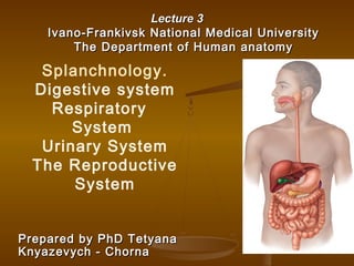
  • 1. Lecture 3Lecture 3 Ivano-Frankivsk National Medical UniversityIvano-Frankivsk National Medical University The Department of Human anatomyThe Department of Human anatomy Prepared by PhD TetyanaPrepared by PhD Tetyana Knyazevych - ChornaKnyazevych - Chorna Splanchnology. Digestive system Respiratory System Urinary System The Reproductive System
  • 2.  Splanchnology-branch of anatomy, which studies the structure of the internal organs.  The internal organs (viscera) are responsible for important functions of the organism: homeostasis, metabolism, water and gases turnover, excretion of metabolic products and reproduction.
  • 3.  ISCOMS – 2009ISCOMS – 2009  ISCOMS – InternationalISCOMS – International Student Congress of MedicalStudent Congress of Medical Sciences – is one of theSciences – is one of the world’s leading studentworld’s leading student conferences in the medicalconferences in the medical sciences.sciences.  the University Medical Center ofthe University Medical Center of GroningenGroningen (the(the NetherlandsNetherlands ))   WithWith Prof. Robin WarrenProf. Robin Warren MD PhD, Nobel PrizeMD PhD, Nobel Prize laureate in 2005laureate in 2005   hehe is credited with the 1979 re-is credited with the 1979 re- discovery of the discovery of the bacteriumbacterium Helicobacter pyloriHelicobacter pylori, together with , together with  Barry MarshallBarry Marshall..
  • 4.  Digestive systemDigestive system is the foodis the food processing system of humanprocessing system of human body. The whole digestivebody. The whole digestive system is in the form of a long,system is in the form of a long, hollow, twisted and turnedhollow, twisted and turned tube, called the alimentarytube, called the alimentary canal, which starts from thecanal, which starts from the oral cavity and ends at theoral cavity and ends at the anus. The overall process ofanus. The overall process of digestion and absorption ofdigestion and absorption of food occurs in this tube. Thefood occurs in this tube. The tube is divided into differenttube is divided into different parts on the basis of structureparts on the basis of structure and function of each part.and function of each part.
  • 5. Necessary Organs Of the digestive system:Necessary Organs Of the digestive system:  ORAL CAVITYORAL CAVITY  ESOPHAGUSESOPHAGUS  STOMACHSTOMACH  SMALL INTESTINESMALL INTESTINE  LARGE INTESTINELARGE INTESTINE  ANUSANUS Accessory Organs of the digestive system:Accessory Organs of the digestive system:  LIVER AND GALL BLADDERLIVER AND GALL BLADDER  PANCREASPANCREAS  SALIVARY GLANDSSALIVARY GLANDS  TEETHTEETH  TONGUETONGUE
  • 6. Functions of digestive system:Functions of digestive system:  IngestionIngestion  Digestion: break down of large particles of foodDigestion: break down of large particles of food  mechanical digestionmechanical digestion  chemical digestionchemical digestion  PropulsionPropulsion  peristalsisperistalsis  segmentationsegmentation  Secretion:Secretion:  digestive enzymesdigestive enzymes  hormoneshormones  Absorption:Absorption:  from external environment into internal environmentfrom external environment into internal environment  across mucosaacross mucosa  Elimination of wastes (defecation)Elimination of wastes (defecation)
  • 7. Oral Cavity (mouth)Oral Cavity (mouth) Entrance to the GI tract.Entrance to the GI tract.  Initial site of digestion:Initial site of digestion:  mechanical digestion (viamechanical digestion (via mastication)mastication)  chemical digestion (viachemical digestion (via enzymes in saliva).enzymes in saliva).  Bounded anteriorly by theBounded anteriorly by the teeth and lipsteeth and lips  Bounded posteriorly by the oropharynx.Bounded posteriorly by the oropharynx.  Superior boundary is formed by theSuperior boundary is formed by the hard and soft palates.hard and soft palates.  Floor, or inferior surface, of the oralFloor, or inferior surface, of the oral cavitycavity  the tonguethe tongue
  • 8.  FaucesFauces represent the opening between the oralrepresent the opening between the oral cavity and the oropharynx.cavity and the oropharynx.  Fauces are bounded by paired muscular folds:Fauces are bounded by paired muscular folds:  glossopalatine arch (anterior fold)glossopalatine arch (anterior fold)  pharyngopalatine arch (posterior fold)pharyngopalatine arch (posterior fold)  Palatine tonsils are housed between the arches.Palatine tonsils are housed between the arches.
  • 9.
  • 10. TeethTeeth  Collectively known as theCollectively known as the dentitiondentition..  Responsible forResponsible for masticationmastication  first part of the mechanicalfirst part of the mechanical digestion.digestion.  A tooth has:A tooth has:  crowncrown  neckneck  one or moreone or more rootsroots  Collectively, the roots, theCollectively, the roots, the dental alveoli, and thedental alveoli, and the periodontal ligament that bindsperiodontal ligament that binds the roots to the alveolarthe roots to the alveolar
  • 11. TOOTH TYPES ARE :TOOTH TYPES ARE : INCISORSINCISORS CANINECANINE PREMOLARSPREMOLARS MOLARSMOLARS COMPONENTS OF THE TOOTHCOMPONENTS OF THE TOOTH INCLUDE:INCLUDE: ENAMEL DENTIN PULP CEMENTUM
  • 12.  20 deciduous teeth, also called “milk teeth,” erupt between 6 months and 30 months after birth.  These teeth are eventually lost and replaced by 32 permanent teeth.
  • 13. THE LARGETHE LARGE SALIVARY GLANDSSALIVARY GLANDS ARE :ARE :  PAROTIDPAROTID  SUBMANDIBULARSUBMANDIBULAR  SUBLINGUALSUBLINGUAL GLANDS PRODUCE SEROUS AND MUCOSALGLANDS PRODUCE SEROUS AND MUCOSAL SECRETIONSSECRETIONS Components of salivaComponents of saliva  Water:Water: makes up 99%makes up 99%  AmylaseAmylase: first step of chemical digestion: first step of chemical digestion  LysozymeLysozyme: antimicrobial: antimicrobial FunctionsFunctions  Moisten foodMoisten food  Food molecules into solution: tasteFood molecules into solution: taste  Form bolus: for swallowingForm bolus: for swallowing  Cleanse oral cavity.Cleanse oral cavity.
  • 14.
  • 15. TONSILS INCLUDETONSILS INCLUDE :: PHARYNGEAL, PALATINE,PHARYNGEAL, PALATINE, LINGUALLINGUAL THETHE PHARYNXPHARYNX INCLUDES :INCLUDES :  NASOPHARYNXNASOPHARYNX  OROPHARYNXOROPHARYNX  LARYNGOPHARYNXLARYNGOPHARYNX
  • 16. SwallowingSwallowing Swallowing involves coordinatedSwallowing involves coordinated activity of tongue, soft palateactivity of tongue, soft palate pharynx and oesophagus.pharynx and oesophagus. The first (buccal) phase isThe first (buccal) phase is voluntary, food being forcedvoluntary, food being forced into the pharynx by the tongue.into the pharynx by the tongue. After this the process is reflex.After this the process is reflex. The tongue blocks the mouth,The tongue blocks the mouth, soft palate closes off the nosesoft palate closes off the nose and the larynx rises so that theand the larynx rises so that the epiglottis closes off theepiglottis closes off the trachea. Food thus moves intotrachea. Food thus moves into the pharynx and onwards bythe pharynx and onwards by peristalsis aided by gravity.peristalsis aided by gravity. If we try to talk whilst swallowingIf we try to talk whilst swallowing food may enter the respiratoryfood may enter the respiratory passages and a cough reflexpassages and a cough reflex expels the bolus.expels the bolus.
  • 17. EsophagusEsophagus  A straight muscular tube about 25-30A straight muscular tube about 25-30 cm longcm long  It begins at the level of the cricoidIt begins at the level of the cricoid cartilage, inferior to the larnyx behindcartilage, inferior to the larnyx behind the trachea and extends through thethe trachea and extends through the chest cavity, pierces the diaphragm atchest cavity, pierces the diaphragm at the esophageal hiatus , and meets withthe esophageal hiatus , and meets with the stomach at an opening called thethe stomach at an opening called the cardiac orifice.cardiac orifice.  It transports food to the stomach andIt transports food to the stomach and secretes mucus, which aids transport.secretes mucus, which aids transport.  The inferior segment is constrictedThe inferior segment is constricted forming the lower esophageal sphincterforming the lower esophageal sphincter which, along with the diaphragm,which, along with the diaphragm, closes to prevent back flow ofcloses to prevent back flow of stomach contentsstomach contents
  • 18. StomachStomach  An enlarged segment of theAn enlarged segment of the tract that functions mainly intract that functions mainly in storing food and mixing it withstoring food and mixing it with gastric juice (creating a pastegastric juice (creating a paste called chyme).called chyme). Other functions of stomachOther functions of stomach include:include:  Chemical digestion of proteinsChemical digestion of proteins  Secretion of intrinsic factor –Secretion of intrinsic factor – a chemical that is necessarya chemical that is necessary for vitamin B12 absorption.for vitamin B12 absorption.  Destruction of ingestedDestruction of ingested bacteria via secretedbacteria via secreted hydrochloric acid.hydrochloric acid.
  • 19. Gross anatomyGross anatomy  CardiaCardia  Cardiac orificeCardiac orifice  FundusFundus  BodyBody  PylorusPylorus  Pyloric sphincterPyloric sphincter  Pyloric orificePyloric orifice  Greater curvatureGreater curvature  Greater omentumGreater omentum  Lesser curvatureLesser curvature  Lesser omemtumLesser omemtum
  • 20.  THE WALL OF THE STOMACHTHE WALL OF THE STOMACH INCLUDESINCLUDES ::  EXTERNAL SEROSAEXTERNAL SEROSA  MUSCLE LAYERMUSCLE LAYER ( CIRCULAR,( CIRCULAR, LONGITUDINAL, OBLIQUE )LONGITUDINAL, OBLIQUE )  SUBMUCOSASUBMUCOSA  INTERIOR MUCOSALINTERIOR MUCOSAL  The mucous membraneThe mucous membrane lining the stomach islining the stomach is thick and vascular. It isthick and vascular. It is thrown into numerousthrown into numerous folds, known as rugae,folds, known as rugae, which are predominantlywhich are predominantly longitudinal in direction.longitudinal in direction. On distention of theOn distention of the stomach, these foldsstomach, these folds flatten out. It contains theflatten out. It contains the glands and the gastricglands and the gastric pits.pits.
  • 21.
  • 22.
  • 23. GASTRIC PITS ARE THEGASTRIC PITS ARE THE OPENINGS INTO THE GASTRICOPENINGS INTO THE GASTRIC GLANDSGLANDS  Mucous neck cellsMucous neck cells – found in the– found in the upper portion of the gland. Secreteupper portion of the gland. Secrete acidic mucus and function as stemacidic mucus and function as stem cells for surface mucous cells.cells for surface mucous cells.  Chief cellsChief cells – primary function is– primary function is the secretion of pepsinogen, anthe secretion of pepsinogen, an inactive form of the protease,inactive form of the protease, pepsin. Pepsinogen is activated bypepsin. Pepsinogen is activated by HCl and by pepsin itself.HCl and by pepsin itself.  Parietal cellsParietal cells – found in the– found in the midportion of the glands. Secretemidportion of the glands. Secrete hydrochloric acid (which gives thehydrochloric acid (which gives the stomach its low pH – usually 1-3)stomach its low pH – usually 1-3) as well as intrinsic factor.as well as intrinsic factor.  Enteroendocrine cellsEnteroendocrine cells – secrete– secrete multiple hormones into the plasma.multiple hormones into the plasma. An example is gastrin, a hormoneAn example is gastrin, a hormone that regulates the stomach’sthat regulates the stomach’s motility and secretory activity.motility and secretory activity.
  • 24. Stomach has three main functions;Stomach has three main functions;  Storage of food:Storage of food:  It stores food for a variable amount It stores food for a variable amount of time (depending on the nature of food) in order toof time (depending on the nature of food) in order to make it appropriate for digestion and absorption in themake it appropriate for digestion and absorption in the small intestine. The storing capacity of stomach in ansmall intestine. The storing capacity of stomach in an average adult is about 1500 ml.average adult is about 1500 ml.  Mixing of food:Mixing of food:  It mixes the food with its own It mixes the food with its own secretions (gastric secretions) to form a semifluidsecretions (gastric secretions) to form a semifluid chyme.chyme.  Controlling the rate of delivery of chyme:Controlling the rate of delivery of chyme:   TheThe stomach also controls the rate of delivery of chyme tostomach also controls the rate of delivery of chyme to the small intestine so that efficient digestion andthe small intestine so that efficient digestion and absorption can take place.absorption can take place.   
  • 25. Small intestineSmall intestine is the longest part of alimentaryis the longest part of alimentary canal. It extends from thecanal. It extends from the pylorus of the stomach to thepylorus of the stomach to the junction between cecum andjunction between cecum and ileum. Much of digestion andileum. Much of digestion and absorption of food takes placeabsorption of food takes place in the small intestine.in the small intestine. IngestedIngested nutrients spend at leastnutrients spend at least 1212 hours in the small intestine.hours in the small intestine. The small intestine is divided into threeThe small intestine is divided into three parts:parts: -- DuodenumDuodenum -- JejunumJejunum -- Ileum.Ileum.
  • 26. Duodenum:Duodenum: It is the first part of smallIt is the first part of small intestine that joins theintestine that joins the stomach. It is a C-stomach. It is a C- shaped tube, about 10shaped tube, about 10 inches longinches long (25,4 sm)(25,4 sm). It. It lies between thelies between the stomach and jejunumstomach and jejunum
  • 27. TThe duodenum is divided into fourhe duodenum is divided into four partsparts::  superiorsuperior  descendingdescending  inferior/horizontainferior/horizonta ll  ascendingascending The organs and structuresThe organs and structures surrounding the duodenumsurrounding the duodenum includes :includes :  Superior (above) – liver andSuperior (above) – liver and gallbladdergallbladder  Anterior (in front) – gallbladder andAnterior (in front) – gallbladder and transverse colontransverse colon  Posterior (behind) – inferior venaPosterior (behind) – inferior vena cava, right psoas major muscle andcava, right psoas major muscle and aortaaorta  Lateral – pancreasLateral – pancreas
  • 28. A A liquid mixtureliquid mixture of food and gastric secretions of food and gastric secretions enters the superior duodenum from the pylorusenters the superior duodenum from the pylorus of the stomach, triggering the release ofof the stomach, triggering the release of pancreas-stimulating hormones from glands inpancreas-stimulating hormones from glands in the duodenal wall. Ducts fromthe duodenal wall. Ducts from the the pancreaspancreas and  and gallbladdergallbladder enter at enter at the descending duodenum, bringing bicarbonatethe descending duodenum, bringing bicarbonate to neutralize the acid in the gastric secretions,to neutralize the acid in the gastric secretions, pancreatic enzymes to further digestion,pancreatic enzymes to further digestion, and and bile saltsbile salts to emulsify fat. The mucous to emulsify fat. The mucous lining of the last two segments of the duodenumlining of the last two segments of the duodenum begins the absorption of nutrients, in particularbegins the absorption of nutrients, in particular iron and calcium, before the food contents enteriron and calcium, before the food contents enter the next part of the small intestine, the the next part of the small intestine, the jejunumjejunum..
  • 29. The Jejunum and IleumThe Jejunum and Ileum  TheThe jejunumjejunum  middle region of the small intestine.middle region of the small intestine.  approximately 2.5 meters (7.5 feet)approximately 2.5 meters (7.5 feet)  makes up approximately two-fifths of the smallmakes up approximately two-fifths of the small intestine’s total length.intestine’s total length.  primary region for chemical digestion andprimary region for chemical digestion and nutrient absorptionnutrient absorption  TheThe ileumileum  is the last region of the small intestine.is the last region of the small intestine.  about 3.6 meters (10.8 feet) in lengthabout 3.6 meters (10.8 feet) in length  forms approximately three-fifths of the smallforms approximately three-fifths of the small intestine.intestine.  terminates at the ileocecal valveterminates at the ileocecal valve  sphincter that controls the entry of materials into thesphincter that controls the entry of materials into the large intestine.large intestine.
  • 30.
  • 31.
  • 32.  Ileocecal valve, as the name indicates, is aIleocecal valve, as the name indicates, is a structure present at the junction of ileum andstructure present at the junction of ileum and cecum. It is a rudimentary valve and consists ofcecum. It is a rudimentary valve and consists of two horizontal folds of mucous membrane thattwo horizontal folds of mucous membrane that project around the opening of the ileum. Ileocecalproject around the opening of the ileum. Ileocecal valve controlvalve controlss the flow of contents from ileum intothe flow of contents from ileum into the large intestine.the large intestine.
  • 33. Large IntestineLarge Intestine  approximate length of 1.5 meters (5approximate length of 1.5 meters (5 feet)feet)  diameter of 6.5 centimeters (2.5diameter of 6.5 centimeters (2.5 inches).inches).  Absorbs most of the waterAbsorbs most of the water andand electrolytes from the remaining digestedelectrolytes from the remaining digested material.material.  Watery material that first enters theWatery material that first enters the large intestine soonlarge intestine soon solidifiessolidifies andand becomesbecomes faeces.faeces.  Stores faecal material until the body isStores faecal material until the body is ready toready to defaecate.defaecate.  Absorbs a very small percentage ofAbsorbs a very small percentage of nutrientsnutrients still remaining in the digestedstill remaining in the digested material.material.  Composed of four segments:Composed of four segments:  thethe caecumcaecum,, coloncolon,, rectumrectum,, analanal canalcanal
  • 34.        Structures Structures         a. cecum with vermiform appendix        a. cecum with vermiform appendix         b. ascending, transverse, descending colon        b. ascending, transverse, descending colon         c. sigmoid colon, rectum        c. sigmoid colon, rectum               Haustra are pouches in wall of large intestine Haustra are pouches in wall of large intestine                     Millions of bacteria colonize the large intestine. TheyMillions of bacteria colonize the large intestine. They breakdown indigestible carbohydrate residues andbreakdown indigestible carbohydrate residues and produce many B vitamins as well as most of the body’sproduce many B vitamins as well as most of the body’s vitamin K supply.vitamin K supply. Typically 100 g of feces are produced per day consisting ofTypically 100 g of feces are produced per day consisting of 75% water and 25% solids. Solids include dead bacteria,75% water and 25% solids. Solids include dead bacteria, fat, inorganic matter, protein, undigested plant fibers,fat, inorganic matter, protein, undigested plant fibers, bile pigments, and shed epithelial cells.  bile pigments, and shed epithelial cells.       
  • 35. The liverThe liver is an accessory digestive organ thatis an accessory digestive organ that has multiple functions including:has multiple functions including:  Carbohydrate metabolism – storage andCarbohydrate metabolism – storage and release of glucoserelease of glucose  Removal of drugs, toxins, and foreignRemoval of drugs, toxins, and foreign chemicals from the plasmachemicals from the plasma  Storage of vitamins (A, D, E, and K) andStorage of vitamins (A, D, E, and K) and minerals (iron and copper)minerals (iron and copper)  Protein metabolismProtein metabolism  Lipid metabolismLipid metabolism  Synthesis of plasma proteins (e.g., albumin,Synthesis of plasma proteins (e.g., albumin, fibrin, etc.)fibrin, etc.)  Phagocytosis of old RBCs and of pathogens.Phagocytosis of old RBCs and of pathogens.  Production of bile (0.5-1 L/day).Production of bile (0.5-1 L/day).
  • 36.  The liverThe liver  composed of four incompletely separatedcomposed of four incompletely separated lobeslobes  supported by two ligamentssupported by two ligaments  Right lobeRight lobe  Left lobeLeft lobe  Falciform ligamentFalciform ligament  Round ligamentRound ligament  Caudate lobeCaudate lobe  Quadrate lobeQuadrate lobe
  • 37.
  • 38.
  • 39.  The gallbladderThe gallbladder is a thin-walledis a thin-walled green muscular sac found on thegreen muscular sac found on the ventral surface of the liver.ventral surface of the liver.  It functions primarily in the storageIt functions primarily in the storage and concentration of bile.and concentration of bile.  The liver continuously produces bile.The liver continuously produces bile. However the hepatopancreaticHowever the hepatopancreatic sphincter is normally closed. Thissphincter is normally closed. This results in bile backing up into theresults in bile backing up into the common bile duct, cystic duct, andcommon bile duct, cystic duct, and ultimately into the gallbladder.ultimately into the gallbladder.  When fatty chyme arrives in the smallWhen fatty chyme arrives in the small intestine, intestinal glands secrete theintestine, intestinal glands secrete the hormone cholecystokinin.hormone cholecystokinin.  CCK causes gallbladder contractionCCK causes gallbladder contraction and relaxation of the hepatopancreaticand relaxation of the hepatopancreatic sphincter, letting bile flow into thesphincter, letting bile flow into the duodenum.duodenum.
  • 40. The pancreasThe pancreas  Mostly retroperitoneal and deep to the greater curvature ofMostly retroperitoneal and deep to the greater curvature of the stomach.the stomach.  TheThe headhead of the pancreas sits next to the duodenum as itof the pancreas sits next to the duodenum as it emanates from and loops away from the pylorus. Theemanates from and loops away from the pylorus. The bodybody extends behind the stomach and itsextends behind the stomach and its tailtail ends at the spleen.ends at the spleen.  The pancreas primarily consists ofThe pancreas primarily consists of aciniacini – small clusters of– small clusters of enzyme secreting cells. These acinar cells empty theirenzyme secreting cells. These acinar cells empty their secretion into small ducts. Small ducts coalesce into largersecretion into small ducts. Small ducts coalesce into larger ducts that empty into theducts that empty into the main pancreatic ductmain pancreatic duct , which, which runs centrally along the long axis of the pancreasruns centrally along the long axis of the pancreas  The major function of the acinar and duct cells is theThe major function of the acinar and duct cells is the secretion ofsecretion of pancreatic juicepancreatic juice (1.5 L/day).(1.5 L/day).  Acinar cells contribute digestive enzymes to the pancreaticAcinar cells contribute digestive enzymes to the pancreatic juice including:juice including: proteases, pancreatic lipase,proteases, pancreatic lipase, pancreatic amylase.pancreatic amylase.  Scattered amidst the pancreatic acini are the hormone-Scattered amidst the pancreatic acini are the hormone- producingproducing islets of Langerhansislets of Langerhans . Their major function is. Their major function is the regulation of blood glucose levels.the regulation of blood glucose levels.
  • 41.  PeritoneumPeritoneum  Parietal peritoneumParietal peritoneum  Visceral peritoneumVisceral peritoneum  Peritoneal cavityPeritoneal cavity  Intraperitoneal organsIntraperitoneal organs  Mesoperitoneal organsMesoperitoneal organs  Retroperitoneal organsRetroperitoneal organs
  • 42.
  • 43.
  • 44. The The respiratory systemrespiratory system is the anatomical system of is the anatomical system of an organism that introduces respiratory gases to thean organism that introduces respiratory gases to the interior and performs gas exchange. In humans theinterior and performs gas exchange. In humans the respiratory system include airways, lungs, and therespiratory system include airways, lungs, and the respiratory muscles. Molecules of oxygen and carbonrespiratory muscles. Molecules of oxygen and carbon dioxide are passively exchanged, by diffusion,dioxide are passively exchanged, by diffusion, between the gaseous external environment andbetween the gaseous external environment and the blood. This exchange process occurs in thethe blood. This exchange process occurs in the alveolar region of the lungsalveolar region of the lungs
  • 45. General Functions of Respiratory System:  1. O2 (oxygenoxygen)) and CO2 (carbon dioxidecarbon dioxide) exchange between blood and air  2. speech and vocalization  3. sense of smell  4. helps control acid base balance of body  5. breathing movements help promote blood and lymph flow
  • 46. Main Organs:  nose  pharynx  larynx  trachea  primary bronchi  lungs:  bronchioles  alveoli/respiratory membrane these organs can also be subdivided into:  upper respiratory tract Nose-pharynx-larynx  lower respiratory tract respiratory organs of the thorax
  • 47.  Functional classifications:Functional classifications:  ConductingConducting portion: transports air.portion: transports air.  RespiratoryRespiratory portion: carries out gas exchange.portion: carries out gas exchange.  respiratory bronchiolesrespiratory bronchioles  alveolar ductsalveolar ducts  air sacs called alveoliair sacs called alveoli  Upper respiratory tract is all conductingUpper respiratory tract is all conducting  Lower respiratory tract has bothLower respiratory tract has both conducting and respiratory portionsconducting and respiratory portions
  • 48. Nose  separated from mouth by hard and soft palate  each nasal cavity is divided into 3 passageways by turbinates (choncae) turbulent passageways to insure that all air makes contact with mucous membranes membranes are heavily vascualrized  remove bacteria, debris and particles  warms and moisturizes air entering lungs  also contains receptors for smell nasolacrimal ducts drain into nasal cavity
  • 49. Paranasal sinuses:Paranasal sinuses:  In four skull bonesIn four skull bones  paired air spacespaired air spaces  decrease skull bone weightdecrease skull bone weight  warm and moisten air  sound resonance  Named for the bonesNamed for the bones in which they are housed.in which they are housed.  frontalfrontal  ethmoidalethmoidal  sphenoidalsphenoidal  maxillarymaxillary  Communicate with the nasal cavity by ducts.Communicate with the nasal cavity by ducts.  Covered with the same pseudostratified ciliatedCovered with the same pseudostratified ciliated columnar epithelium as the nasal cavity.columnar epithelium as the nasal cavity.
  • 50. Pharynx (throat) - Is situated from base of skull to junction with esophagus and trachea - made of muscle and lined with mucous membrane - junction between digestive and respiratory systems divided into three regions: a. Nasopharynx  behind nose to level of soft palate  includes uvula, tonsils (adenoids)  auditory tube (eustachian tube) b. Oropharynx  behind mouth  from soft palate to level of hyoid bone  palatine and lingual tonsils c. Laryngopharynx  from hyoid bone to esophagus/larynx
  • 51. Larynx (voice box)  enlarged beginning portion of trachea  composed of cartilage and muscles  opening into larynx = glottis (prevent food from entering lower respiratory system sound)  9 cartilages:  three individual piecesthree individual pieces  Thyroid cartilageThyroid cartilage  Cricoid cartilageCricoid cartilage  EpiglottisEpiglottis  three cartilage pairsthree cartilage pairs  Arytenoids: on cricoidArytenoids: on cricoid  Corniculates: attach toCorniculates: attach to arytenoidsarytenoids  Cuniforms:in aryepiglottic foldCuniforms:in aryepiglottic fold
  • 52. Sound ProductionSound Production  Two pairs of ligamentsTwo pairs of ligaments  Inferior ligaments, calledInferior ligaments, called vocal ligamentsvocal ligaments  covered by a mucous membranecovered by a mucous membrane  vocal folds: ligament and mucosa.vocal folds: ligament and mucosa.  are “true vocal cords”are “true vocal cords”  they produce sound when air passes between themthey produce sound when air passes between them  Superior ligaments, calledSuperior ligaments, called vestibular ligamentsvestibular ligaments  Covered by mucosaCovered by mucosa  vestibular folds: ligament and mucosavestibular folds: ligament and mucosa  Are “false vocal cords”Are “false vocal cords”  no function in sound productionno function in sound production  protect the vocal folds.protect the vocal folds.  The vestibular folds attach to the corniculateThe vestibular folds attach to the corniculate cartilagescartilages ..
  • 53.  The tension, length,The tension, length, and position of theand position of the vocal folds determine the quality of thevocal folds determine the quality of the sound.sound.  Longer vocal folds produce lower soundsLonger vocal folds produce lower sounds  More taunt, higher pitchMore taunt, higher pitch  Loudness based on force of airLoudness based on force of air  Rima glottidisRima glottidis: opening between the vocal: opening between the vocal foldsfolds  GlottisGlottis: rima glottidis and the vocal folds: rima glottidis and the vocal folds
  • 54.
  • 55.
  • 56.
  • 57. Trachea  extends from larynx to bronchi (immediately anterior to the esophagus,immediately anterior to the esophagus, inferior to the larynx, superior to theinferior to the larynx, superior to the primary bronchi of the lungs.primary bronchi of the lungs. )  surrounded by 15 to 2015 to 20 “C” – shaped bands of cartilage ends joined by bands of muscle tissue (holds walls open, prevents collapse)  lined by pseudostratified ciliated columnar epithelium
  • 58.
  • 59.  At the level of theAt the level of the sternal anglesternal angle, the, the trachea bifurcates into two smaller tubes,trachea bifurcates into two smaller tubes, called thecalled the right and left primary bronchiright and left primary bronchi..  Each primary bronchus projects laterallyEach primary bronchus projects laterally toward each lung.toward each lung.  The most inferior tracheal cartilageThe most inferior tracheal cartilage separates the primary bronchi at theirseparates the primary bronchi at their origin and forms an internal ridge calledorigin and forms an internal ridge called thethe carina.carina.
  • 60. Bronchi  bronchi resemble trachea in structure (also supported by C-shaped cartilages)  also have lots of elastic connective tissue  Right primary bronchusRight primary bronchus  shorter, wider, and more vertically orientedshorter, wider, and more vertically oriented than the left primary bronchus.than the left primary bronchus.  Foreign particles are more likely to lodge inForeign particles are more likely to lodge in the right primary bronchus.the right primary bronchus.  each bronchus enters lung and continues to divide into smaller and smaller branches = bronchi, then into microscopic bronchioles  because of the extensive branching it formes bronchial tree
  • 61.  Primary bronchiPrimary bronchi  enter the hilum of each lungenter the hilum of each lung  Also entering hilum:Also entering hilum:  pulmonary vesselspulmonary vessels  lymphatic vesselslymphatic vessels  nerves.nerves.  Secondary bronchi (or lobar bronchi)Secondary bronchi (or lobar bronchi)  Branch of primary bronchusBranch of primary bronchus  left lung:left lung:  two lobestwo lobes  two secondary bronchitwo secondary bronchi  right lungright lung  three lobesthree lobes  three secondary bronchi.three secondary bronchi.  Tertiary bronchi (or segmental bronchi)Tertiary bronchi (or segmental bronchi)  Branch of secondary bronchiBranch of secondary bronchi  left lung is supplied by 8 to 10 tertiary bronchi.left lung is supplied by 8 to 10 tertiary bronchi.  right lung is supplied by 10 tertiary bronchiright lung is supplied by 10 tertiary bronchi  supply a part of the lung called a bronchopulmonarysupply a part of the lung called a bronchopulmonary segment.segment.
  • 62. Bronchioles  smallest branches of “respiratory tree”  <1mm diameter  no cartilage rings (but larger branches may have small patches of cartilage)  lined with ciliated cuboidal epithelium and layer of smooth muscle  asthma affects the smallest terminal bronchioles
  • 63.
  • 64. Respiratory Bronchioles, AlveolarRespiratory Bronchioles, Alveolar Ducts, and AlveoliDucts, and Alveoli  Contain small saccular outpocketings calledContain small saccular outpocketings called alveoli.alveoli.  An alveolus is about 0.25 to 0.5 millimeter inAn alveolus is about 0.25 to 0.5 millimeter in diameter.diameter.  Its thin wall is specialized to promote diffusionIts thin wall is specialized to promote diffusion of gases between the alveolus and the blood inof gases between the alveolus and the blood in the pulmonary capillaries.the pulmonary capillaries.  Gas exchange can take place in the respiratoryGas exchange can take place in the respiratory bronchioles and alveolar ducts as well as in thebronchioles and alveolar ducts as well as in the lungs, which contain approximately 300–400lungs, which contain approximately 300–400 million alveoli.million alveoli.  The spongy natureThe spongy nature of the lung is due to theof the lung is due to the packing of millions of alveoli together.packing of millions of alveoli together.
  • 65.
  • 66.
  • 67.
  • 68. Gross Anatomy of the LungsGross Anatomy of the Lungs  Each lung has a conical shape.Each lung has a conical shape.  Its wide, concave base rests upon the muscular diaphragm.Its wide, concave base rests upon the muscular diaphragm.  Its relatively blunt superior region, called the apex orIts relatively blunt superior region, called the apex or (cupola), projects superiorly to a point that is slightly(cupola), projects superiorly to a point that is slightly superior and posterior to the clavicle.superior and posterior to the clavicle.  Both lungs are bordered by the thoracic wall anteriorly,Both lungs are bordered by the thoracic wall anteriorly, laterally, and posteriorly, and supported by the rib cage.laterally, and posteriorly, and supported by the rib cage.  Toward the midline, the lungs are separated from eachToward the midline, the lungs are separated from each other by the mediastinum.other by the mediastinum.  The relatively broad, rounded surface in contact with theThe relatively broad, rounded surface in contact with the thoracic wall is called the costal surface of the lung.thoracic wall is called the costal surface of the lung.
  • 69.
  • 70.
  • 71.
  • 72.
  • 73.
  • 74. Pleura and Pleural CavitiesPleura and Pleural Cavities  The outer surface of each lung and the adjacentThe outer surface of each lung and the adjacent internal thoracic wall are lined by a serousinternal thoracic wall are lined by a serous membrane called pleura, which is formed frommembrane called pleura, which is formed from simple squamous epithelium.simple squamous epithelium.  The outer surface of each lung is tightlyThe outer surface of each lung is tightly covered by the visceral pleura, while thecovered by the visceral pleura, while the internal thoracic walls, the lateral surfaces ofinternal thoracic walls, the lateral surfaces of the mediastinum, and the superior surface ofthe mediastinum, and the superior surface of the diaphragm are lined by the parietal pleura.the diaphragm are lined by the parietal pleura.  The parietal and visceral pleural layers areThe parietal and visceral pleural layers are continuous at the hilum of each lung.continuous at the hilum of each lung.
  • 75. Pleura and Pleural CavitiesPleura and Pleural Cavities  The potential space between theseThe potential space between these serous membrane layers is a pleuralserous membrane layers is a pleural cavity.cavity.  The pleural membranes produce a thin,The pleural membranes produce a thin, serous fluid that circulates in the pleuralserous fluid that circulates in the pleural cavity and acts as a lubricant, ensuringcavity and acts as a lubricant, ensuring minimal friction during breathing.minimal friction during breathing.
  • 76.
  • 77.
  • 78.
  • 79.
  • 80.
  • 81.
  • 82.
  • 83. 27-83 General Structure and FunctionsGeneral Structure and Functions of the Urinary Systemof the Urinary System  Urinary System organsUrinary System organs  remove waste products from the bloodremove waste products from the blood (there are toxic and accumulate in blood)(there are toxic and accumulate in blood)  then from the bodythen from the body  Major homeostatic systemMajor homeostatic system
  • 84. 27-84 General Structure and FunctionsGeneral Structure and Functions of the Urinary Systemof the Urinary System  Organs of the Urinary System:Organs of the Urinary System:  KidneysKidneys  UretersUreters  Urinary BladderUrinary Bladder  UrethraUrethra  Primary organs: kidneysPrimary organs: kidneys  filter waste products from the bloodstreamfilter waste products from the bloodstream  convert the filtrate into urine.convert the filtrate into urine.  The Urinary Tract:The Urinary Tract:  Includes:Includes:  uretersureters  urinary bladderurinary bladder  urethraurethra  Because they transport the urine out of the body.Because they transport the urine out of the body.
  • 85. 85
  • 86. 86
  • 87. 87
  • 88. 27-88 Functions of the Urinary SystemFunctions of the Urinary System  Removing waste products from the bloodstream.Removing waste products from the bloodstream.  Storage of urine.Storage of urine.  the urinary bladder is an expandable, muscular sac that canthe urinary bladder is an expandable, muscular sac that can store as much as 1 liter of urinestore as much as 1 liter of urine  Excretion of urine.Excretion of urine.  Blood volume regulation.Blood volume regulation.  the kidneys control the volume of interstitial fluid and bloodthe kidneys control the volume of interstitial fluid and blood under the direction of certain hormonesunder the direction of certain hormones  Regulation of erythrocyte production.Regulation of erythrocyte production.  as the kidneys filter the blood, they are also indirectlyas the kidneys filter the blood, they are also indirectly measuring the oxygen level in the bloodmeasuring the oxygen level in the blood  Erythropoietin (EPO): hormone produced by kidneyErythropoietin (EPO): hormone produced by kidney  Released if blood oxygen levels fallReleased if blood oxygen levels fall  Stimulates RBC production in red bone marrowStimulates RBC production in red bone marrow
  • 89. 27-89 Kidneys: Gross and SectionalKidneys: Gross and Sectional AnatomyAnatomy  RetroperitonealRetroperitoneal  Anterior surface covered with peritoneumAnterior surface covered with peritoneum  Posterior surface against posterior abdominalPosterior surface against posterior abdominal wallwall  Superior pole: T-12Superior pole: T-12  Inferior pole: L-3Inferior pole: L-3  Right kidney ~ 2cm lower than leftRight kidney ~ 2cm lower than left  Adrenal gland on superior poleAdrenal gland on superior pole
  • 90. 90
  • 91. 27-91 Kidneys: Gross and SectionalKidneys: Gross and Sectional AnatomyAnatomy  Hilum: concave medial borderHilum: concave medial border  Renal sinus: internal spaceRenal sinus: internal space  Houses blood vessels, lymphatic vessels,Houses blood vessels, lymphatic vessels, nervesnerves  Houses renal pelvis, renal calycesHouses renal pelvis, renal calyces  Also fatAlso fat
  • 92. 27-92 Kidneys: Gross and SectionalKidneys: Gross and Sectional AnatomyAnatomy  Surrounding tissues, from deep to superficial:Surrounding tissues, from deep to superficial:  Fibrous capsule (renal capsule)Fibrous capsule (renal capsule)  Dense irregular CTDense irregular CT  Covers outer surfaceCovers outer surface  Perinephric fat (adipose capsule)Perinephric fat (adipose capsule)  Also called perirenal fatAlso called perirenal fat  Completely surrounds kidneyCompletely surrounds kidney  Cushioning and insulationCushioning and insulation  Renal fasciaRenal fascia  Dense irregular CTDense irregular CT  Anchors kidney to posterior wall and peritoneumAnchors kidney to posterior wall and peritoneum  Paranephric fatParanephric fat  Between renal fascia and peritoneumBetween renal fascia and peritoneum
  • 93. 93
  • 94. 27-94 Kidneys: Gross and SectionalKidneys: Gross and Sectional AnatomyAnatomy  Sectioned on a coronal plane:Sectioned on a coronal plane:  Renal CortexRenal Cortex  Renal archesRenal arches  Renal columnsRenal columns  Renal MedullaRenal Medulla  Divided into renal pyramids (8 to 15 per kidney)Divided into renal pyramids (8 to 15 per kidney)  Base against cortexBase against cortex  Apex called renal papillaApex called renal papilla
  • 95. 27-95 Kidneys: Gross and SectionalKidneys: Gross and Sectional AnatomyAnatomy  Minor calyx:Minor calyx:  Funnel shapedFunnel shaped  Receives renal papillaReceives renal papilla  8 to 15 per kidney, one per pyramid8 to 15 per kidney, one per pyramid  Major calyxMajor calyx  Fusion of minor calycesFusion of minor calyces  2 to 3 per kidney2 to 3 per kidney  Major calyces merge to form renal pelvisMajor calyces merge to form renal pelvis  Renal LobeRenal Lobe  Pyramid plus some cortical tissuePyramid plus some cortical tissue  8 to 15 per kidney8 to 15 per kidney
  • 96. 96
  • 97. 97
  • 98. 27-98 Blood Supply to the KidneyBlood Supply to the Kidney  About 20 to 25% of cardiac output toAbout 20 to 25% of cardiac output to kidneyskidneys  Path:Path:  Renal artery to segmental arteries toRenal artery to segmental arteries to interlobar arteries to arcuate arteries tointerlobar arteries to arcuate arteries to interlobular arteries to:interlobular arteries to:  Afferent arteriole to glomerulus to efferentAfferent arteriole to glomerulus to efferent arteriole to peritubular capilaries and vasaarteriole to peritubular capilaries and vasa rectarecta
  • 99. 99
  • 100. 27-100 Blood Supply to the KidneyBlood Supply to the Kidney  Blood plasma is filtered across the glomerulusBlood plasma is filtered across the glomerulus into the glomerular space.into the glomerular space.  Once the blood plasma is filteredOnce the blood plasma is filtered  blood leaves the glomerulusblood leaves the glomerulus  enters an efferent arteriole.enters an efferent arteriole.  efferent arteriole is still carrying oxygenatedefferent arteriole is still carrying oxygenated bloodblood  a gas and nutrient exchange with the kidney tissuesa gas and nutrient exchange with the kidney tissues has not yet occurred.has not yet occurred.
  • 101. 27-101 Blood Supply to the KidneyBlood Supply to the Kidney  The efferent arterioles branch into one of twoThe efferent arterioles branch into one of two types of capillary networks:types of capillary networks:  peritubular capillariesperitubular capillaries  vasa rectavasa recta  these capillary networks are responsiblethese capillary networks are responsible for the actual exchange of gases andfor the actual exchange of gases and nutrientsnutrients  Peritubular capillaries:Peritubular capillaries: primarily in cortexprimarily in cortex  Vasa recta:Vasa recta: surround the thin tubes thatsurround the thin tubes that project into the medulla.project into the medulla.
  • 102. 27-102 Blood Supply to the KidneyBlood Supply to the Kidney  Path for veins:Path for veins:  Interlobar veins to arcuate veins to interlobarInterlobar veins to arcuate veins to interlobar veins to the renal veinveins to the renal vein
  • 103. 103
  • 104. 27-104 NephronsNephrons  The functional filtration unit in the kidney.The functional filtration unit in the kidney.  Consists of the following:Consists of the following:  Renal corpuscleRenal corpuscle  GlomerulusGlomerulus  Glomerular capsule (Bowman’s capsule)Glomerular capsule (Bowman’s capsule)  Proximal convoluted tubule (PCT)Proximal convoluted tubule (PCT)  Nephron loop (loop of Henle)Nephron loop (loop of Henle)  Ascending loop of HenleAscending loop of Henle  Descending loop of HenleDescending loop of Henle  Distal convoluted tubule (DCT)Distal convoluted tubule (DCT)  collectively called thecollectively called the renal tubulerenal tubule  In both kidneys: approximatelyIn both kidneys: approximately 2.5 million nephrons.2.5 million nephrons.  Are microscopic: measure aboutAre microscopic: measure about 5 centimeters5 centimeters inin length.length.
  • 105. 27-105 NephronsNephrons  Cortical NephronsCortical Nephrons  Near peripheral edge of cortexNear peripheral edge of cortex  Short nephron loopsShort nephron loops  Have peritubular capillariesHave peritubular capillaries  Juxtamedullary nephronsJuxtamedullary nephrons  Near corticomedullary borderNear corticomedullary border  Long nephron loopsLong nephron loops  Have vasa rectaHave vasa recta
  • 106. 106
  • 107. 27-107 Urine FormationUrine Formation  Three processesThree processes  FiltrationFiltration  Renal corpuscle: forms filtrateRenal corpuscle: forms filtrate  From blood to tubuleFrom blood to tubule  ReabsorptionReabsorption  Mostly PCTMostly PCT  Water and salt: rest of nephronWater and salt: rest of nephron  From tubule to bloodFrom tubule to blood  SecretionSecretion  From blood to tubuleFrom blood to tubule
  • 108. 108
  • 109. 27-109 Urinary Tract : UretersUrinary Tract : Ureters  long, fibromuscular tubeslong, fibromuscular tubes  conduct urine from the kidneys to the urinary bladder.conduct urine from the kidneys to the urinary bladder.  average 25 centimeters in lengthaverage 25 centimeters in length  retroperitoneal.retroperitoneal.  ureters originate at the renal pelvisureters originate at the renal pelvis  extend inferiorly to enter the posterolateral wall of theextend inferiorly to enter the posterolateral wall of the base of the urinary bladder.base of the urinary bladder.  wall is composed of three concentric tunics.wall is composed of three concentric tunics.  mucosamucosa  muscularismuscularis  adventitia.adventitia.
  • 110. 27-110 Urinary Tract – Urinary BladderUrinary Tract – Urinary Bladder  The urinary bladder:The urinary bladder:  expandable, muscular containerexpandable, muscular container  serves as a reservoir for urineserves as a reservoir for urine  positioned immediately superior and posterior to the pubicpositioned immediately superior and posterior to the pubic symphysis.symphysis.  in femalesin females  the urinary bladder is in contact with the uterus posterosuperiorlythe urinary bladder is in contact with the uterus posterosuperiorly and with the vagina posteroinferiorly.and with the vagina posteroinferiorly.  in malesin males  it is in contact with the rectum posterosuperiorly and is immediatelyit is in contact with the rectum posterosuperiorly and is immediately superior to the prostate gland.superior to the prostate gland.  is a retroperitoneal organ.is a retroperitoneal organ.  when empty exhibits an upside-down pyramidal shape.when empty exhibits an upside-down pyramidal shape.  Filling with urine distends it superiorly until it assumes an ovalFilling with urine distends it superiorly until it assumes an oval shape.shape.
  • 111. 27-111 Urinary Tract – Urinary BladderUrinary Tract – Urinary Bladder  TrigoneTrigone  posteroinferior triangular area of the urinary bladder wallposteroinferior triangular area of the urinary bladder wall  formed by imaginary linesformed by imaginary lines  connect the two posterior ureteral openingsconnect the two posterior ureteral openings  and the anterior urethral opening.and the anterior urethral opening.  The trigone remains immovable as the urinaryThe trigone remains immovable as the urinary bladder fills and evacuates.bladder fills and evacuates.  It functions as a funnelIt functions as a funnel  directs urine into the urethra as the bladder wall contractsdirects urine into the urethra as the bladder wall contracts  four tunicsfour tunics  mucosamucosa  submucosasubmucosa  Muscularis: called the detrusor muscleMuscularis: called the detrusor muscle  adventitia.adventitia.  Internal urethral sphincter (smooth muscle)Internal urethral sphincter (smooth muscle)
  • 112. 112
  • 113. 113
  • 114. 114
  • 115. 28-115 Human AnatomyHuman Anatomy The Reproductive SystemThe Reproductive System
  • 116. 28-116
  • 117. 28-117 Comparison of the Female andComparison of the Female and Male Reproductive SystemsMale Reproductive Systems  Female typically produces and releases aFemale typically produces and releases a singlesingle oocyte monthly.oocyte monthly.  MaleMale producesproduces 100,000,000’s100,000,000’s of (sperm)of (sperm) daily.daily.  male gametes aremale gametes are storedstored for a shortfor a short timetime  if they are not expelled from the bodyif they are not expelled from the body within that period, they arewithin that period, they are resorbedresorbed
  • 118. 28-118 Anatomy of the MaleAnatomy of the Male Reproductive SystemReproductive System  Internal organs: testes, spermaticInternal organs: testes, spermatic cord, ductus deferens, seminalcord, ductus deferens, seminal glands, prostate.glands, prostate.  External organs include:External organs include:  The penisThe penis  The scrotumThe scrotum  The male urethraThe male urethra
  • 119. 28-119 TestesTestes  Small, oval organSmall, oval organ  Housed in the scrotumHoused in the scrotum  Produces:Produces:  SpermSperm  Androgens (Androgens (testosteronetestosterone).).  CoveringsCoverings  Serous membrane called tunica vaginalisSerous membrane called tunica vaginalis  Parietal layerParietal layer  Visceral layer.Visceral layer.  Tunica albugineaTunica albuginea  Forms internal septaForms internal septa  250 lobules per testis250 lobules per testis  Each lobule has up to 4 seminiferousEach lobule has up to 4 seminiferous tubulestubules  Two types of cellTwo types of cell  Sustentacular cellsSustentacular cells  Germ cellsGerm cells  Interior is called mediastinum testis.Interior is called mediastinum testis.
  • 120. 28-120
  • 121. 28-121 TestesTestes  Sperm develop in the semineferous tubulesSperm develop in the semineferous tubules  Interstitial spaces: surroundInterstitial spaces: surround the seminiferous tubules.the seminiferous tubules.  Contain interstitial (Leydig) cellsContain interstitial (Leydig) cells  produce hormones calledproduce hormones called androgensandrogens..  Several types ofSeveral types of androgensandrogens  most common one ismost common one is testosteronetestosterone..  the adrenal cortex secretes a small amount of androgensthe adrenal cortex secretes a small amount of androgens  the vast majority of androgen release is viathe vast majority of androgen release is via interstitialinterstitial cellscells in the testisin the testis  These hormones cause males to develop the classicThese hormones cause males to develop the classic characteristics:characteristics:  axillary and pubic hairaxillary and pubic hair  deeper voicedeeper voice  sperm production.sperm production.
  • 122. 28-122
  • 123. 28-123 TestesTestes  Series of tubes:Series of tubes:  SeminiferousSeminiferous tubulestubules  Straight ductsStraight ducts  Rete testisRete testis  Efferent ductuleEfferent ductule  EpididymisEpididymis  Ductus deferensDuctus deferens
  • 124. 28-124 The testicular tunicsThe testicular tunics 1- testis; (orchis) 2- epididymis 3- ductus deferens 4- tunica vaginalis testis 5- cavum vaginalis testis 6- fascia spermatica interna 7- m. cremaster 8- fascia cremasterica 9- fascia spermatica externa 10-tunica dartos 11-cutis
  • 125. 28-125 Spermatic CordSpermatic Cord  The blood vesselsThe blood vessels and nerves to theand nerves to the testis travel fromtestis travel from within the abdomenwithin the abdomen to the scrotum in ato the scrotum in a multilayeredmultilayered structure called thestructure called the spermatic cord.spermatic cord.
  • 126. 28-126 Developmemt of spermDevelopmemt of sperm  Called spermatogenesisCalled spermatogenesis  Occurs in the seminiferous tubulesOccurs in the seminiferous tubules  Process:Process:  SpermatogoniumSpermatogonium  Primary spermatocytePrimary spermatocyte  Secondary spermatocyteSecondary spermatocyte  SpermatidSpermatid  SpermiogenesisSpermiogenesis  Spermatid matures into spermatozoonSpermatid matures into spermatozoon
  • 127. 28-127
  • 128. 28-128
  • 129. 28-129 EpididymisEpididymis  The epididymis is a comma-shaped structure composed ofThe epididymis is a comma-shaped structure composed of anan internal ductinternal duct and an external covering of connectiveand an external covering of connective tissue.tissue.  ItsIts headhead lies on the superior surface of the testis, while thelies on the superior surface of the testis, while the bodybody andand tailtail are posterior to the testis.are posterior to the testis.  Internally, the epididymis contains a long, convolutedInternally, the epididymis contains a long, convoluted ductduct of the epididymisof the epididymis, which is approximately, which is approximately 4 to 5 meters4 to 5 meters inin length.length.  Sperm must reside in the epididymis for a period of time toSperm must reside in the epididymis for a period of time to becomebecome maturemature andand fully motilefully motile..  If they are expelledIf they are expelled too soontoo soon, they, they lack the motilitylack the motility necessary to travel through the female reproductive tractnecessary to travel through the female reproductive tract and fertilize an oocyte.and fertilize an oocyte.  If sperm are not ejected from the male reproductive systemIf sperm are not ejected from the male reproductive system in a timely manner,in a timely manner, the old sperm degeneratethe old sperm degenerate in thein the epididymis.epididymis.
  • 130. 28-130 Ductus DeferensDuctus Deferens  When sperm leave the epididymis,When sperm leave the epididymis, they enter thethey enter the ductus deferensductus deferens,, also called thealso called the vas deferensvas deferens..  The ductus deferens is a thick-The ductus deferens is a thick- walled tube that travels within thewalled tube that travels within the spermatic cordspermatic cord, through the, through the inguinal canal, and within theinguinal canal, and within the pelvic cavity before it reaches thepelvic cavity before it reaches the prostate gland.prostate gland.  TheThe ampullaampulla of the ductusof the ductus deferens unites with the proximaldeferens unites with the proximal region of the seminal vesicle toregion of the seminal vesicle to form the terminal portion of theform the terminal portion of the reproductive duct system, calledreproductive duct system, called thethe ejaculatory ductejaculatory duct..
  • 131. 28-131 1 -1 - lobula testislobula testis 2 -2 - septula testisseptula testis 3 -3 - tubuli seminiferitubuli seminiferi contorticontorti 4 -4 - tubuli seminiferi rectitubuli seminiferi recti 5 -5 - rete testisrete testis 6 -6 - ductuli efferentesductuli efferentes testistestis 7 -7 - ductus epididymidisductus epididymidis 8 -8 - ductus deferensductus deferens 9 -9 - canalis inguinaliscanalis inguinalis 10 -10 - vesicula seminalisvesicula seminalis 11 -11 - ductus excretoriusductus excretorius 12 -12 - ductus ejaculatoriusductus ejaculatorius
  • 132. 28-132 UrethraUrethra  Transports semen from theTransports semen from the ejaculatory duct to the outside ofejaculatory duct to the outside of the body.the body.  Subdivided into:Subdivided into:  prostatic urethraprostatic urethra that extendsthat extends through the prostate glandthrough the prostate gland  membranous urethramembranous urethra thatthat travels through the urogenitaltravels through the urogenital diaphragmdiaphragm  penile urethrapenile urethra that endsthat ends through the penisthrough the penis  Sperm leave the body throughSperm leave the body through the urethra.the urethra.
  • 133. 28-133 Accessory GlandsAccessory Glands  The vagina has aThe vagina has a highly acidichighly acidic environment to prevent bacterialenvironment to prevent bacterial growth.growth.  SpermSperm cannot survivecannot survive in this type of environment, so anin this type of environment, so an alkalinealkaline secretion calledsecretion called seminal fluidseminal fluid is needed tois needed to lessen the aciditylessen the acidity of theof the vagina and bring pH values closer to neutral.vagina and bring pH values closer to neutral.  As the sperm travel through the reproductive tract (a process that canAs the sperm travel through the reproductive tract (a process that can take several days), they aretake several days), they are nourishednourished by nutrients within the seminalby nutrients within the seminal fluid.fluid.  The components ofThe components of seminal fluidseminal fluid are produced by accessory glands:are produced by accessory glands:  seminal vesiclesseminal vesicles  prostate glandprostate gland  bulbourethral glandsbulbourethral glands
  • 134. 28-134
  • 135. 28-135 Seminal VesiclesSeminal Vesicles  TheThe paired seminal vesiclespaired seminal vesicles are located on the posterior surface ofare located on the posterior surface of the urinary bladder adjacent to the ampulla of the ductus deferens.the urinary bladder adjacent to the ampulla of the ductus deferens.  Each seminal vesicle is an elongated, pouchlike hollow organEach seminal vesicle is an elongated, pouchlike hollow organ approximately 5–8 centimeters long.approximately 5–8 centimeters long.  It is the proximal portion of each seminal vesicle that merges with aIt is the proximal portion of each seminal vesicle that merges with a ductus deferens to form theductus deferens to form the ejaculatory ductejaculatory duct..  The seminal vesicles secrete aThe seminal vesicles secrete a viscous, whitish-yellow alkaline fluidviscous, whitish-yellow alkaline fluid containing bothcontaining both fructosefructose and prostaglandins.and prostaglandins.  The fructose is aThe fructose is a sugarsugar that nourishes the sperm as they travelthat nourishes the sperm as they travel through the female reproductive tract, while the prostaglandinsthrough the female reproductive tract, while the prostaglandins promote thepromote the widening and slight dilationwidening and slight dilation of the external os of theof the external os of the cervix.cervix.
  • 136. 28-136 Prostate GlandProstate Gland  A compact encapsulated organ that weighs about 20 grams and isA compact encapsulated organ that weighs about 20 grams and is shaped like a walnut, measuring approximately 2 cm by 3 cm by 4shaped like a walnut, measuring approximately 2 cm by 3 cm by 4 cm.cm.  Located immediatelyLocated immediately inferiorinferior to the bladder.to the bladder.  Secretes aSecretes a slightly milky fluidslightly milky fluid that is weakly acidic and rich in citricthat is weakly acidic and rich in citric acid, seminalplasmin, and prostate-specific antigen (PSA).acid, seminalplasmin, and prostate-specific antigen (PSA).  citric acid is a nutrient forcitric acid is a nutrient for sperm healthsperm health  seminalplasmin is an antibiotic thatseminalplasmin is an antibiotic that combats urinary tractcombats urinary tract infectionsinfections  PSA acts as an enzyme to help liquefy semen followingPSA acts as an enzyme to help liquefy semen following ejaculationejaculation
  • 137. 28-137 Bulbourethral GlandsBulbourethral Glands  Paired, pea-shapedPaired, pea-shaped  Also called Cowper’s glandsAlso called Cowper’s glands  Location:Location:  within the urogenital diaphragmwithin the urogenital diaphragm  on each side of the membranous urethra.on each side of the membranous urethra.  Each gland has a short ductEach gland has a short duct  projects into the base of the penisprojects into the base of the penis  enters the spongy urethra.enters the spongy urethra.  secretory productsecretory product  clear, viscous mucin (forms mucus when mixed with water).clear, viscous mucin (forms mucus when mixed with water).  mucin protects the urethramucin protects the urethra  serves as a lubricant during sexual intercourse.serves as a lubricant during sexual intercourse.
  • 138. 28-138 ScrotumScrotum a skin-covered sac that houses:a skin-covered sac that houses:  male gonadsmale gonads  first portion of the duct systemfirst portion of the duct system  Testes exposed to elevated temperaturesTestes exposed to elevated temperatures  Skin of the scrotal sac becomesSkin of the scrotal sac becomes thinthin  TheThe cremaster musclecremaster muscle relaxesrelaxes  The testes temperature becomesThe testes temperature becomes lessless than normalthan normal body temperature.body temperature.  TheThe oppositeopposite occurs if the testes are exposedoccurs if the testes are exposed toto coldcold..
  • 139. 28-139
  • 140. 28-140
  • 141. 28-141 SemenSemen  Combination of seminal fluid from theCombination of seminal fluid from the accessory glands and sperm.accessory glands and sperm.  Called theCalled the ejaculateejaculate (when released during(when released during ejaculation)ejaculation)  normally about 3 to 5 millilitersnormally about 3 to 5 milliliters  contains approximatelycontains approximately 200 to 500 million200 to 500 million spermatozoaspermatozoa..  Average transit time: aboutAverage transit time: about 2 weeks2 weeks  from release of sperm into the lumen of thefrom release of sperm into the lumen of the seminiferous tubules, passage through the ductseminiferous tubules, passage through the duct system, and appearance in the ejaculate.system, and appearance in the ejaculate.
  • 142. 28-142 Anatomy of the FemaleAnatomy of the Female Reproductive SystemReproductive System INTERNAL organsINTERNAL organs of the female are:of the female are: the ovaries, uterine tubes, vaginathe ovaries, uterine tubes, vagina EXTERNALEXTERNAL organs comprise the strructuresorgans comprise the strructures associated with the pudendal cleftassociated with the pudendal cleft
  • 143. 28-143
  • 144. 28-144
  • 145. 28-145
  • 146. 28-146 OvaryOvary (ovarium, oophoron)(ovarium, oophoron)  The principal gland of female genial system,The principal gland of female genial system, which produces the oocytes and hormoneswhich produces the oocytes and hormones (estrogen and progesterone)(estrogen and progesterone)  It is a paired flattend ovoid organ sized 1x2x3It is a paired flattend ovoid organ sized 1x2x3 cmcm  The principal substance of ovary is divided intoThe principal substance of ovary is divided into the ovarian cortex and ovarian medullathe ovarian cortex and ovarian medulla
  • 147. 28-147 Ovarian FolliclesOvarian Follicles  Within the cortex areWithin the cortex are thousands of ovarianthousands of ovarian follicles.follicles.  Several different kinds ofSeveral different kinds of ovarian follicles, eachovarian follicles, each representing a differentrepresenting a different stage of development.stage of development.  Oogenesis:Oogenesis:  maturation of a primarymaturation of a primary oocyte to a secondaryoocyte to a secondary oocyte.oocyte.
  • 148. 28-148
  • 149. 28-149
  • 150. 28-150 Before BirthBefore Birth  The process ofThe process of oogenesisoogenesis occurs in a female fetusoccurs in a female fetus beforebefore birthbirth. At this time, the ovary contains. At this time, the ovary contains primordial germprimordial germ cellscells calledcalled oogoniaoogonia, which are, which are diploiddiploid cells, meaningcells, meaning they have 23 pairs of chromosomes.they have 23 pairs of chromosomes.  During the fetal period, theDuring the fetal period, the oogoniaoogonia start the process ofstart the process of meiosismeiosis , but they are, but they are stopped at prophase Istopped at prophase I . At this. At this point, the cells are calledpoint, the cells are called primary oocytesprimary oocytes ..  At birth, the ovary of a female child is estimated toAt birth, the ovary of a female child is estimated to contain approximatelycontain approximately 1.5 to 2 million primordial follicles1.5 to 2 million primordial follicles within its cortex.within its cortex.  The primary oocytes in the primordial follicles remainThe primary oocytes in the primordial follicles remain arrested inarrested in prophase Iprophase I until afteruntil after pubertypuberty ..
  • 151. 28-151 From Puberty to MenopauseFrom Puberty to Menopause  During childhood ovaries areDuring childhood ovaries are inactiveinactive , and, and no folliclesno follicles developdevelop..  Atresia occursAtresia occurs , in which some primordial follicles regress, in which some primordial follicles regress or break down.or break down.  By the time she reachesBy the time she reaches pubertypuberty only about 400,000only about 400,000 primordial follicles remain.primordial follicles remain.  At puberty, the hypothalamus releasesAt puberty, the hypothalamus releases GnRHGnRH (gonadotropin-releasing hormone), which stimulates the(gonadotropin-releasing hormone), which stimulates the anterior pituitary to releaseanterior pituitary to release FSHFSH (follicle-stimulating(follicle-stimulating hormone) andhormone) and LHLH (luteinizing hormone).(luteinizing hormone).  TheThe levelslevels of FSH and LHof FSH and LH varyvary in a cyclical pattern andin a cyclical pattern and produce aproduce a monthly ovarian cycle.monthly ovarian cycle.  TheThe threethree phases of the ovarian cycle: arephases of the ovarian cycle: are the follicularthe follicular phasephase,, ovulationovulation, and the, and the luteal phase.luteal phase.
  • 152. 28-152 The Three Phases of the Ovarian CycleThe Three Phases of the Ovarian Cycle  Follicular phaseFollicular phase occupies days 1–13 of an approximate 28-dayoccupies days 1–13 of an approximate 28-day ovarian cycle.ovarian cycle.  OvulationOvulation occurs onoccurs on day 14day 14 of a 28-day ovarian cycle and isof a 28-day ovarian cycle and is defined as the release of the secondary oocyte from a vesiculardefined as the release of the secondary oocyte from a vesicular follicle.follicle.  only one ovary ovulates each monthonly one ovary ovulates each month  Luteal phaseLuteal phase occurs duringoccurs during days 15–28days 15–28 when the remainingwhen the remaining follicle cells in the ruptured vesicular follicle turn into a corpusfollicle cells in the ruptured vesicular follicle turn into a corpus luteum.luteum.  secretes progesterone and estrogen that stabilize and buildsecretes progesterone and estrogen that stabilize and build up theup the uterine lininguterine lining, and prepare for possible implantation, and prepare for possible implantation of a fertilized oocyteof a fertilized oocyte  has a life span of about 10–13 days if the secondary oocytehas a life span of about 10–13 days if the secondary oocyte is not fertilizedis not fertilized  it regresses and becomes a corpus albicansit regresses and becomes a corpus albicans  the uterine lining to be shed as menstruationthe uterine lining to be shed as menstruation  menarchemenarche
  • 153. 28-153 Regions of the UterusRegions of the Uterus  FundusFundus  BodyBody  IsthmusIsthmus  CervixCervix  Cervical canalCervical canal  Internal ostiumInternal ostium  External ostiumExternal ostium
  • 154. 28-154 Wall of the UterusWall of the Uterus  Composed ofComposed of three concentric tunics:three concentric tunics:  PerimetriumPerimetrium  MyometriumMyometrium  EndometriumEndometrium  TheThe outer tunicouter tunic of most of the uterus is aof most of the uterus is a serosaserosa called thecalled the perimetriumperimetrium..  continuous with the broad ligamentcontinuous with the broad ligament  TheThe myometriummyometrium is the thick,is the thick, middlemiddle tunic of the uterine walltunic of the uterine wall formed fromformed from three intertwining layers of smooth musclethree intertwining layers of smooth muscle..  in thein the nonpregnantnonpregnant uterus, the muscle cells are less thanuterus, the muscle cells are less than 0.25 millimeters in length0.25 millimeters in length  during the course of a pregnancy, smooth muscle cellsduring the course of a pregnancy, smooth muscle cells increase both in size and in numberincrease both in size and in number
  • 155. 28-155
  • 156. 28-156 The Uterus Serves FourThe Uterus Serves Four FunctionsFunctions  Site for implantation.Site for implantation.  pre-embryo implants into the inner uterine wall and becomespre-embryo implants into the inner uterine wall and becomes connected to the uterine liningconnected to the uterine lining  Supports, protects, and nourishes the developing embryo/fetusSupports, protects, and nourishes the developing embryo/fetus  forms a vascular connection with the mother’s uterine wall thatforms a vascular connection with the mother’s uterine wall that later develops into thelater develops into the placentaplacenta  Ejects the fetus at birthEjects the fetus at birth after maternal oxytocin levels increase toafter maternal oxytocin levels increase to initiate theinitiate the uterine contractions of labor.uterine contractions of labor.  Site for menstruation.Site for menstruation.  if an oocyte is not fertilized or after a baby is expelled, theif an oocyte is not fertilized or after a baby is expelled, the muscular wall of the uterus contracts and sheds its inner liningmuscular wall of the uterus contracts and sheds its inner lining as menstruationas menstruation
  • 157. 28-157 After MenopauseAfter Menopause  The time when a woman is nearing menopause is calledThe time when a woman is nearing menopause is called perimenopause.perimenopause.  estrogen levels begin to dropestrogen levels begin to drop, and, and  a woman may experience irregular periods,a woman may experience irregular periods, skip some periodsskip some periods, or have, or have very light periodsvery light periods  When a woman hasWhen a woman has stopped having monthly menstrualstopped having monthly menstrual cycles for 1 year and is not pregnantcycles for 1 year and is not pregnant , she is said to be in, she is said to be in menopause.menopause.  The age at onset typically is betweenThe age at onset typically is between 45 and 55 years45 and 55 years  follicles stop maturing, and significant amounts of estrogen andfollicles stop maturing, and significant amounts of estrogen and progesterone are no longer being secretedprogesterone are no longer being secreted  a woman’sa woman’s endometrial lining does not growendometrial lining does not grow, and she, and she no longer has ano longer has a menstrual periodmenstrual period
  • 158. 28-158 Uterine TubesUterine Tubes  TheThe uterine tubesuterine tubes, also called the, also called the fallopian tubesfallopian tubes oror oviductsoviducts,, extend laterally from both sides of the uterus toward the ovaries.extend laterally from both sides of the uterus toward the ovaries.  In these tubes, theIn these tubes, the secondary oocyte is fertilizedsecondary oocyte is fertilized, and the, and the pre-pre- embryoembryo begins to develop as it travels toward the uterus.begins to develop as it travels toward the uterus.  Usually it takes the pre-embryo aboutUsually it takes the pre-embryo about 5 to 6 days5 to 6 days to reach theto reach the lumen of the uterus.lumen of the uterus.  Parts: lined with mucosa (simple ciliated columnar ep),Parts: lined with mucosa (simple ciliated columnar ep), muscularis, serosamuscularis, serosa  InfundibulumInfundibulum  AmpullaAmpulla  IsthmusIsthmus  Interstitial segmentInterstitial segment
  • 159. 28-159
  • 160. 28-160
  • 161. 28-161 Uterine (Menstrual) Cycle andUterine (Menstrual) Cycle and MenstruationMenstruation  The menstrual phaseThe menstrual phase occurs approximately duringoccurs approximately during days 1–5days 1–5 of the cycle. This phase is marked byof the cycle. This phase is marked by sloughing of the functionalsloughing of the functional layerlayer and lasts through the period ofand lasts through the period of menstrual bleedingmenstrual bleeding..  The proliferative phaseThe proliferative phase follows, spanning approximatelyfollows, spanning approximately days 6–14days 6–14. The initial development of the functional layer of the. The initial development of the functional layer of the endometrium overlaps the time ofendometrium overlaps the time of follicle growthfollicle growth andand estrogenestrogen secretion.secretion.  The last phase is the secretory phaseThe last phase is the secretory phase , which occurs at, which occurs at approximatelyapproximately days 15–28days 15–28. During the secretary phase,. During the secretary phase, increased progesterone secretion from the corpus luteumincreased progesterone secretion from the corpus luteum results inresults in increased vascularization and development of uterineincreased vascularization and development of uterine glandsglands..  If the oocyte isIf the oocyte is notnot fertilized, the corpus luteum degenerates,fertilized, the corpus luteum degenerates, and the progesterone level drops dramatically.and the progesterone level drops dramatically.  Without progesterone, the functional layer lining sloughs off, andWithout progesterone, the functional layer lining sloughs off, and thethe next menstrual phase beginsnext menstrual phase begins..
  • 163. 28-163 VaginaVagina  The vagina isThe vagina is  thick-walled, fibromuscular tubethick-walled, fibromuscular tube  forms the inferior-most region of the female reproductive tractforms the inferior-most region of the female reproductive tract  measures about 10 centimeters in length in an adult female.measures about 10 centimeters in length in an adult female.  The vagina connects the uterus with the outside of the bodyThe vagina connects the uterus with the outside of the body anteroventrallyanteroventrally  functions as the birth canal.functions as the birth canal.  Also the copulatory organ of the femaleAlso the copulatory organ of the female  Serves as the passageway for menstruation.Serves as the passageway for menstruation.  The vaginal wall is heavily invested with both blood vessels andThe vaginal wall is heavily invested with both blood vessels and lymphatic vessels.lymphatic vessels.  The vagina’s relatively thin, distensible wall consists of threeThe vagina’s relatively thin, distensible wall consists of three tunics:tunics:  an inner mucosa, a middle muscularis, and an outer adventitiaan inner mucosa, a middle muscularis, and an outer adventitia
  • 164. 28-164 External GenitaliaExternal Genitalia  The external sex organs of the female, are collectively called theThe external sex organs of the female, are collectively called the vulvavulva..  TheThe mons pubismons pubis is an expanse of skin and subcutaneousis an expanse of skin and subcutaneous connective tissue immediately anterior to the pubic symphysis.connective tissue immediately anterior to the pubic symphysis.  covered withcovered with pubic hairpubic hair in postpubescent femalesin postpubescent females  labia majoralabia majora  labia minoralabia minora  Contain the vestibuleContain the vestibule  Urethral orificeUrethral orifice  Vaginal orificeVaginal orifice  Clitoris located at the anterior regions of theClitoris located at the anterior regions of the labia minoralabia minora  glansglans  prepuce−prepuce−an external fold of the labia minora thatan external fold of the labia minora that forms a hoodlike covering over the clitoris.forms a hoodlike covering over the clitoris.
  • 165. 28-165 DevelopmentDevelopment  The reproductive organs are developed from the The reproductive organs are developed from the  intermediate mesodermintermediate mesoderm. The permanent organs. The permanent organs of the adult are preceded by a set of structuresof the adult are preceded by a set of structures which are purely embryonic, and which with thewhich are purely embryonic, and which with the exception of the ducts disappear almost entirelyexception of the ducts disappear almost entirely before the end of fetal life. These embryonicbefore the end of fetal life. These embryonic structures are the structures are the WolffianWolffian and  and Müllerian ductsMüllerian ducts,, also known as mesonephric andalso known as mesonephric and paramesonephric ducts, respectively. Theparamesonephric ducts, respectively. The Wolffian duct remains as the duct in males, andWolffian duct remains as the duct in males, and the Müllerian as that of the female.the Müllerian as that of the female.
  • 166. 28-166 Development of male gonadsDevelopment of male gonads From indifferent primordia developsFrom indifferent primordia develops seminiferous tubules, which connects to theseminiferous tubules, which connects to the mesonephros and its duct to form semenmesonephros and its duct to form semen transporting pathways. The mesonephrictransporting pathways. The mesonephric duct eventually transforms into the ductusduct eventually transforms into the ductus deferens while the paramesonephric ductdeferens while the paramesonephric duct disappear.disappear.
  • 167. 28-167 Development of female gonadsDevelopment of female gonads  The primordia tissue differentiates into theThe primordia tissue differentiates into the cortex and medulla. Each ovary containscortex and medulla. Each ovary contains approximately 1 million of primordial cells.approximately 1 million of primordial cells. Each oogonium surrounded by a singleEach oogonium surrounded by a single layer of epithelial cells forms a primarylayer of epithelial cells forms a primary follicle.follicle.
  • 168. 28-168 Descent of the gonadsDescent of the gonads  The gonadal primordia appear at the levelThe gonadal primordia appear at the level of L4-L5. However, neither ovaries norof L4-L5. However, neither ovaries nor testes remain at the place of origin buttestes remain at the place of origin but descend caudally.descend caudally.  By the 3By the 3rdrd month of development, the testismonth of development, the testis reaches the pelvic cavity and by the 7reaches the pelvic cavity and by the 7thth month – the deep inguinal ring.month – the deep inguinal ring.
  • 169. 28-169
  • 170. 28-170
  • 171. 28-171
  • 173. 28-173
  • 174. 28-174 Congenital anomalies:Congenital anomalies:  EpispadiasEpispadias: Urethral opening on: Urethral opening on the dorsum of the penis.the dorsum of the penis.  HypospadiasHypospadias: Urethra opens on: Urethra opens on the ventral aspect of the penis orthe ventral aspect of the penis or scrotum.scrotum.  Exstrophy of bladder withExstrophy of bladder with epispadias.epispadias.  MicropenisMicropenis..  Bifid penis & double penisBifid penis & double penis :: Occurs if the genital tubercle splits.Occurs if the genital tubercle splits. EpispadiassHypospadias Hypospadias Exstrophy & Epispadias
  • 175. 28-175 Thank you for attention!Thank you for attention!