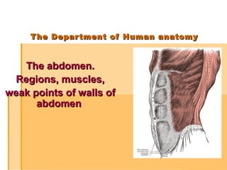
The abdomen. Regions, muscles, weak points of walls of abdomen
•Download as PPT, PDF•
46 likes•12,606 views

1. Abdominal regions. 2. Walls of abdomen. 3. Muscles of abdomen: classification. 4. Superficial and internal inguinal rings. Abdominal press. 5. Fasciae of abdomen. 6. Weak points of walls of abdomen.

Recommended
More Related Content
What's hot
What's hot (20)
Similar to The abdomen. Regions, muscles, weak points of walls of abdomen
Similar to The abdomen. Regions, muscles, weak points of walls of abdomen (20)
More from Eneutron
More from Eneutron (20)
Recently uploaded
Recently uploaded (20)
The abdomen. Regions, muscles, weak points of walls of abdomen
- 1. The Department of Human anatomyThe Department of Human anatomy The abdomen.The abdomen. Regions, muscles,Regions, muscles, weak points of walls ofweak points of walls of abdomenabdomen
- 2. PLANPLAN Abdominal regions.Abdominal regions. Walls of abdomen.Walls of abdomen. Muscles of abdomen: classification.Muscles of abdomen: classification. Superficial and internal inguinalSuperficial and internal inguinal rings. Abdominal press.rings. Abdominal press. Fasciae of abdomen.Fasciae of abdomen. Weak points of walls of abdomen.Weak points of walls of abdomen.
- 3. Abdomen is theAbdomen is the region of bodyregion of body lying betweenlying between the diaphragmthe diaphragm above and pelvisabove and pelvis below. It containsbelow. It contains the contents ofthe contents of digestive anddigestive and urinary systems.urinary systems.
- 4. Two horizontal andTwo horizontal and two vertical linestwo vertical lines divide the abdomendivide the abdomen into nine "regions."into nine "regions."
- 5. I. Epigastrium:I. Epigastrium: 1 (regio epigastrica)1 (regio epigastrica) 2. (regio hypocondrica dexter)2. (regio hypocondrica dexter) 3. (regio hypochondrica sinistra)3. (regio hypochondrica sinistra) ІІ. Mesogastrium:ІІ. Mesogastrium: 4. (regio umbilicalis)4. (regio umbilicalis) 5. (regio lateralis dextra)5. (regio lateralis dextra) 6. (regio lateralis sinistra)6. (regio lateralis sinistra) ІІІ. Hypogastrium:ІІІ. Hypogastrium: 7. (regio pubica)7. (regio pubica) 8. (regio inguinalis dextra)8. (regio inguinalis dextra) 9. (regio inguinalis sinister)9. (regio inguinalis sinister) ("hypo" means "below" and "epi" means("hypo" means "below" and "epi" means "above", "chond" means "cartilage""above", "chond" means "cartilage" (in this case, the cartilage of the rib)(in this case, the cartilage of the rib) and "gast" means stomach).and "gast" means stomach).
- 6. Walls of abdomen:Walls of abdomen: Superior Wall:Superior Wall: The superior wall of abdomen is formed by theThe superior wall of abdomen is formed by the diaphragm. It is a strong muscular sheath that has a vital role indiaphragm. It is a strong muscular sheath that has a vital role in respiratory system.respiratory system. Inferior Wall:Inferior Wall: the conditional line to the entry of the pelvicthe conditional line to the entry of the pelvic cavity.cavity. Anterior Wall:Anterior Wall: The anterior wall of abdomen is formed above byThe anterior wall of abdomen is formed above by the lower part of thoracic cage. Below it is formed by Rectusthe lower part of thoracic cage. Below it is formed by Rectus abdominis, External oblique, Internal oblique and Transversusabdominis, External oblique, Internal oblique and Transversus abdominis muscles along with their fasciae.abdominis muscles along with their fasciae. Posterior Wall:Posterior Wall: The posterior wall of abdomen is formed in theThe posterior wall of abdomen is formed in the midline by the five lumbar vertebrae and their intervertebralmidline by the five lumbar vertebrae and their intervertebral discs. Laterally, it is formed by the twelfth rib, psoas muscle,discs. Laterally, it is formed by the twelfth rib, psoas muscle, quadratus lumborum muscle and the border of bony pelvis. Thequadratus lumborum muscle and the border of bony pelvis. The aponeurosis of origin of transversus abdominis muscle is alsoaponeurosis of origin of transversus abdominis muscle is also involved in the formation of posterior wall of the abdomen.involved in the formation of posterior wall of the abdomen.
- 7. The layers of the abdominal wall are (fromThe layers of the abdominal wall are (from superficial to deep):superficial to deep): SkinSkin FasciaFascia Camper's fascia - fatty superficial layer.Camper's fascia - fatty superficial layer. Scarpa's fascia - deep fibrous layer.Scarpa's fascia - deep fibrous layer. MuscleMuscle Rectus abdominisRectus abdominis External oblique muscleExternal oblique muscle Internal oblique muscleInternal oblique muscle Transverse abdominal muscleTransverse abdominal muscle Fascia transversalisFascia transversalis PeritoneumPeritoneum
- 8. Abdominal muscles The muscles of anterior wall The muscles of posterior wall The muscles of laterar wall Pyramidalis Obliquus externus abdominis Rectus Abdominus Obliquus internus abdominis Transversus abdominis Quadratus lumborum
- 9. The muscles of anterior wall Rectus Abdominus Origin 5, 6, 7 costal cartilages,5, 6, 7 costal cartilages, xiphoid processxiphoid process InsertionInsertion Pubic crest and pubic symphysis ActionAction Flexes trunk, aids forcedFlexes trunk, aids forced expiration and raise intra-expiration and raise intra- abdominal pressureabdominal pressure
- 10. Pyramidalis Origin Pubic crest Insertion Lower linea alba ActionAction Is a tensor of the lineaIs a tensor of the linea albaalba Reinforces lower rectusReinforces lower rectus sheathsheath
- 11. The muscles of lateral wall Obliquus externus Abdominis Origin Anterior angles of lower eight ribs InsertionInsertion Outer anterior half of iliac crest,Outer anterior half of iliac crest, inguinal lig, pubic tubercle andinguinal lig, pubic tubercle and crest, and aponeurosis ofcrest, and aponeurosis of anterior rectus sheathanterior rectus sheath Action Supports abdominal wall, assists forced expiration, aids raising intraabdominal pressure and, with muscles of opposite side, abducts and rotates trunk
- 12. Obliquus internus abdominis Origin Lumbar fascia, ant. two thirds of iliac crest and lateral two thirds of inguinal ligament InsertionInsertion Costal margin, aponeurosis ofCostal margin, aponeurosis of rectus sheath (ant. and post. ),rectus sheath (ant. and post. ), conjoint tendon to pubic crestconjoint tendon to pubic crest and pectineal lineand pectineal line ActionAction Supports abdominal wall,Supports abdominal wall, assists forced respiration, aidsassists forced respiration, aids raising intraabdominal pressureraising intraabdominal pressure & , with muscles of other side ,& , with muscles of other side , abducts and rotates trunk.abducts and rotates trunk. Conjoint tendon supportsConjoint tendon supports posterior wall of inguinal canalposterior wall of inguinal canal
- 13. Transversus abdominis Origin Costal margin , lumbar fascia, ant two thirds of iliac crest and lateral half of inguinal ligament InsertionInsertion Aponeurosis of posterior andAponeurosis of posterior and anterior rectus sheath andanterior rectus sheath and conjoint tendon to pubic crestconjoint tendon to pubic crest and pectineal lineand pectineal line ActionAction Supports abdominal wall, aidsSupports abdominal wall, aids forced expiration and raisingforced expiration and raising intraabdominal pressure.intraabdominal pressure. Conjoint tendon supportsConjoint tendon supports posterior wall of inguinal canalposterior wall of inguinal canal
- 14. The muscles of posterior wall Quadratus lumborum Origin Inferior border of 12th rib Insertion Transverse processes of L1-4, iliolumbar ligament and post. 1/3 of iliac crest ActionAction Fixes 12th rib duringFixes 12th rib during respiration and lateralrespiration and lateral Flexes of trunkFlexes of trunk
- 15. Rectus sheath above and below the umbilical chordRectus sheath above and below the umbilical chord 1 – (m. rectus abdominis);1 – (m. rectus abdominis); 2 – (m. obliguus externus abdominis);2 – (m. obliguus externus abdominis); 3 – (m. obliguus internus abdominis);3 – (m. obliguus internus abdominis); 4 – (m. transversus abdominis);4 – (m. transversus abdominis); а) (aponeurosis m. obliguus externus abdominis);а) (aponeurosis m. obliguus externus abdominis); b) (lamina anterior m. obliguus internus abdominis);b) (lamina anterior m. obliguus internus abdominis); c) (lamina posterior m. obliguus internus abdominis);c) (lamina posterior m. obliguus internus abdominis); d) (aponeurosis m. transverses abdominis);d) (aponeurosis m. transverses abdominis); e) (fascia transversalis).e) (fascia transversalis).
- 16. weak points of walls of abdomenweak points of walls of abdomen 1) canalis inquinalis1) canalis inquinalis 2) linea alba2) linea alba 3) anulus umbilicalis3) anulus umbilicalis 4) trigonum lumbale Petiti4) trigonum lumbale Petiti 5) spatium tendinosum lumbale5) spatium tendinosum lumbale 6) trigonum hypochondriacum6) trigonum hypochondriacum 7) linea semilunaris Spigelii7) linea semilunaris Spigelii 8) trigonum sternocostale dext. et sin.8) trigonum sternocostale dext. et sin. trigonum costolumbalis dext. et sin.trigonum costolumbalis dext. et sin. 9) canalis obturatorius9) canalis obturatorius 10) Foramen supra- et infrapiriformis10) Foramen supra- et infrapiriformis 11) canalis femoralis11) canalis femoralis
- 18. Inguinal canal is an obliqueInguinal canal is an oblique passage through the lower partpassage through the lower part of anterior abdominal wall. It isof anterior abdominal wall. It is present in both males andpresent in both males and females.females.
- 19. Functions of inguinal canal:Functions of inguinal canal: In males, the inguinal canal allows structures ofIn males, the inguinal canal allows structures of the spermatic cord to pass to and from the testisthe spermatic cord to pass to and from the testis to the abdomen. This allows the testes to leaveto the abdomen. This allows the testes to leave the abdominal cavity. The importance of thisthe abdominal cavity. The importance of this phenomenon can be appreciated by remindingphenomenon can be appreciated by reminding the fact that spermatogenesis takes place only ifthe fact that spermatogenesis takes place only if the testes leave the abdominal cavity to go to athe testes leave the abdominal cavity to go to a cooler environment in the scrotum.cooler environment in the scrotum. In the females, the inguinal canal is smaller asIn the females, the inguinal canal is smaller as compared to males. It transmits the roundcompared to males. It transmits the round ligament of uterus to pass from the uterus toligament of uterus to pass from the uterus to labium majus.labium majus. In both males and females, the inguinal canal alsoIn both males and females, the inguinal canal also transmits the ilioinguinal nerve.transmits the ilioinguinal nerve.
- 20. Structure of inguinal canal:Structure of inguinal canal: Inguinal canal is about 4-5 cm long in adults. ItInguinal canal is about 4-5 cm long in adults. It extends fromextends from deepdeep inguinal ringinguinal ring, downward and, downward and medially, to themedially, to the superficial inguinal ringsuperficial inguinal ring. The deep. The deep inguinal ring is an oval opening in fasciainguinal ring is an oval opening in fascia transversalis, while the superficial inguinal ring is atransversalis, while the superficial inguinal ring is a triangular defect in the aponeuorsis of externaltriangular defect in the aponeuorsis of external oblique muscle.oblique muscle. The inguinal canal lies immediately above andThe inguinal canal lies immediately above and parallel to the inguinal ligament. However, inparallel to the inguinal ligament. However, in infants, the deep inguinal ring lies almost posteriorinfants, the deep inguinal ring lies almost posterior to the superficial, so that the canal is shorter. As ato the superficial, so that the canal is shorter. As a result of growth in following years, the deepresult of growth in following years, the deep inguinal ring moves laterally and the canalinguinal ring moves laterally and the canal elongates.elongates.
- 21. Walls of inguinal canal:Walls of inguinal canal:
- 22. Anterior wall:Anterior wall: TheThe anterior wall is formedanterior wall is formed along its entire lengthalong its entire length by the aponeuorsis ofby the aponeuorsis of external obliqueexternal oblique muscle. In its lateralmuscle. In its lateral part, it is reinforced bypart, it is reinforced by the origin of internalthe origin of internal oblique from theoblique from the inguinal ligament.inguinal ligament.
- 23. Posterior wall:Posterior wall: TheThe posterior wall of inguinalposterior wall of inguinal canal is formed along itscanal is formed along its entire length by theentire length by the fascia transversalis.fascia transversalis.
- 24. Floor of inguinal canalFloor of inguinal canal (inferior wall):(inferior wall): The floor ofThe floor of the canal is formed by thethe canal is formed by the inguinal ligament. Thisinguinal ligament. This ligament is in fact the rolled-ligament is in fact the rolled- under inferior edge ofunder inferior edge of aponeuorsis of externalaponeuorsis of external oblique muscle. The medialoblique muscle. The medial part of this wall is formed bypart of this wall is formed by the lacunar ligament.the lacunar ligament. Roof of inguinal canalRoof of inguinal canal (superior wall):(superior wall): The roof ofThe roof of the canal is formed by thethe canal is formed by the lowest fibers of internallowest fibers of internal oblique and transversusoblique and transversus abdominis muscles.abdominis muscles.
- 25. Internal surface of anterior abdominalInternal surface of anterior abdominal wallwall
- 27. Inguinal canal as a potential weakness inInguinal canal as a potential weakness in abdominal wall:abdominal wall: The presence of inguinal canal in lower partThe presence of inguinal canal in lower part of anterior abdominal wall presents aof anterior abdominal wall presents a potential weakness the structure of the wall.potential weakness the structure of the wall. The canal is oblique in its form. This causesThe canal is oblique in its form. This causes the weaker parts of it to lie at some distancethe weaker parts of it to lie at some distance apart, thus reducing chances of any type ofapart, thus reducing chances of any type of damage.damage.
- 28. The weak inguinal canal can be exploitedThe weak inguinal canal can be exploited during forceful activities like coughing,during forceful activities like coughing, sneezing, straining, parturition, defecationsneezing, straining, parturition, defecation etc. To prevent this, the arching lowestetc. To prevent this, the arching lowest fibers of internal oblique and transversusfibers of internal oblique and transversus abdominis muscles contract and flatten outabdominis muscles contract and flatten out the arched roof so that it is loweredthe arched roof so that it is lowered towards the floor. In this condition, thetowards the floor. In this condition, the canal is virtually closed.canal is virtually closed.
- 29. AnAn inguinal herniainguinal hernia is a protrusionis a protrusion of abdominal-of abdominal- cavity contentscavity contents through the inguinalthrough the inguinal canal. They are verycanal. They are very common (lifetimecommon (lifetime risk 27% for men,risk 27% for men, 3% for women), and3% for women), and their repair is one oftheir repair is one of the most frequentlythe most frequently performed surgicalperformed surgical operations.operations.
- 30. There are two types ofThere are two types of inguinal hernia,inguinal hernia, directdirect andand indirectindirect, which are, which are defined by their relationship to the inferiordefined by their relationship to the inferior epigastric vessels. Direct inguinal hernias occurepigastric vessels. Direct inguinal hernias occur medial to the inferior epigastric vessels whenmedial to the inferior epigastric vessels when abdominal contents herniate through a weak spotabdominal contents herniate through a weak spot in the fascia of the posterior wall of the inguinalin the fascia of the posterior wall of the inguinal canal, which is formed by the transversalis fascia.canal, which is formed by the transversalis fascia. Indirect inguinal hernias occur when abdominalIndirect inguinal hernias occur when abdominal contents protrude through the deep inguinal ring,contents protrude through the deep inguinal ring, lateral to the inferior epigastric vessels; this maylateral to the inferior epigastric vessels; this may be caused by failure of embryonic closure ofbe caused by failure of embryonic closure of the processus vaginalis.the processus vaginalis.
- 31. Inguinal hernias, in turn, belongInguinal hernias, in turn, belong to groin hernias, which alsoto groin hernias, which also includes femoral hernias. A femoral herniaincludes femoral hernias. A femoral hernia is not via the inguinal canal, but via theis not via the inguinal canal, but via the femoral canal, which normally allowsfemoral canal, which normally allows passage of the common femoral artery andpassage of the common femoral artery and vein from the pelvis to the leg.vein from the pelvis to the leg. In Amyand's hernia, the content of theIn Amyand's hernia, the content of the hernial sac is the vermiform appendix.hernial sac is the vermiform appendix. In Littre's hernia, the content of the hernialIn Littre's hernia, the content of the hernial sac contains a Meckel's Diverticulum.sac contains a Meckel's Diverticulum.
- 32. Thank you for attention!Thank you for attention!