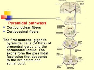
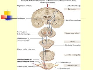
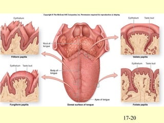
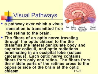
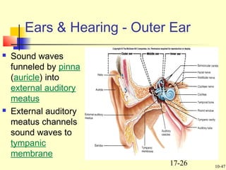
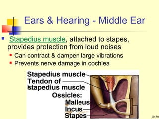
1
Ivano-Frankivsk National Medical University
The Department of Human anatomy
Descending (efferent) pathways.
Pathways of visual, auditory
analyzers, taste and smell.
Topography of pathways of spinal
cord on the transverse section.
Prepared by PhDPrepared by PhD
Tetyana Knyazevych - ChornaTetyana Knyazevych - Chorna
17-2
Descending (efferent) pathways:
 Pyramidal pathways conduct voluntary
motor inpulses from the cerebral cortex
 Extrapyramidal pathways deals with the
basal nuclei, maintain interrelations and
act as a single unit
17-3
Pyramidal pathways
 Corticonuclear fibers
 Corticospinal fibers
The first neurons- gigantic
pyramidal cells (of Betz) of
precentral gyrus and the
paracentral lobule. The
axons form the pyramidal
fasciculus that descends
to the brainstem and
spinal cord.
17-4
 Some fibers decussate yet in the
brainstem and terminate on the
motor nuclei of the cranial nerves
(III and IV pairs in midbrain; V-VIII
in pons; IX-XII in medulla) and form
the corticonuclear fibers.
 The motor cells of the nuclei of
cranial nerves are the second
neurons. Their axons quit the
brainstem and reach the areas of
the responsibility
17-5
17-6
 The larger portion of the
fibers descends to the
pyramids of the medulla as
a corticospinal fibers.
 80% decussate and enter
the lateral funiculus to form
the lateral corticospinal
tract. The rest of fibers
proceed to the anterior
funiculus to form the
anterior corticospinal tract.
17-7
 Injury of the
pyramidal tracts
results in central
paralysis of both
limbs while the
diaphragm and the
muscles of trunk
manifest little
malfunctioning.
17-8
Extrapyramidal pathways
 the extrapyramidal system is a neural
network that causes involuntary reflexes
and movement, and modulation of
movement. Extrapyramidal tracts are chiefly
found in the reticular formation of
the pons and medulla, and target neurons
in the spinal cord involved in reflexes,
locomotion, complex movements, and
postural control.
17-9
 These tracts are in turn modulated by
various parts of the central nervous
system, including the nigrostriatal
pathway, the basal ganglia, the 
cerebellum, the vestibular nuclei, and
different sensory areas of the cerebral
cortex. All of these regulatory components
can be considered part of the
extrapyramidal system, in that they
modulate motor activity without directly
innervating motor neurons.
17-10
Extrapyramidal pathways
 The rubrospinal tract
 The tectospinal tract
 The vestibulospinal tract
 The olivospinal tract
 The reticulospinal tract
17-11
17-12
 I. funiculus posterior.
 II. funiculus lateralis.
 III. funiculus anterior.
 1. (fasciculus gracilis (Golli).
 2. (fasciculus cuneatus (Burdach).
 3. (fasciculus proprius posterior).
 4. (tractus spinocerebellaris posterior (Flechsig).
 5. (tractus spinocerebellaris anterior (Gowers).
 6. (tractus olivospinalis).
 7. (tractus corticospinalis lateralis).
 8. ( tractus rubrospinalis (Monakov).
17-13
 9. (tractus spinothalamicus lateralis).
 10. (tractus spinotectalis).
 11. (fasciculus proprius lateralis).
 12. (fasciculus longitudinalis medialis).
 13. (tractus tectospinalis).
 14. (tractus corticospinalis anterior).
 15. (tractus reticulospinalis).
 16. (tractus spinothalamicus anterior).
 17. (fasciculus proprius anterius).
 18. (tractus vestibulospinalis).
17-14
Olfaction – Sense of Smell
 The olfactory system represents
one of the oldest sensory
modalities in the phylogenetic
history of mammals. As a chemical
sensor, the olfactory system
detects food and influences social
and sexual behavior.
17-15
 The olfactory area related to the superior
nasal concha and adherent portion of
nasal septum. The olfactory part
comprise 3 types of cells: receptor cells,
supporting cells, basal cells.
 The axons of receptor cells pass the
cribriform plate of ethmoid bone as the
olfactory nerve, they reach the olfactory
bulb to synapse with the second
neurons.
17-16
 The second neurons form the
olfactory tract, which terminates
within the olfactory trigone, the
anterior perforated substance,
septum pellucidum, where the 3-d
neuron stay.
17-17
 Axons of 3-d neuron
run to amygdaloid
body, the olfacrory
cortex of
parahippocampal
gyrus, uncus.
 It has the
connections with the
mammilary bodies,
reticular formation,
nuclei of cranial
nerves.
17-18
17-19
Gustation – Sense of Taste
 Gustatory receptors are housed in specialized taste buds
on the surface of the tongue.
 4 types of papillae:
 filiform
 fungiform
 vallate
 foliate
The tongue detects five basic taste sensations:
 salty
 sweet
 sour
 bitter
 umami
17-20
17-21
 The first neurons are the
pseudounipolar neurons of
the sensory ganglia of facial
and glossopharyngeal
nerves.
 Lingual nerve of facial
cranial nerve (from anterior
2/3 of tongue) and lingual
branches of
glossopharyngeal nerve
(from posterior 1/3 of
tongue) terminates the
nucleus of solitary tract,
where the bodes of 2-d
neurons stay.
 The axons of 2-nd neurons
decussate and join the
medial lemniscus that
ascends to the thalamus.
 The axons of 3-d neurons of
thalamus reach the uncus of
17-22
17-23
Visual Pathways
 a pathway over which a visual
sensation is transmitted from
the retina to the brain.
 The fibers of an optic nerve traveling
through the optic chiasm to the the
thalamus,the lateral geniculate body and
superior coliculi, and optic radiations
terminating in an occipital lobe (sulcus
calcarinus). Each optic nerve contains
fibers from only one retina. The fibers from
the middle parts of the retinas cross to the
opposite side of the brain at the optic
chiasm.
17-24
 The fibers from the
lateral part of each eye
do not cross at the
optic chiasm, pass
through the lateral
geniculate body on the
same side of the brain,
and continue back to
the occipital lobe. If the
right optic tract were
destroyed, a person
would lose partial
vision in both eyes -
the right medial and the
left lateral fields of
vision.
17-25
17-26
 Sound waves
funneled by pinna
(auricle) into
external auditory
meatus
 External auditory
meatus channels
sound waves to
tympanic
membrane
Ears & Hearing - Outer Ear
10-47
17-27
 Malleus (hammer) is
attached to tympanic
membrane
 Carries vibrations to
incus (anvil)
 Stapes (stirrup)
receives vibrations
from incus, transmits
to oval window
Ears & Hearing - Middle Ear
10-49
17-28
 Stapedius muscle, attached to stapes,
provides protection from loud noises
 Can contract & dampen large vibrations
 Prevents nerve damage in cochlea
Ears & Hearing - Middle Ear
10-50
17-29
Ears & Hearing - Cochlea
 Cochlea consists of a tube that looks like snail
shell
10-51
17-30
Ears & Hearing - Cochlea
 Tube is divided
into 3 fluid-filled
chambers
 Scala vestibuli,
cochlear duct,
scala tympani
10-52
17-31
Ears & Hearing - Cochlea
 Oval window attached to scala vestibuli (at base of
cochlea)
 Vibrations at oval window induce pressure waves in
perilymph fluid of scala vestibuli
 Scalas vestibuli & tympani are continuous at apex
 So waves in vestibuli pass to tympani & displace round
window (at base of cochlea)

Necessary because fluids are incompressible & waves would not be
possible without round window
10-53
17-32
Ears & Hearing - Cochlea
 Low frequencies can travel all way thru vestibuli & back in
tympani
 As frequencies increase they travel less before passing directly
through vestibular & basilar membranes to tympani
Fig 10.20
10-54
17-33
Ears & Hearing - Cochlea
 High
frequencies
produce
maximum
stimulation of
Spiral Organ
closer to base
of cochlea &
lower
frequencies
stimulate
closer to apex
10-55
17-34
Spiral Organ (Organ of Corti)
 Spiral Organ is
where sound is
transduced
 Sensory hair cells
located on the
basilar membrane
 1 row of inner
cells extend
length of basilar
membrane
 Multiple rows of
outer hair cells
are embedded in
tectorial
membrane
10-56
17-35
Spiral Organ (Organ of Corti)
 Pressure waves moving thru cochlear duct
create shearing forces between basilar &
tectorial membranes, moving & bending
stereocilia
10-57
17-36
Neural Pathway for Hearing
 Info from 8th
nerve goes to
pons, then to
inferior
colliculus, then
to thalamus, &
to auditory
cortex
10-58
17-37
Neural Pathways for Hearing
 Neurons in
different
regions of
cochlea
stimulate
neurons in
corresponding
areas of
auditory cortex
 Each area of
cortex
represents
different part of
cochlea & thus
a different pitch
10-59
17-38
Vestibular Apparatus
 Provides sense of
equilibrium
 =orientation to gravity
 Vestibular apparatus &
cochlea form inner ear
 V. apparatus consists
of otolith organs
(utricle & saccule) &
semicircular canals
10-35
17-39
Semicircular Canals
 Provide information
about rotational
acceleration
 Project in 3
different planes
 Each contains a
semicircular duct
 At base is crista
ampullaris where
sensory hair cells
are located
Fig 10.12
10-42
17-40
 Utricle and saccule provide info about linear
acceleration
 Semicircular canals, oriented in 3 planes, give sense of
angular acceleration
Vestibular Apparatus
10-37
17-41
 Hair cells are receptors for equilibrium
 Each contains 20-50 hair-like extensions called stereocilia

1 of these is a kinocilium
Vestibular Apparatus
10-38
17-42
 When stereocilia are bent toward kinocilium, hair cell
depolarizes & releases NT that stimulates 8th nerve
 When bent away from kinocilium, hair cell hyperpolarizes
 In this way, frequency of APs in hair cells carries information about
movement
Vestibular Apparatus
10-39
17-43
Utricle & Saccule
 Have a macula containing hair cells
 Hair cells embedded in gelatinous otolithic membrane

Which contains calcium carbonate crystals (=otoliths) that resist
change in movement
10-40
17-44
Utricle & Saccule
 Utricle sensitive to
horizontal
acceleration
 Hairs pushed
backward during
forward
acceleration
 Saccule sensitive
to vertical
acceleration

Hairs pushed
upward when
person descends
10-41
17-45
Semicircular Canals
 Provide information
about rotational
acceleration
 Project in 3
different planes
 Each contains a
semicircular duct
 At base is crista
ampullaris where
sensory hair cells
are located
10-42
17-46
17-47
Thank you for attention!

Pathways 2

  • 1.
    1 Ivano-Frankivsk National MedicalUniversity The Department of Human anatomy Descending (efferent) pathways. Pathways of visual, auditory analyzers, taste and smell. Topography of pathways of spinal cord on the transverse section. Prepared by PhDPrepared by PhD Tetyana Knyazevych - ChornaTetyana Knyazevych - Chorna
  • 2.
    17-2 Descending (efferent) pathways: Pyramidal pathways conduct voluntary motor inpulses from the cerebral cortex  Extrapyramidal pathways deals with the basal nuclei, maintain interrelations and act as a single unit
  • 3.
    17-3 Pyramidal pathways  Corticonuclearfibers  Corticospinal fibers The first neurons- gigantic pyramidal cells (of Betz) of precentral gyrus and the paracentral lobule. The axons form the pyramidal fasciculus that descends to the brainstem and spinal cord.
  • 4.
    17-4  Some fibersdecussate yet in the brainstem and terminate on the motor nuclei of the cranial nerves (III and IV pairs in midbrain; V-VIII in pons; IX-XII in medulla) and form the corticonuclear fibers.  The motor cells of the nuclei of cranial nerves are the second neurons. Their axons quit the brainstem and reach the areas of the responsibility
  • 5.
  • 6.
    17-6  The largerportion of the fibers descends to the pyramids of the medulla as a corticospinal fibers.  80% decussate and enter the lateral funiculus to form the lateral corticospinal tract. The rest of fibers proceed to the anterior funiculus to form the anterior corticospinal tract.
  • 7.
    17-7  Injury ofthe pyramidal tracts results in central paralysis of both limbs while the diaphragm and the muscles of trunk manifest little malfunctioning.
  • 8.
    17-8 Extrapyramidal pathways  the extrapyramidalsystem is a neural network that causes involuntary reflexes and movement, and modulation of movement. Extrapyramidal tracts are chiefly found in the reticular formation of the pons and medulla, and target neurons in the spinal cord involved in reflexes, locomotion, complex movements, and postural control.
  • 9.
    17-9  These tractsare in turn modulated by various parts of the central nervous system, including the nigrostriatal pathway, the basal ganglia, the  cerebellum, the vestibular nuclei, and different sensory areas of the cerebral cortex. All of these regulatory components can be considered part of the extrapyramidal system, in that they modulate motor activity without directly innervating motor neurons.
  • 10.
    17-10 Extrapyramidal pathways  Therubrospinal tract  The tectospinal tract  The vestibulospinal tract  The olivospinal tract  The reticulospinal tract
  • 11.
  • 12.
    17-12  I. funiculusposterior.  II. funiculus lateralis.  III. funiculus anterior.  1. (fasciculus gracilis (Golli).  2. (fasciculus cuneatus (Burdach).  3. (fasciculus proprius posterior).  4. (tractus spinocerebellaris posterior (Flechsig).  5. (tractus spinocerebellaris anterior (Gowers).  6. (tractus olivospinalis).  7. (tractus corticospinalis lateralis).  8. ( tractus rubrospinalis (Monakov).
  • 13.
    17-13  9. (tractusspinothalamicus lateralis).  10. (tractus spinotectalis).  11. (fasciculus proprius lateralis).  12. (fasciculus longitudinalis medialis).  13. (tractus tectospinalis).  14. (tractus corticospinalis anterior).  15. (tractus reticulospinalis).  16. (tractus spinothalamicus anterior).  17. (fasciculus proprius anterius).  18. (tractus vestibulospinalis).
  • 14.
    17-14 Olfaction – Senseof Smell  The olfactory system represents one of the oldest sensory modalities in the phylogenetic history of mammals. As a chemical sensor, the olfactory system detects food and influences social and sexual behavior.
  • 15.
    17-15  The olfactoryarea related to the superior nasal concha and adherent portion of nasal septum. The olfactory part comprise 3 types of cells: receptor cells, supporting cells, basal cells.  The axons of receptor cells pass the cribriform plate of ethmoid bone as the olfactory nerve, they reach the olfactory bulb to synapse with the second neurons.
  • 16.
    17-16  The secondneurons form the olfactory tract, which terminates within the olfactory trigone, the anterior perforated substance, septum pellucidum, where the 3-d neuron stay.
  • 17.
    17-17  Axons of3-d neuron run to amygdaloid body, the olfacrory cortex of parahippocampal gyrus, uncus.  It has the connections with the mammilary bodies, reticular formation, nuclei of cranial nerves.
  • 18.
  • 19.
    17-19 Gustation – Senseof Taste  Gustatory receptors are housed in specialized taste buds on the surface of the tongue.  4 types of papillae:  filiform  fungiform  vallate  foliate The tongue detects five basic taste sensations:  salty  sweet  sour  bitter  umami
  • 20.
  • 21.
    17-21  The firstneurons are the pseudounipolar neurons of the sensory ganglia of facial and glossopharyngeal nerves.  Lingual nerve of facial cranial nerve (from anterior 2/3 of tongue) and lingual branches of glossopharyngeal nerve (from posterior 1/3 of tongue) terminates the nucleus of solitary tract, where the bodes of 2-d neurons stay.  The axons of 2-nd neurons decussate and join the medial lemniscus that ascends to the thalamus.  The axons of 3-d neurons of thalamus reach the uncus of
  • 22.
  • 23.
    17-23 Visual Pathways  apathway over which a visual sensation is transmitted from the retina to the brain.  The fibers of an optic nerve traveling through the optic chiasm to the the thalamus,the lateral geniculate body and superior coliculi, and optic radiations terminating in an occipital lobe (sulcus calcarinus). Each optic nerve contains fibers from only one retina. The fibers from the middle parts of the retinas cross to the opposite side of the brain at the optic chiasm.
  • 24.
    17-24  The fibersfrom the lateral part of each eye do not cross at the optic chiasm, pass through the lateral geniculate body on the same side of the brain, and continue back to the occipital lobe. If the right optic tract were destroyed, a person would lose partial vision in both eyes - the right medial and the left lateral fields of vision.
  • 25.
  • 26.
    17-26  Sound waves funneledby pinna (auricle) into external auditory meatus  External auditory meatus channels sound waves to tympanic membrane Ears & Hearing - Outer Ear 10-47
  • 27.
    17-27  Malleus (hammer)is attached to tympanic membrane  Carries vibrations to incus (anvil)  Stapes (stirrup) receives vibrations from incus, transmits to oval window Ears & Hearing - Middle Ear 10-49
  • 28.
    17-28  Stapedius muscle,attached to stapes, provides protection from loud noises  Can contract & dampen large vibrations  Prevents nerve damage in cochlea Ears & Hearing - Middle Ear 10-50
  • 29.
    17-29 Ears & Hearing- Cochlea  Cochlea consists of a tube that looks like snail shell 10-51
  • 30.
    17-30 Ears & Hearing- Cochlea  Tube is divided into 3 fluid-filled chambers  Scala vestibuli, cochlear duct, scala tympani 10-52
  • 31.
    17-31 Ears & Hearing- Cochlea  Oval window attached to scala vestibuli (at base of cochlea)  Vibrations at oval window induce pressure waves in perilymph fluid of scala vestibuli  Scalas vestibuli & tympani are continuous at apex  So waves in vestibuli pass to tympani & displace round window (at base of cochlea)  Necessary because fluids are incompressible & waves would not be possible without round window 10-53
  • 32.
    17-32 Ears & Hearing- Cochlea  Low frequencies can travel all way thru vestibuli & back in tympani  As frequencies increase they travel less before passing directly through vestibular & basilar membranes to tympani Fig 10.20 10-54
  • 33.
    17-33 Ears & Hearing- Cochlea  High frequencies produce maximum stimulation of Spiral Organ closer to base of cochlea & lower frequencies stimulate closer to apex 10-55
  • 34.
    17-34 Spiral Organ (Organof Corti)  Spiral Organ is where sound is transduced  Sensory hair cells located on the basilar membrane  1 row of inner cells extend length of basilar membrane  Multiple rows of outer hair cells are embedded in tectorial membrane 10-56
  • 35.
    17-35 Spiral Organ (Organof Corti)  Pressure waves moving thru cochlear duct create shearing forces between basilar & tectorial membranes, moving & bending stereocilia 10-57
  • 36.
    17-36 Neural Pathway forHearing  Info from 8th nerve goes to pons, then to inferior colliculus, then to thalamus, & to auditory cortex 10-58
  • 37.
    17-37 Neural Pathways forHearing  Neurons in different regions of cochlea stimulate neurons in corresponding areas of auditory cortex  Each area of cortex represents different part of cochlea & thus a different pitch 10-59
  • 38.
    17-38 Vestibular Apparatus  Providessense of equilibrium  =orientation to gravity  Vestibular apparatus & cochlea form inner ear  V. apparatus consists of otolith organs (utricle & saccule) & semicircular canals 10-35
  • 39.
    17-39 Semicircular Canals  Provideinformation about rotational acceleration  Project in 3 different planes  Each contains a semicircular duct  At base is crista ampullaris where sensory hair cells are located Fig 10.12 10-42
  • 40.
    17-40  Utricle andsaccule provide info about linear acceleration  Semicircular canals, oriented in 3 planes, give sense of angular acceleration Vestibular Apparatus 10-37
  • 41.
    17-41  Hair cellsare receptors for equilibrium  Each contains 20-50 hair-like extensions called stereocilia  1 of these is a kinocilium Vestibular Apparatus 10-38
  • 42.
    17-42  When stereociliaare bent toward kinocilium, hair cell depolarizes & releases NT that stimulates 8th nerve  When bent away from kinocilium, hair cell hyperpolarizes  In this way, frequency of APs in hair cells carries information about movement Vestibular Apparatus 10-39
  • 43.
    17-43 Utricle & Saccule Have a macula containing hair cells  Hair cells embedded in gelatinous otolithic membrane  Which contains calcium carbonate crystals (=otoliths) that resist change in movement 10-40
  • 44.
    17-44 Utricle & Saccule Utricle sensitive to horizontal acceleration  Hairs pushed backward during forward acceleration  Saccule sensitive to vertical acceleration  Hairs pushed upward when person descends 10-41
  • 45.
    17-45 Semicircular Canals  Provideinformation about rotational acceleration  Project in 3 different planes  Each contains a semicircular duct  At base is crista ampullaris where sensory hair cells are located 10-42
  • 46.
  • 47.