1. 1
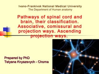
Ivano-Frankivsk National Medical University
The Department of Human anatomy
Pathways of spinal cord and
brain, their classification.
Associative, comissural and
projection ways. Ascending
projection ways.
Prepared by PhDPrepared by PhD
Tetyana Knyazevych - ChornaTetyana Knyazevych - Chorna
2. 17-2
A neural pathway connects one part of
the nervous system with another and
usually consists of bundles of
elongated, myelin-insulated neurons,
known collectively as white matter.
Neural pathways serve to connect
relatively distant areas of the brain or
nervous system, compared to the local
communication of grey matter.
4. 17-4
Association fibers connect areas within the same hemisphere.
Short association fibers connect areas that are located in
the same lobe (arcuate fibers).
Long association fibers connect areas that are located in
different lobes of the brain:
- Superior longitudinal fasciculus
- Inferior longitudinal fasciculus
- The cingulum
- The uncinate fasciculus
7. 17-7
Exteroceptive pathways
Transmit the impulses from the skin receptors,
the retina, the internal ear and the tongue
- Lateral spinothalamic tract (pain and
temperature)
- Anterior spinothalamic tract (light touch (crude
touch), pressure, tickle, itch)
- The pathways of sense cranial nerves (visual,
auditory, taste)
8. 17-8
Lateral spinothalamic tract (pain
and temperature)
The first neurons
(pseudounipolar
cells) reside within
the spinal ganglia.
The dendrites run
to the skin, the
axons form the
posterior root.
9. 17-9
The second neurons
reside in the nucleus
proprius. The axons
decussate and enter the
lateral funiculus to form
the lateral spinothalamic
tract. The tract traverses
the medulla oblongata,
pons, midbrain and ends
in thalamus.
10. 17-10
The axons of
the third
neurons pass
through the
posterior limb
of internal
capsule, join
the corona
radiata and
terminate in
the
poscentral
gyrus.
11. 17-11
Anterior spinothalamic tract
(touch, pressure)
The first neurons
are the
pseudounipolar cells
of the spinal ganglia.
The dendrites run to
the skin, the axons
form the posterior
root.
12. 17-12
The second neurons reside in
the gelatinous substance of
the posterior gray column. The
axons decussate and enter the
anterior funiculus to form the
anterior spinothalamic tract.
The tract traverses the
medulla oblongata, pons,
midbrain and ends in
thalamus.
The axons of the third neurons
terminate in the poscentral
gyrus.
14. 17-14
Proprioceptive pathways
Transmit the information from the
muscles, fascia, joints.
Provides spatial sensation of body
posture and muscle tonus.
The proprioceptive pathways divided into:
- The pathways to the cerebral cortex
- The pathways to the cerebellum
15. 17-15
The proprioceptive tract to the
cerebral cortex
(tr.bulbothalamicus)
The body of the first
neurons (pseudounipolar
cells) reside in the spinal
and the cranial ganglia. The
dendrites form the
receptors muscles, fascia,
tendons, joints. The axons
form the posterior root of
the spinal cord (or the
sensory root of the cranial
nerves). Then it form the
cuneate and gracile
fasciculi.
16. 17-16
tr.bulbothalamicus
The gracile fasciculus (Goll’s
tract) carries the impulses
from the lower limbs and the
lower portion of the body. The
cuneate fasciculus (Burdach’s
tract) carries the impulses
from the upper limbs, the
upper portion of the body and
the neck.
17. 17-17
tr.bulbothalamicus
The second neurons give off
the external arcuate fibers
(form the proprioceptive tract
to the cerebellum) and the
internal arcuate fibers, which
decussate, form the medial
lemniscus (runs through the
medulla oblongata, pons,
midbrain and ends in
thalamus).
18. 17-18
The axons of the
third neurons
(runs through the
posterior limb of
the internal
capsule, corona
radiata)
terminate in the
poscentral gyrus.
20. 17-20
The proprioceptive tract to the
cerebellum
The anterior (ventral) spinocerebellar
tract (Gowers’ tract)
The posterior (dorsal) spinocerebellar
tract (Flechsig’ tract)
21. 17-21
The anterior spinocerebellar tract
(Gowers’ tract)
The first neurons - pseudounipolar cells, reside
in the spinal ganglia. The axons form the
posterior roots and reach the grey matter to
synapse with the cells of the intermediomedial
nucleus.
The axons of the second neurons form
decussation and reach the lateral funiculus. It
passes the medulla oblongata, pons and
reaches the superior medullary velum. Upon
entering the velum the fibers decussate again
and turn back to enter the superior cerebellar
peduncle.
The fibers terminate in the cortex of vermis.
22. 17-22
The posterior spinocerebellar
tract (Flechsig’ tract)
The first neurons - pseudounipolar cells,
reside in the spinal ganglia. The axons
form the posterior roots and reach the
grey matter to synapse with the cells of
the thoracic nucleus.
The axons of the second neurons do not
decussate and proceed to the lateral
funiculus on the same side. It passes the
medulla oblongata and enter the
cerebellum via inferior cerebellar
peduncle.
The fibers terminate in the cortex of
24. 17-24
Interoceptive pathways
Transmit the impulses from the internal
organs, glands and smooth muscles to
the brain.
The afferent fibers of vegetative NS,
spinal nerves, some cranial nerves
(V,VII,IX,X) are the conductive part.
25. 17-25
Ascending Pathway LesionsAscending Pathway Lesions
Unilateral lesion usually causes
contralateral anaesthesia (loss of pain
and temperature). Anaesthesia will
normally begin 1-2 segments below the
level of lesion, affecting all caudal body
areas. This is clinically tested by using
pin pricks.
26. 17-26
Ascending PathwayAscending Pathway
LesionsLesions
If lesion is hemisection (halfway across the spinal
cord) (causing hemiplegia)) it is known as
Brown-Séquard syndrome.
Brown-Séquard syndrome may be caused by a
spinal cord tumour, trauma (such as a gunshot
wound or puncture wound to the neck or back),
ischemia (obstruction of a blood vessel), or
infectious or inflammatory diseases such as
tuberculosis, or multiple sclerosis.
Any presentation of spinal injury which is an incomplete
lesion can be called a partial Brown-Séquard or incomplete
Brown-Séquard syndrome, so long as it is characterized by
features of a motor loss on the same side of the spinal
injury and loss of sensation on the opposite side.