Myocarditis
Dr. Shinjan Patra
Definition
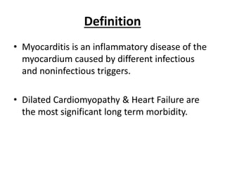
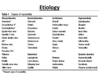
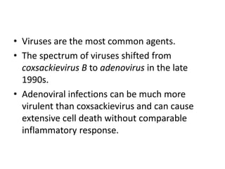
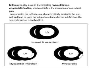
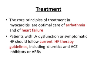
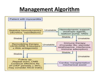
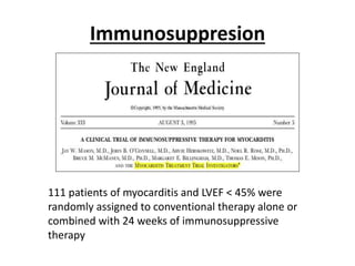
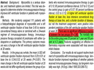
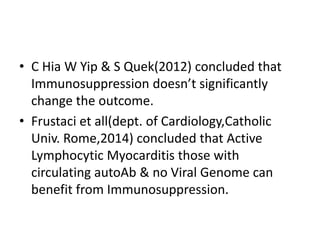
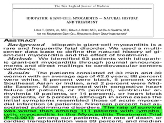
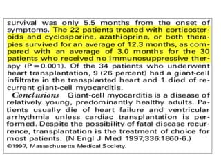
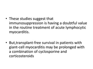
• Myocarditis is an inflammatory disease of the
myocardium caused by different infectious
and noninfectious triggers.
• Dilated Cardiomyopathy & Heart Failure are
the most significant long term morbidity.
Etiology
• Viruses are the most common agents.
• The spectrum of viruses shifted from
coxsackievirus B to adenovirus in the late
1990s.
• Adenoviral infections can be much more
virulent than coxsackievirus and can cause
extensive cell death without comparable
inflammatory response.
Statistics
• Bowles & coworkers analyzed biopsy
specimens from 624 patients with PCR and
found overall viral positivity was 38%
(239/624).
• On analysis,22.8% tested positive for
adenovirus, 13.6% for enterovirus and 1% for
parvovirus.
Bowles et al, JACC 42:466,2003
Pathophysiology
Viral Infection
Inflammation and Injury
Decreased Myocardial Contractility
Heart Enlarges:
 Cardiac Output
 Sympathetic Tone
CHF
 LAP
Pulm.
edema
Scarring
Dysrhythmias
Clinical Features
• Acute Viral Myocarditis often presents with
Heart Failure with preceding fever & myalgias.
• Chest Pain(due to Pericarditis or myocardial
injury & destruction)
• Palpitations(due to Arrhythmia's)
• Rapid progressions from a febrile respiratory
syndrome to Cardiogenic Shock(discharged
from Urgent care settings for Viral illness)
• Sudden death (in young adults, myocarditis
causes up to 20% of all cases of sudden
death).
Important types of Myocarditis
1. Chagas Diseases
• Most common Infective cause.
• Endemic in Rural areas of South & Central
America
• Chronic infection leads to Conduction system
anomaly, AF, Ventricular Tachyarrhythmia.
• Treatment- HF medications & benznidazole-
Nifurtimox.
2.Granulomatous Myocarditis
• Sarcoidosis
• Rapid onset HF & ventricular Tachyarrhythmia’s
• Conduction block
• Giant cell Myocarditis
• Typically with Rapidly progressive HF &
ventricular Tachycardia
• Diffuse Granulomatous lesion surrounded by
extensive inflammatory infiltrate in
endomyocardial biopsy
Diagnosis
• ECG- changes widely variable & non-specific-
1. Non specific ST segment & T wave changes
2. Sinus tachycardia
3. Ventricular arrhythmia's
4. AV conduction defects
Cardiac Biomarkers
• Cardiac Troponins more sensitive than CPK-
MB.
• Largely nonspecific
Echocardiography
• Rule out non-inflammatory cardiac disease
such as valve disease and to monitor changes
in cardiac chamber size, wall thickness,
ventricular function, and pericardial effusions.
• Global ventricular dysfunction, regional wall
motion abnormalities may occur in
myocarditis.
MRI of Heart
MRI is emerging as an important tool
for the diagnosis and follow-up of
patients with acute myocarditis
MRI can also play a role in discriminating myocarditis from
myocardial infarction, which can help in the evaluation of acute chest
pain.
In myocarditis the infiltrates are characteristically located in the mid-
wall and tend to spare the sub-endocardium,whereas in infarction, the
sub-endocardium is involved first.
Gold Standard
• Endomyocardial Biopsy(Dallas Criteria)
• At least 5 separate biopsy specimens
• First Biopsy- Myocyte necrosis or
degeneration or both associated with an
inflammatory infiltrate adjacent to the
degenerating or necrotic myocyte.
Subsequent Changes
• Ongoing(persisting) Myocarditis with or
without fibrosis
• Resolving(healing) Myocarditis with or
without Fibrosis
• Resolved(healed) Myocarditis with or without
Fibrosis
Histological Picture
• Chow and McManus demonstrated that with a
single EMB sample, histologic myocarditis
could be demonstrated in only 25% of cases.
Even with 5 random samples, correct
diagnosis by classic Dallas criteria could be
reached in only about 2/3rd of subjects.
Clinical Classification
1. Possible subclinical Acute Myocarditis
 Typical Viral syndrome without cardiac
symptoms with one or more of following-
• Elevated Cardiac Biomarkers
• ECG findings suggestive of Acute Injury
• Reduced LVEF
• Abnormality in Cardiac Imaging
2. Probable Acute Myocarditis
• Previous criteria accompanied by Cardiac
symptoms.
3. Definite Myocarditis
• Previous criteria plus histological evidence
Treatment
• The core principles of treatment in
myocarditis are optimal care of arrhythmia
and of heart failure
• Patients with LV dysfunction or symptomatic
HF should follow current HF therapy
guidelines, including diuretics and ACE
inhibitors or ARBs
Management Algorithm
Immunosuppresion
111 patients of myocarditis and LVEF < 45% were
randomly assigned to conventional therapy alone or
combined with 24 weeks of immunosuppressive
therapy
• C Hia W Yip & S Quek(2012) concluded that
Immunosuppression doesn’t significantly
change the outcome.
• Frustaci et all(dept. of Cardiology,Catholic
Univ. Rome,2014) concluded that Active
Lymphocytic Myocarditis those with
circulating autoAb & no Viral Genome can
benefit from Immunosuppression.
• These studies suggest that
immunosuppression is having a doubtful value
in the routine treatment of acute lymphocytic
myocarditis.
• But,transplant-free survival in patients with
giant-cell myocarditis may be prolonged with
a combination of cyclosporine and
corticosteroids
Myocarditis

Myocarditis

  • 1.
  • 2.
    Definition • Myocarditis isan inflammatory disease of the myocardium caused by different infectious and noninfectious triggers. • Dilated Cardiomyopathy & Heart Failure are the most significant long term morbidity.
  • 3.
  • 4.
    • Viruses arethe most common agents. • The spectrum of viruses shifted from coxsackievirus B to adenovirus in the late 1990s. • Adenoviral infections can be much more virulent than coxsackievirus and can cause extensive cell death without comparable inflammatory response.
  • 5.
    Statistics • Bowles &coworkers analyzed biopsy specimens from 624 patients with PCR and found overall viral positivity was 38% (239/624). • On analysis,22.8% tested positive for adenovirus, 13.6% for enterovirus and 1% for parvovirus. Bowles et al, JACC 42:466,2003
  • 6.
  • 7.
    Viral Infection Inflammation andInjury Decreased Myocardial Contractility Heart Enlarges:  Cardiac Output  Sympathetic Tone CHF  LAP Pulm. edema Scarring Dysrhythmias
  • 8.
    Clinical Features • AcuteViral Myocarditis often presents with Heart Failure with preceding fever & myalgias. • Chest Pain(due to Pericarditis or myocardial injury & destruction) • Palpitations(due to Arrhythmia's)
  • 9.
    • Rapid progressionsfrom a febrile respiratory syndrome to Cardiogenic Shock(discharged from Urgent care settings for Viral illness) • Sudden death (in young adults, myocarditis causes up to 20% of all cases of sudden death).
  • 10.
    Important types ofMyocarditis 1. Chagas Diseases • Most common Infective cause. • Endemic in Rural areas of South & Central America • Chronic infection leads to Conduction system anomaly, AF, Ventricular Tachyarrhythmia. • Treatment- HF medications & benznidazole- Nifurtimox.
  • 11.
    2.Granulomatous Myocarditis • Sarcoidosis •Rapid onset HF & ventricular Tachyarrhythmia’s • Conduction block • Giant cell Myocarditis • Typically with Rapidly progressive HF & ventricular Tachycardia • Diffuse Granulomatous lesion surrounded by extensive inflammatory infiltrate in endomyocardial biopsy
  • 12.
    Diagnosis • ECG- changeswidely variable & non-specific- 1. Non specific ST segment & T wave changes 2. Sinus tachycardia 3. Ventricular arrhythmia's 4. AV conduction defects
  • 13.
    Cardiac Biomarkers • CardiacTroponins more sensitive than CPK- MB. • Largely nonspecific
  • 14.
    Echocardiography • Rule outnon-inflammatory cardiac disease such as valve disease and to monitor changes in cardiac chamber size, wall thickness, ventricular function, and pericardial effusions. • Global ventricular dysfunction, regional wall motion abnormalities may occur in myocarditis.
  • 16.
    MRI of Heart MRIis emerging as an important tool for the diagnosis and follow-up of patients with acute myocarditis
  • 17.
    MRI can alsoplay a role in discriminating myocarditis from myocardial infarction, which can help in the evaluation of acute chest pain. In myocarditis the infiltrates are characteristically located in the mid- wall and tend to spare the sub-endocardium,whereas in infarction, the sub-endocardium is involved first.
  • 18.
    Gold Standard • EndomyocardialBiopsy(Dallas Criteria) • At least 5 separate biopsy specimens • First Biopsy- Myocyte necrosis or degeneration or both associated with an inflammatory infiltrate adjacent to the degenerating or necrotic myocyte.
  • 19.
    Subsequent Changes • Ongoing(persisting)Myocarditis with or without fibrosis • Resolving(healing) Myocarditis with or without Fibrosis • Resolved(healed) Myocarditis with or without Fibrosis
  • 20.
  • 21.
    • Chow andMcManus demonstrated that with a single EMB sample, histologic myocarditis could be demonstrated in only 25% of cases. Even with 5 random samples, correct diagnosis by classic Dallas criteria could be reached in only about 2/3rd of subjects.
  • 22.
    Clinical Classification 1. Possiblesubclinical Acute Myocarditis  Typical Viral syndrome without cardiac symptoms with one or more of following- • Elevated Cardiac Biomarkers • ECG findings suggestive of Acute Injury • Reduced LVEF • Abnormality in Cardiac Imaging
  • 23.
    2. Probable AcuteMyocarditis • Previous criteria accompanied by Cardiac symptoms. 3. Definite Myocarditis • Previous criteria plus histological evidence
  • 24.
    Treatment • The coreprinciples of treatment in myocarditis are optimal care of arrhythmia and of heart failure • Patients with LV dysfunction or symptomatic HF should follow current HF therapy guidelines, including diuretics and ACE inhibitors or ARBs
  • 25.
  • 26.
    Immunosuppresion 111 patients ofmyocarditis and LVEF < 45% were randomly assigned to conventional therapy alone or combined with 24 weeks of immunosuppressive therapy
  • 28.
    • C HiaW Yip & S Quek(2012) concluded that Immunosuppression doesn’t significantly change the outcome. • Frustaci et all(dept. of Cardiology,Catholic Univ. Rome,2014) concluded that Active Lymphocytic Myocarditis those with circulating autoAb & no Viral Genome can benefit from Immunosuppression.
  • 31.
    • These studiessuggest that immunosuppression is having a doubtful value in the routine treatment of acute lymphocytic myocarditis. • But,transplant-free survival in patients with giant-cell myocarditis may be prolonged with a combination of cyclosporine and corticosteroids