This document discusses current trends in the diagnosis and treatment of myocarditis. It begins by defining myocarditis and noting that debates remain around its classification, diagnosis, and management. While endomyocardial biopsy has improved understanding, clinical presentations are extremely diverse, ranging from nonspecific symptoms to sudden death. Recent data implicate myocarditis in 8.6-12% of sudden cardiac deaths in young adults. The document then reviews limitations of endomyocardial biopsy for diagnosis, proposed clinicopathological classification systems, evolving understanding of viral etiologies through molecular techniques, and associations between myocarditis and conditions like HIV.


![have proven to be a more reliable predictor of myocardial
injury than levels of creatine kinase.73 ECG changes sugges-
tive of acute myocardial ischemia typically may include
ST-segment elevation in Ն2 contiguous leads (54%), T-wave
inversions (27%), widespread ST-segment depressions
(18%), and pathological Q waves (18% to 27%).9,71 Segmen-
tal or global echocardiographic wall motion abnormalities are
frequently evident despite angiographically normal coronary
anatomy.71 Sarda et al,72 using myocardial indium111
-labeled
antimyosin antibody and rest thallium imaging, identified 35
of 45 patients (78%) who presented with acute chest pain,
ischemic ECG abnormalities, and elevated cardiac biomark-
ers as having myocarditis. However, biopsy verification of
actual myocarditis was not undertaken in this series. In
another series of 34 patients with known normal coronary
anatomy presenting with symptoms and ECG changes con-
sistent with an acute coronary syndrome, 11 (32%) of the
patients were found to have myocarditis on biopy.9 Clinicians
should consider acute myocarditis in younger patients who
present with acute coronary syndromes when coronary risk
factors are absent, ECG abnormalities extend beyond a single
coronary artery territory, or global rather than segmental left
ventricular dysfunction is evident on echocardiography.
Myocarditis can produce variable effects on the cardiac
conduction system. Ventricular tachycardia is an uncommon
initial manifestation of myocarditis but often develops during
long-term follow-up.74 The Giant Cell Myocarditis Study
Group reported an initial incidence of ventricular tachycardia
of Ͻ5% in a multicenter cohort.70 Ventricular tachycardia due
to either lymphocytic or granulomatous myocarditis may
infrequently result in sudden cardiac death.75
Diagnostic Evaluation
Biopsy
The Dallas criteria10 have standardized the histopathological
definition of myocarditis. Despite its considerable limitations,
yielding diagnostic information in only 10% to 20% of
cases,76 EMB findings remain the gold standard for unequiv-
ocally establishing the diagnosis. The largest case series of
patients with an unexplained cardiomyopathy used biopsy
findings to diagnose 111 of 1230 patients (9%) with myocar-
ditis.68 Fewer than 10% of 2233 patients with idiopathic heart
failure referred to the Myocarditis Treatment Trial6 had
EMBs deemed positive by the Dallas criteria. However,
multiple investigators have described strong clinical, ven-
triculographic, and laboratory evidence of myocarditis among
patients with negative biopsies.7,69 Biopsies performed within
weeks of symptom onset have a higher yield than those
undertaken when symptoms have been more longstanding.
Current American College of Cardiology/American Heart
Association (ACC/AHA) guidelines for the treatment of heart
failure77 describe EMB as a class IIb recommendation.
Biopsy is generally reserved for patients with rapidly pro-
gressive cardiomyopathy refractory to conventional therapeu-
tic management or an unexplained cardiomyopathy that is
associated with progressive conduction system disease or
life-threatening ventricular arrhythmias. It should also be
considered when cardiovascular signs or symptoms develop
in a patient with a systemic disease known to cause left
ventricular dysfunction78 (Table 2).
Use of Cardiac Biomarkers
Serum cardiac biomarkers (creatine kinase [CK], troponin I
and T) are routinely measured when myocarditis is suspected.
CK or its isoform (CK-MB) is not generally useful for
noninvasive screening because of its low predictive value.
Lauer et al79 reported that only 28 of 80 patients (35%) with
suspected myocarditis had elevated troponin levels. Using a
serum troponin T cutoff Ͼ0.1 ng/mL, these investigators
reported a sensitivity for detecting myocarditis of 53%, a
specificity of 94%, a positive predictive value of 93%, and a
negative predictive value of 56%.79 Smith and coworkers73
also examined the value of troponin I in a subgroup of the
Multicenter Myocarditis Treatment trial. Although the sensi-
tivity of an elevated troponin I for the entire group was low
(34%), its specificity was high (89%). Not surprisingly, a
short duration of symptoms (Ͻ4 weeks) was associated with
a significantly higher sensitivity for detecting biopsy-proven
disease.73 More importantly, the positive predictive value was
acceptable at 82%. Most clinicians now routinely measure
either troponin T or I whenever a clinical diagnosis of
myocarditis is considered.80
An early trial used the erythrocyte sedimentation rate to
characterize a population with “reactive” myocardial disease
but found its sensitivity and specificity to be extremely low.81
Other serum immunologic biomarkers have included comple-
ment,82 cytokines,49 and anti-heart antibodies.83 None of these
approaches has been prospectively validated to accurately
screen for biopsy-proven myocarditis.
Immunologic Approaches
Advances in immunology have expanded the diagnostic
capabilities of the EMB. Immunohistochemical staining has
enabled more precise characterization of infiltrating lympho-
cytes subtypes84 and can accurately define and help quantify
upregulation of major histocompatibility (MHC) antigens.
Some investigators have adopted myocyte-specific MHC
expression as an essential criterion for diagnosing inflamma-
tory cardiomyopathy. This approach has greater sensitivity
TABLE 2. Indications for Endomyocardial Biopsy*
Exclusion of potential common etiologies of dilated cardiomyopathy (familial;
ischemic; alcohol; postpartum; cardiotoxic exposures) and the following:
Subacute or acute symptoms of heart failure refractory to standard
management
Substantial worsening of EF despite optimized pharmacological therapy
Development of hemodynamically significant arrhythmias, particularly
progressive heart block and ventricular tachycardia
Heart failure with concurrent rash, fever, or peripheral eosinophilia
History of collagen vascular disease such as systemic lupus
erythematosus, scleroderma, or polyarteritis nodosum
New-onset cardiomyopathy in the presence of known amyloidosis,
sarcoidosis, or hemachromatosis
Suspicion for giant cell myocarditis (young age, new subacute heart
failure, or progressive arrhythmia without apparent etiology)
Adapted with permission from Wu et al.78
880 Circulation February 14, 2006
by guest on January 24, 2015http://circ.ahajournals.org/Downloaded from](https://image.slidesharecdn.com/magnani2006myocarditis-180228131053/85/Magnani2006-myocarditis-5-320.jpg)
![than the Dallas criteria and has reopened the discussion
concerning the true incidence of myocarditis among patients
with “idiopathic” dilated cardiomyopathy.85 Herskowitz et
al86 compared quantitative MHC antigen expression in 13
active myocarditis patients with 8 control patients with other
forms of cardiac disease. MHC class I and II expression was
increased by 10-fold in the myocarditis cohort. Eleven of 13
myocarditis patients (85%) had either myocyte or microvas-
cular endothelial MHC class I or class II expression com-
pared with only 1 of 8 controls (12%). The sensitivity and
specificity of any MHC expression for detecting biopsy-
proven myocarditis were 80% and 85%, respectively. This
methodology was more recently evaluated in a larger cohort
of 83 patients with clinically suspected myocarditis.17 Sur-
prisingly, these investigators found no correlation between
MHC immunostaining and histopathological findings of ac-
tive myocarditis by Dallas criteria. As discussed by the
investigators, MHC expression could represent a more
chronic form of myocardial injury and may not be responsible
for the patients’ clinical presentation. The discordance be-
tween these findings is currently unexplained because the
staining methods and patient populations appeared to be
similar. Despite these shortcomings, biopsy assessment of
MHC expression has recently been used to guide therapy of
patients with inflammatory cardiomyopathy (see below).87
Myocardial Imaging
Noninvasive diagnostic myocardial imaging techniques for
detection of myocarditis may include echocardiography, nu-
clear imaging with gallium67
- or indium111
-labeled antimyosin
antibodies, and MRI.
Echocardiography is currently recommended in the initial
diagnostic evaluation of all patients with suspected myocar-
ditis. Several studies have specifically evaluated the role of
transthoracic echocardiography for diagnosing myocardi-
tis.88,89 Pinamonti et al88 retrospectively analyzed echocardio-
graphic findings among 42 patients with biopsy-proven myo-
carditis. Left ventricular dysfunction was commonly
observed (69%), but left ventricular cavity enlargement was
frequently minimal or absent, consistent with other forms of
acute dilated cardiomyopathy. Right ventricular dysfunction
was present in only 23% of this cohort. Not surprisingly,
patients who presented with chest pain or heart block rather
than heart failure almost always had preserved ventricular
size and function. Segmental wall motion abnormalities were
observed in 64% of patients and included hypokinetic,
akinetic, or frankly dyskinetic regions. Reversible left ven-
tricular hypertrophy was noted in 15% of patients and
typically resolved over several months. Thus, echocardio-
graphic findings can be varied but relatively nonspecific.
Serial studies have been shown to be useful in assessing the
response to treatment of several forms of myocarditis. Reso-
lution of marked concentric left ventricular hypertrophy in
eosinophilic myocarditis after corticosteroid treatment has
been reported.90
Although anatomic features on echocardiography (ie,
chamber dimensions, ejection fraction [EF], wall motion
abnormalities) are insufficient to differentiate myocarditis
from other forms of cardiomyopathy, ultrasonic tissue char-
acterization may prove to be more useful. Transmission and
reflection of ultrasound energy depends on tissue density,
elasticity, and acoustical impedance. Changes in 1 or more of
these factors lead to different ultrasonic backscatter and an
altered image texture. Lieback et al91 evaluated mean gray-
scale values (indicative of average brightness) in 52 patients
with biopsy-proven myocarditis; 12 patients had persistent
myocarditis, 9 patients had healed myocarditis but lacked
fibrosis, and 17 patients had healed myocarditis and fibrosis.
Tissue characterization was highly effective in differentiating
myocarditis from healthy control myocardium, with sensitiv-
ity and specificity values of 100% and 90%, respectively.91
However, ultrasonic tissue characterization could not accu-
rately differentiate between idiopathic dilated cardiomyopa-
thy and active myocarditis. More recent techniques, particu-
larly tissue Doppler imaging and myocardial velocity
measurements, are better able to characterize tissue changes
in acute myocarditis and to monitor changes in these param-
eters over time. Additional validation studies will be required
to determine their clinical utility.
Indium111
-labeled monoclonal antibody fragments (directed
against heavy chain myosin) bind to cardiac myocytes that
have lost the integrity of their sarcolemmal membranes and
have exposed their intracellular myosin to the extracellular
fluid space. Unlike gallium67
, which detects the extent of
myocardial inflammation, antimyosin cardiac uptake reflects
the extent of myocyte necrosis. Dec et al92 evaluated the
utility of antimyosin imaging in a large cohort of patients
with clinically suspected myocarditis. On the basis of EMB,
antimyosin uptake was found to be highly sensitive (83%) but
only moderately specific (53%) for detecting myocardial
necrosis. However, the predictive value of a negative scan
was high at 92%. More recently, Margari et al93 have reported
that the presence of both a positive antimyosin scan and a
nondilated left ventricular cavity (left ventricular end-diastol-
ic dimension Յ62 mm) was highly predictive for detecting
myocarditis on biopsy.
Contrast-enhanced MRI appears to be the most promising
technique for diagnosing myocardial inflammation and myo-
cyte injury on the basis of small, observational clinical
studies. Besides providing anatomic and morphological in-
formation, MRI can provide accurate tissue characterization
by measuring T1 and T2 relaxation times and spin densities.
Because active myocarditis is typically associated with myo-
cyte injury, including edema and cellular swelling, assess-
ment of relaxation times provides a sensitive measure for its
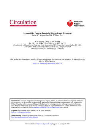
detection.94 Friedrich et al95 evaluated the diagnostic utility of
contrast-enhanced cardiac MRI in 19 patients with suspected
myocarditis. Early after presentation, myocardial enhance-
ment was generally focal in distribution (Figure 4A). Global
enhancement became prominent during later imaging times
(Figure 4B) and returned to baseline within 90 days. Unfor-
tunately, the study did not examine the ability of MRI to
differentiate viral myocarditis from other causes of acute
dilated cardiomyopathy.
Roditi et al96 evaluated 20 patients with T1 spin-echo cine
MR angiography and gadolinium-enhanced spin-echo imag-
ing. Focal myocardial enhancement was associated with
regional wall motion abnormalities in 10 of the 12 patients
Magnani and Dec Diagnosis and Treatment of Myocarditis 881
by guest on January 24, 2015http://circ.ahajournals.org/Downloaded from](https://image.slidesharecdn.com/magnani2006myocarditis-180228131053/85/Magnani2006-myocarditis-6-320.jpg)
![with suspected or proven myocarditis. The authors concluded
that focal myocardial enhancement combined with regional
wall motion abnormalities (hypokinesis, akinesis, or dyski-
nesis) strongly supported a diagnosis of myocarditis. The
ability of contrast-enhanced MRI techniques to diagnose
other forms of inflammatory heart disease, particularly car-
diac sarcoidosis, has also been validated recently.97
New contrast MR techniques using segmented inversion
recovery gradient-echo pulse sequences and both early and
late gadolinium enhancement provide substantial improve-
ment in contrast between diseased and normal myocardium.98
Mahrholt et al99 recently used this new technique to perform
gadolinium-enhanced MRI-guided biopsy of the right and left
ventricles in 32 patients with suspected myocarditis. Left
ventricular biopsy was generally performed from the region
showing the most marked contrast enhancement. Biopsy of
these specific myocardial regions resulted in positive and
negative predictive values for detecting myocarditis of 71%
and 100%, respectively. MRI may not only be useful in
identifying those patients who should undergo biopsy but can
also facilitate a guided approach to the abnormal region of
myocardium. It is hoped that this focused methodology will
improve the sensitivity of EMB for establishing a correct
histological diagnosis. Serial MRI studies have also shown
promise for tracking the natural history of the disease and
could, in the near future, allow noninvasive reassessment of
the myocardial response to therapy.
Natural History of Myocarditis
The natural history of myocarditis is as varied as its clinical
presentations. Myocarditis masquerading as myocardial in-
farction almost universally results in a full recovery of
cardiovascular status in previously healthy adults.9,71,72 Indi-
viduals with smallpox vaccine–associated myocarditis have
also been shown to have rapid resolution of clinical, labora-
tory, and echocardiographic abnormalities.100 Patients who
present with heart failure may have mildly compromised
ventricular function (left ventricular ejection fraction [LVEF]
of 40% to 50%) and typically improve within weeks to
months. Alternatively, a smaller cohort of patients will
present with more advanced left ventricular dysfunction
(LVEF Ͻ35%, left ventricular end-diastolic dimension
Ͼ60 mm). Among this group, 50% of patients will develop
chronic ventricular dysfunction, and 25% of patients will
progress to transplantation or death; however, the remaining
25% of patients will have spontaneous improvement in their
ventricular function.67,69 A small minority of these patients
will present with cardiogenic shock requiring mechanical
circulatory support as a bridge to cardiac recovery or trans-
plantation.101 Somewhat surprisingly, fulminant myocarditis
has been described in 1 published series as having the best
long-term prognosis with a Ͼ90% event-free survival rate.5
The Myocarditis Treatment Trial reported mortality rates
for biopsy-verified myocarditis of 20% and 56% at 1 year and
4.3 years, respectively.6 These outcomes are similar to the
Mayo Clinic’s observational data of 5-year survival rates that
approximate 50%.4 Survival with giant cell myocarditis is
substantially lower, with Ͻ20% of patients surviving 5
years70 (Figure 5).
Predicting prognosis for the individual patient with newly
diagnosed cardiomyopathy due to myocarditis remains prob-
lematic. Fuse et al102 evaluated a variety of clinical, hemo-
dynamic, and laboratory parameters in patients with biopsy-
proven acute myocarditis. Clinical variables were unable to
Figure 4. A, T1-weighted MRI cross-
sectional views at the midventricular
level in a patient with acute myocarditis.
Left, Unenhanced view obtained on day
2 after onset of symptoms. There are
small foci of increased signal intensity in
the subepicardial parts of the posterior
myocardium and in the basal septum,
which were more evident (right) after
administration of gadopentetate dimeglu-
mine (arrows). Reproduced with permis-
sion from Circulation. 1998;97:1805. B,
T1-weighed MRI images obtained before
(left) and after enhancement (right) in the
same patient as shown in A at 14 days
after presentation. More diffuse enhance-
ment of myocardium after gadopentetate
dimeglumine, including the apical part of
the septum and visible areas of the right
ventricle. Reproduced with permission
from Circulation. 1998;97:1805. © Copy-
right 1998, American Heart Association.
882 Circulation February 14, 2006
by guest on January 24, 2015http://circ.ahajournals.org/Downloaded from](https://image.slidesharecdn.com/magnani2006myocarditis-180228131053/85/Magnani2006-myocarditis-7-320.jpg)

![of myocytes. Its precise characterization and natural history
have been limited by the extraordinary variability of its
clinical presentations, laboratory findings, and the diversity
of etiologies. The relatively low incidence and difficulties in
unequivocally establishing a diagnosis have limited the con-
duct of large-scale, randomized clinical trials to evaluate
treatment strategies.
ECG, echocardiography, measurement of serum troponin,
and noninvasive cardiac MRI are warranted for initial diag-
nostic evaluation. Patients presenting with ST elevations,
elevated cardiac markers, and ischemic symptoms should
undergo prompt coronary angiography. Myocarditis should
be considered in patients who lack evidence of coronary
atherosclerosis or other pathophysiological etiologies such as
stress-induced cardiomyopathy (takotsubo syndrome). Endo-
myocardial biopsy should be considered for a highly selected
group (Ͻ5%) of patients, particularly those with increased
myocardial enhancement on cardiac MRI, rapidly progressive
cardiomyopathy due to suspected giant cell myocarditis or
sarcoidosis, suspected allergic myocarditis, or unexplained
ventricular dysfunction in the presence of an autoimmune
disease known to affect the myocardium. More precise biopsy
localization with the use of MRI targeting combined with
more sophisticated analysis of myocardial specimens with the
use of immunostaining for HLA expression and detection of
viral genomic material by PCR will undoubtedly lead to
reconsideration of the diagnostic role of biopsy in unex-
plained cardiomyopathy in the near future.
Treatment of myocarditis in 2006 remains largely support-
ive. Immunosuppression has not been shown to be effective
as routine treatment for acute lymphocytic myocarditis. Early
trials of antiviral therapies, such as interferons, suggest a
potential therapeutic role but require further investigation.
Currently, the standard of care from acute cardiomyopathy
remains hemodynamic and cardiovascular support, including
use of ventricular assist devices and transplantation when
necessary. Pharmacological therapy should consist of a heart
failure regimen demonstrated to improve hemodynamics and
symptoms. Although the high rate of spontaneous improve-
ment in acute myocarditis and cardiomyopathy provides
some optimism, patients who progress to chronic dilated
cardiomyopathy experience 5-year survival rates Ͻ50%.
Ongoing clinical trials should help to clarify whether
immune-modulating strategies can improve this prognosis.
Acknowledgments
The authors would like to acknowledge James Stone, MD, PhD, who
prepared and contributed the photomicrographs for Figures 1, 2, and 3.
Disclosures
None.
References
1. Fabre A, Sheppard MN. Sudden adult death syndrome and other non
ischaemic causes of sudden cardiac death: a UK experience. Heart. 2005
[Epub ahead of print].
2. Doolan A, Langlois N, Semsarian C. Causes of sudden cardiac death in
young Australians. Med J Aust. 2004;180:110–112.
3. Felker GM, Hu W, Hare JM, Hruban RH, Baughman KL, Kasper EK.
The spectrum of dilated cardiomyopathy: the Johns Hopkins experience
with 1,278 patients. Medicine (Baltimore). 1999;78:270–283.
4. Grogan M, Redfield MM, Bailey KR, Reeder GS, Gersh RJ, Edwards
WD, Rodeheffer RJ. Long-term outcome of patients with biopsy-proved
myocarditis: comparison with idiopathic dilated cardiomyopathy. J Am
Coll Cardiol. 1995;26:80–84.
5. McCarthy RE, Boehmer JP, Hruban RH, Hutchins GM, Kasper EK,
Hare JM, Baughman KL. Long-term outcome of fulminant myocarditis
as compared with acute (non-fulminant) myocarditis. N Engl J Med.
2000;342:690–695.
6. Mason JW, O’Connell JB, Herskowitz A, Rose NR, McManus BM,
Billingham ME, Moon TE, for the Myocarditis Treatment Trial Inves-
tigators. A clinical trial of immunosuppressive therapy for myocarditis.
N Engl J Med. 1995;333:269–275.
7. Dec GW, Palacios IF, Fallon JT, Aretz HT, Mills J, Lee DC, Johnson
RA. Active myocarditis in the spectrum of acute dilated cardiomyopa-
thies: clinical features, histologic correlates, and clinical outcome.
N Engl J Med. 1985;312:885–890.
8. Heart Failure Society of America. HFSA guidelines for the management
of patients with heart failure due to left ventricular systolic dysfunction:
pharmacological approaches. Congest Heart Fail. 2000;6:11–39.
9. Dec GW, Waldman H, Southern J, Fallon JT, Hutter AM, Palacios I.
Viral myocarditis mimicking acute myocardial infarction. J Am Coll
Cardiol. 1992;20:85–89.
10. Aretz HT, Billingham ME, Edwards WD, Parker MM, Factor SM,
Fallon JT, Fenoglio JJ. Myocarditis: a histopathologic definition and
classification. Am J Cardiovasc Pathol. 1987;1:3–14.
11. Magnani JW, Suk-Danik HJ, Dec GW, DiSalvo TG. Survival in biopsy-
proven myocarditis: a long-term retrospective analysis of the his-
topathologic, clinical, and hemodynamic predictors. Am Heart J. In
press.
12. Hauck AJ, Kearney DL, Edwards WD. Evaluation of postmortem en-
domyocardial biopsy specimens from 38 patients with lymphocytic
myocarditis: implications for role of sampling error. Mayo Clin Proc.
1989;64:1235–1245.
13. Shanes JG, Ghali J, Billingham ME, Ferrans VJ, Fenoglio JJ, Edwards
WD, Tsai CC, Saffitz JE, Isner J, Forner S. Interobserver variability in
the pathologic interpretation of endomyocardial biopsy results. Circu-
lation. 1987;75:401–405.
14. Parrillo JE. Inflammatory cardiomyopathy (myocarditis): which patients
should be treated with anti-inflammatory therapy? Circulation. 2001;
104:4–6.
15. Lieberman EB, Hutchins GM, Herskowitz A, Rose NR, Baughman KL.
Clinicopathologic description of myocarditis. J Am Coll Cardiol. 1991;
18:1617–1626.
16. Baboonian C, Treasure T. Meta-analysis of the association of entero-
viruses with human heart disease. Heart. 1997;78:539–543.
17. Wojnicz R, Nowalany-Kozielska E, Wodniecki J, Szczurek-Katanski S,
Nozynski J, Zembala M, Rozek MM. Immunohistological diagnosis of
myocarditis: potential role of sarcolemmal induction of the MHC and
ICAM-1 in the detection of autoimmune mediated myocyte injury. Eur
Heart J. 1998;19:1564–1572.
18. Griffiths PD, Hannington G, Booth JC. Coxsackie B virus infections and
myocardial infarction: results from a prospective, epidemiologically
controlled study. Lancet. 1980;1:1387–1389.
19. Keeling PJ, Poloniecki LA, Caforio AL, Davies MJ, Booth JC,
McKenna WJ. A prospective case-control study of antibodies to Cox-
sackie B virus in idiopathic dilated cardiomyopathy. J Am Coll Cardiol.
1994;23:593–598.
20. Grumbach IM, Heim A, Pring-Akerblom P, Vonhof S, Hein WJ, Muller
G, Figulla HR. Adenoviruses and enteroviruses as pathogens in myo-
carditis and dilated cardiomyopathy. Acta Cardiol. 1999;54:83–88.
21. Archard LC, Khan MA, Soteriou BA, Zhang H, Why HJ, Robinson NM,
Richardson PJ. Characterization of Coxsackie B virus RNA in myocar-
dium from patients with dilated cardiomyopathy by nucleotide
sequencing of reverse transcription-nested polymerase chain reaction
products. Hum Pathol. 1998;29:578–584.
22. Why HJ, Meany BT, Richardson PJ, Olsen EG, Bowles NE,
Cunningham L, Freeke CA, Archard LC. Clinical and prognostic sig-
nificance of detection of enteroviral RNA in the myocardium of patients
with myocarditis or dilated cardiomyopathy. Circulation. 1994;89:
2582–2589.
23. Pauschinger M, Bowles NE, Fuentes-Garcia FJ, Pham V, Kuhl U,
Schwimmbeck PL, Schultheiss HP, Towbin JA. Detection of adenoviral
genome in the myocardium of adult patients with idiopathic left ven-
tricular dysfunction. Circulation. 1999;99:1348–1354.
Magnani and Dec Diagnosis and Treatment of Myocarditis 887
by guest on January 24, 2015http://circ.ahajournals.org/Downloaded from](https://image.slidesharecdn.com/magnani2006myocarditis-180228131053/85/Magnani2006-myocarditis-12-320.jpg)