MYOCARDITIS
By
Shipra
Layers of the Heart Muscle
Coronary artery with
branch into myocardium
Pericardium
Heart muscle
(ventricular wall)
Endocardium
(inner lining)
Myocardium
(heart muscle)
Epicardium
(outer surface)
Inflammatory Disorders of the Heart
• Pericarditis
• Myocarditis
• Endocarditis
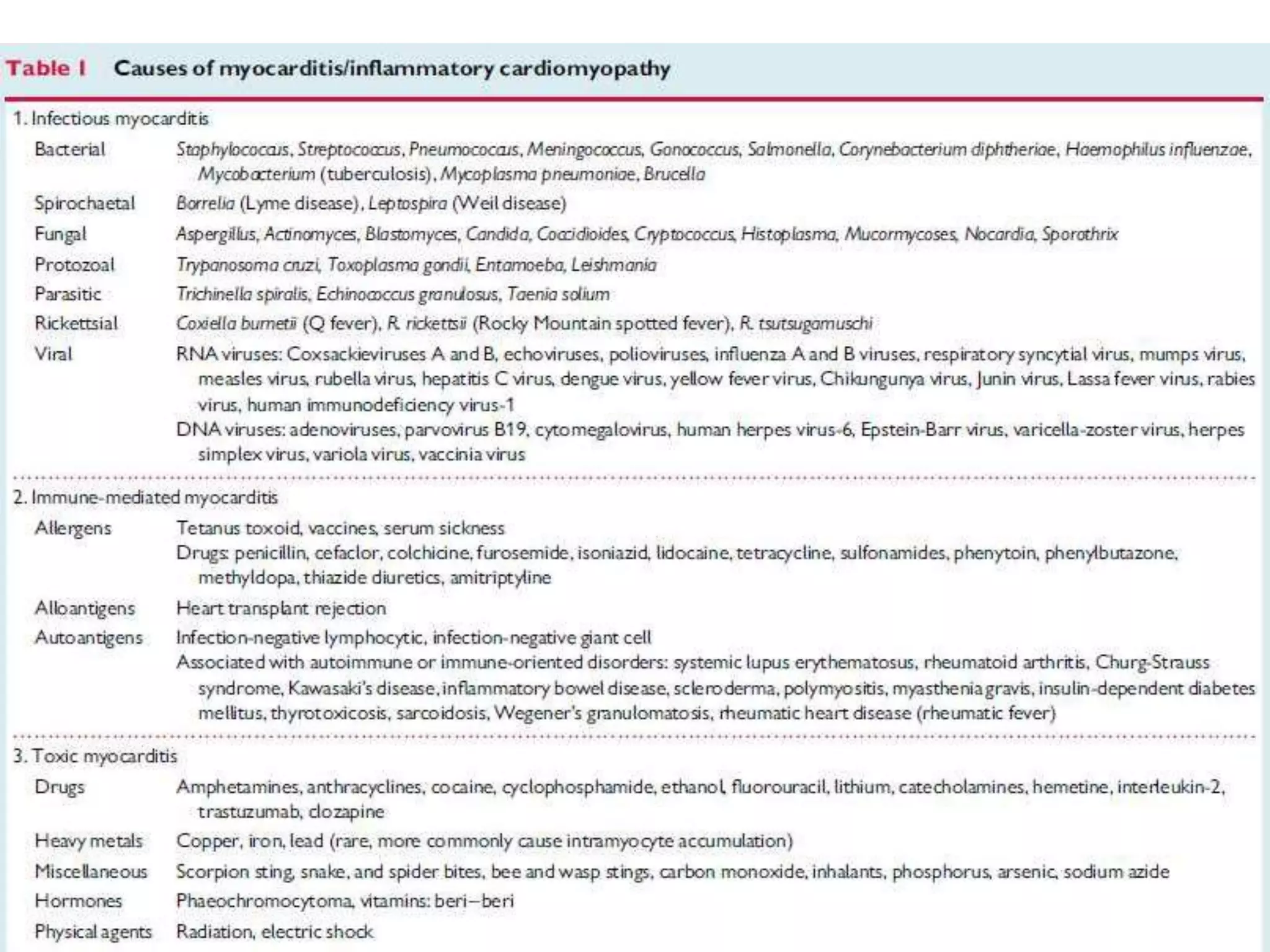
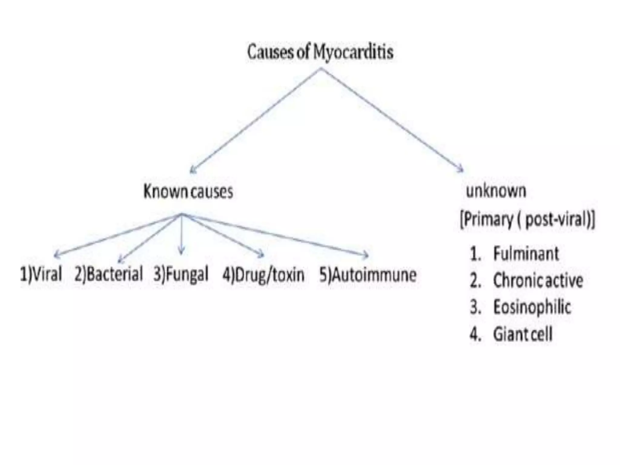
Myocarditis is an inflammatory
disease of the myocardium
caused by different infectious and
noninfectious triggers
Important types of Myocarditis
1. Chagas Diseases:
• Most common Infective cause.
• Endemic in Rural areas of South & Central America
• Chronic infection leads to Conduction system anomaly, AF,
ventricular Tachyarrhythmia.
• Treatment- HF medications & benznidazole-Nifurtimox.
2. Granulomatous Myocarditis:
• Sarcoidosis
 Rapid onset HF & ventricular Tachyarrhythmia’s
 Conduction block
• Giant cell Myocarditis
 • Typically with Rapidly progressive HF & ventricular Tachycardia
 • Diffuse Granulomatous lesion surrounded by extensive
inflammatory infiltrate in endomyocardial biopsy.
Viruses That Have Been Shown to
Cause Myocarditis
• Common
- Coxsackievirus A
- Coxsackievirus B
- Echovirus
- Human immunodeficiency virus
- Influenza
• Less Common
- Adenovirus family
- Arbovirus
- Epstein-Barr virus
- Herpes simplex virus type 1
- Human cytomegalovirus
- Measles virus
- Rubella virus
- Varicella-zoster virus
New England Journal of Medicine 343:1391 2000
Time course of viral myocarditis in 3 phases
Scarring of
tissue
Endomyocardial biopsy in acute myocarditis:
Arrow shows a collection of lymphocytes infiltrating the cardiac muscle
in response to a viral infection..
The arrowhead shows an area of cardiac muscle damage induced by
the virus directly or to the cytotoxic immune response to the viral infection.
Histopathology
Fulminant myocarditis
Acute myocarditis
Fulminant myocarditis is characterized by more
extensive and diffuse lympocytic infiltration and
myocyte necrosis than acute myocarditis
Signs and symptoms
• Chest pain (often described as "stabbing" in character).
• CHF(leading to edema,breathlessness and hepatic congestion).
• Palpitations (due to arrhythmias).
• Sudden death (in young adults, myocarditis causes up to 20% of all
cases of sudden death).
• Fever (especially when infectious)
• Since myocarditis is often due to a viral illness, many patients give a
history of symptoms consistent with a recent viral infection,
including fever, diarrhea, joint pains, and easy fatigueability.
• Myocarditis is often associated with pericarditis, and many patients
present with signs and symptoms that suggest concurrent myocarditis
and pericarditis.
Diagnosis
Myocarditis is a challenging diagnosis due to
the heterogeneity of clinical presentations.
Clinical presentation
Myocarditis presents in many different ways, ranging from
mild symptoms of chest pain and palpitations associated
with transient ECG changes to life-threatening cardiogenic
shock and ventricular arrhythmia
Diagnostic Tests
• ECG- Non-specific T-wave abnormalities
• CK-MB and Troponin may be elevated
• Chest X-Ray- Variable (Normal to Cardiomegaly)
• Echocardiogram
• Cardiovascular Magnetic Resonace
• A safe and sensitive noninvasive diagnostic test to confirm the
diagnosis is not available
• Endomyocardial biopsy- there are risks and not used for every
case but is definitive for myocarditis
Biomarkers
Inflammatory markers:
ESR and CRP levels are often raised in myocarditis, but they do not
confirm the diagnosis and are often increased in acute pericarditis
While cardiac troponins are more sensitive of myocyte
injury in patients with clinically suspected
myocarditis than creatine phospho kinase levels, they
are nonspecific and when normal do not exclude
myocarditis.
ECG in Myocarditis:
ECG changes can be variable and include:
•Sinus tachycardia
•QRS / QT prolongation
•Diffuse T wave inversion
•Ventricular arrhythmias
•AV conduction defects
•With inflammation of the adjacent pericardium, ECG features of pericarditis can
also been seen (myopericarditis)
NB. The most common abnormality seen in myocarditis is sinus tachycardia with non-
specific ST segment and T wave changes
Myocarditis mimicking acute
myocardial infarction:
Occasionally, a pseudo infarct pattern and
ischemic changes are seen.
ST segment elevation is commonly seen, but ST
segment depression, T wave inversion, poor R
wave progression,and Q waves have also been
described
Echocardiography
•Echocardiography helps to rule out non-inflammatory
cardiac disease such as valve disease and to monitor changes
in cardiac chamber size, wall thickness, ventricular function,
and pericardial effusions.
• Global ventricular dysfunction, regional wall motion
abnormalities,and diastolic dysfunction with preserved EF
may occur in myocarditis.
• Histologically proven myocarditis may resemble dilated,
hypertrophic, and restrictive cardiomyopathy and can mimic
ischaemic heart disease.
Echocardiogram
Echocardiogram
markedly dilated heart
with ejection fraction
of 15 %, mural
thrombus was present
Echocardiographic Findings in
Fulminant and Acute Myocarditis
Fulminant myocarditis
Acute myocarditis
Fulminant myocarditis often presents with a non-dilated, thickened, and
hypocontractile left ventricle as the intense inflammatory response results in
interstitial oedema and loss of ventricular contractility
Chest Radiograph
Endomyocardial biopsy (Dallas Criteria)
• The gold standard in diagnosis
of myocarditis is still the EMB.
• At least 5 separate biopsy
specimens
RV-EMB:THETECHNIQUE(jugularapproach)
Normal myocardial*fibres
Damaged* ' ,
myocyte5-
Interstitial inflammatory infiltrate
Viral myocarditis:
*N.B. established histological Dallas criteria defined as
follows:histological evidence of inflammatory infiltrates within the
myocardium associated with myocyte degeneration and necrosis of
nonischaemic origin
MRI is emerging as an important tool
for the diagnosis and follow-up of
patients with acute myocarditis
MRI can also play a role in discriminating myocarditis from
myocardial infarction, which can help in the evaluation of acute
chest pain.
In myocarditis the infiltrates are characteristically located in the
mid-wall and tend to spare the sub-endocardium,whereas in
infarction, the sub-endocardium is involved first.
cine-SSFP images are shown in diastole and systole
and suggest absence of any wall motion abnormality
T2-weighted edema images
demonstrate the presence of
patchy focal edema in the
subepicardium of the inferolateral
wall.
T1-weighted LGE images
demonstrate presence of
subepicardially distributed
LGE which is typical for
acute myocarditis.
When is a heart attack not a heart attack?
Viral myocarditis may have various
clinical presentations, sometimes
mimicking acute myocardial
infarction or ischaemia with:
• Chest pain
• ECG Changes
• Elevated Cardiac Enzymes
Disproportionate thickening,
increased echogenicity, and
dyskinesis of the inferolateral wall
relative to the septum; findings
are consistent with tissue edema.
Diffuse ST-segment elevation in
precordial and limb leads. Hyperacute
T waves are seen in leads V2 and V3
(A) asymmetric
thickening consistent
with extensive
myocardial oedema in
the inferior and
inferolateral segments of
the left ventricle.
(B) extensive
enhancement of mid-
MANAGEMENT ALGORITHM
Treatment
Acute myocarditis resolves in about 50% of cases in the
first 2-4 weeks, but about 25% will develop persistent
cardiac dysfunction and 12-25% may acutely deteriorate
and either die or progress to end-stage DCM with a need
for heart transplantation.
The core principles of treatment in myocarditis are
optimal care of arrhythmia and of heart failure
Treatment
* Patients with LV dysfunction or symptomatic HF
should follow current HF therapy guidelines,
including diuretics and ACE inhibitors or ARBs
*Beta-blockers can be used cautiously in the acute
setting.
*Digoxin should be avoided in patients suffering
from acute HF induced by viral myocarditis
Diet and Lifestyle
• Restrict salt intake to 2-3g of sodium per day
• Exercise especially during the acute phase of virus
myocarditis enhances viral replication rate, enhances
immune mechanisms and increases inflammatory
lesions and necrosis.
• Resumption of physical activity can take place within
2 months of the acute disease.
Investigational treatment options:
Because mechanism-based therapy of myocarditis is not
proven, different approaches have been investigated in
clinical studies in recent years.
More than 20 treatment trials have been reported, using
immunosuppressive, immunomodulating, or
antiinflammatory agents as well as immunoadsorption
therapy
Conclusions
Acute myocarditis presents multiple
challenges in diagnosis and treatment.
Myocarditis by Shipra Shekhar

Myocarditis by Shipra Shekhar

  • 1.
  • 2.
    Layers of theHeart Muscle Coronary artery with branch into myocardium Pericardium Heart muscle (ventricular wall) Endocardium (inner lining) Myocardium (heart muscle) Epicardium (outer surface)
  • 3.
    Inflammatory Disorders ofthe Heart • Pericarditis • Myocarditis • Endocarditis
  • 4.
    Myocarditis is aninflammatory disease of the myocardium caused by different infectious and noninfectious triggers
  • 7.
    Important types ofMyocarditis 1. Chagas Diseases: • Most common Infective cause. • Endemic in Rural areas of South & Central America • Chronic infection leads to Conduction system anomaly, AF, ventricular Tachyarrhythmia. • Treatment- HF medications & benznidazole-Nifurtimox. 2. Granulomatous Myocarditis: • Sarcoidosis  Rapid onset HF & ventricular Tachyarrhythmia’s  Conduction block • Giant cell Myocarditis  • Typically with Rapidly progressive HF & ventricular Tachycardia  • Diffuse Granulomatous lesion surrounded by extensive inflammatory infiltrate in endomyocardial biopsy.
  • 8.
    Viruses That HaveBeen Shown to Cause Myocarditis • Common - Coxsackievirus A - Coxsackievirus B - Echovirus - Human immunodeficiency virus - Influenza • Less Common - Adenovirus family - Arbovirus - Epstein-Barr virus - Herpes simplex virus type 1 - Human cytomegalovirus - Measles virus - Rubella virus - Varicella-zoster virus
  • 11.
    New England Journalof Medicine 343:1391 2000
  • 12.
    Time course ofviral myocarditis in 3 phases
  • 13.
  • 14.
    Endomyocardial biopsy inacute myocarditis: Arrow shows a collection of lymphocytes infiltrating the cardiac muscle in response to a viral infection.. The arrowhead shows an area of cardiac muscle damage induced by the virus directly or to the cytotoxic immune response to the viral infection. Histopathology
  • 15.
    Fulminant myocarditis Acute myocarditis Fulminantmyocarditis is characterized by more extensive and diffuse lympocytic infiltration and myocyte necrosis than acute myocarditis
  • 17.
    Signs and symptoms •Chest pain (often described as "stabbing" in character). • CHF(leading to edema,breathlessness and hepatic congestion). • Palpitations (due to arrhythmias). • Sudden death (in young adults, myocarditis causes up to 20% of all cases of sudden death). • Fever (especially when infectious) • Since myocarditis is often due to a viral illness, many patients give a history of symptoms consistent with a recent viral infection, including fever, diarrhea, joint pains, and easy fatigueability. • Myocarditis is often associated with pericarditis, and many patients present with signs and symptoms that suggest concurrent myocarditis and pericarditis.
  • 18.
    Diagnosis Myocarditis is achallenging diagnosis due to the heterogeneity of clinical presentations. Clinical presentation Myocarditis presents in many different ways, ranging from mild symptoms of chest pain and palpitations associated with transient ECG changes to life-threatening cardiogenic shock and ventricular arrhythmia
  • 19.
    Diagnostic Tests • ECG-Non-specific T-wave abnormalities • CK-MB and Troponin may be elevated • Chest X-Ray- Variable (Normal to Cardiomegaly) • Echocardiogram • Cardiovascular Magnetic Resonace • A safe and sensitive noninvasive diagnostic test to confirm the diagnosis is not available • Endomyocardial biopsy- there are risks and not used for every case but is definitive for myocarditis
  • 20.
    Biomarkers Inflammatory markers: ESR andCRP levels are often raised in myocarditis, but they do not confirm the diagnosis and are often increased in acute pericarditis While cardiac troponins are more sensitive of myocyte injury in patients with clinically suspected myocarditis than creatine phospho kinase levels, they are nonspecific and when normal do not exclude myocarditis.
  • 21.
    ECG in Myocarditis: ECGchanges can be variable and include: •Sinus tachycardia •QRS / QT prolongation •Diffuse T wave inversion •Ventricular arrhythmias •AV conduction defects •With inflammation of the adjacent pericardium, ECG features of pericarditis can also been seen (myopericarditis) NB. The most common abnormality seen in myocarditis is sinus tachycardia with non- specific ST segment and T wave changes
  • 22.
    Myocarditis mimicking acute myocardialinfarction: Occasionally, a pseudo infarct pattern and ischemic changes are seen. ST segment elevation is commonly seen, but ST segment depression, T wave inversion, poor R wave progression,and Q waves have also been described
  • 23.
    Echocardiography •Echocardiography helps torule out non-inflammatory cardiac disease such as valve disease and to monitor changes in cardiac chamber size, wall thickness, ventricular function, and pericardial effusions. • Global ventricular dysfunction, regional wall motion abnormalities,and diastolic dysfunction with preserved EF may occur in myocarditis. • Histologically proven myocarditis may resemble dilated, hypertrophic, and restrictive cardiomyopathy and can mimic ischaemic heart disease.
  • 24.
    Echocardiogram Echocardiogram markedly dilated heart withejection fraction of 15 %, mural thrombus was present
  • 25.
    Echocardiographic Findings in Fulminantand Acute Myocarditis Fulminant myocarditis Acute myocarditis Fulminant myocarditis often presents with a non-dilated, thickened, and hypocontractile left ventricle as the intense inflammatory response results in interstitial oedema and loss of ventricular contractility
  • 26.
  • 27.
    Endomyocardial biopsy (DallasCriteria) • The gold standard in diagnosis of myocarditis is still the EMB. • At least 5 separate biopsy specimens
  • 28.
  • 29.
    Normal myocardial*fibres Damaged* ', myocyte5- Interstitial inflammatory infiltrate Viral myocarditis: *N.B. established histological Dallas criteria defined as follows:histological evidence of inflammatory infiltrates within the myocardium associated with myocyte degeneration and necrosis of nonischaemic origin
  • 30.
    MRI is emergingas an important tool for the diagnosis and follow-up of patients with acute myocarditis
  • 31.
    MRI can alsoplay a role in discriminating myocarditis from myocardial infarction, which can help in the evaluation of acute chest pain. In myocarditis the infiltrates are characteristically located in the mid-wall and tend to spare the sub-endocardium,whereas in infarction, the sub-endocardium is involved first.
  • 32.
    cine-SSFP images areshown in diastole and systole and suggest absence of any wall motion abnormality
  • 33.
    T2-weighted edema images demonstratethe presence of patchy focal edema in the subepicardium of the inferolateral wall. T1-weighted LGE images demonstrate presence of subepicardially distributed LGE which is typical for acute myocarditis.
  • 34.
    When is aheart attack not a heart attack? Viral myocarditis may have various clinical presentations, sometimes mimicking acute myocardial infarction or ischaemia with: • Chest pain • ECG Changes • Elevated Cardiac Enzymes
  • 35.
    Disproportionate thickening, increased echogenicity,and dyskinesis of the inferolateral wall relative to the septum; findings are consistent with tissue edema. Diffuse ST-segment elevation in precordial and limb leads. Hyperacute T waves are seen in leads V2 and V3 (A) asymmetric thickening consistent with extensive myocardial oedema in the inferior and inferolateral segments of the left ventricle. (B) extensive enhancement of mid-
  • 36.
  • 37.
    Treatment Acute myocarditis resolvesin about 50% of cases in the first 2-4 weeks, but about 25% will develop persistent cardiac dysfunction and 12-25% may acutely deteriorate and either die or progress to end-stage DCM with a need for heart transplantation. The core principles of treatment in myocarditis are optimal care of arrhythmia and of heart failure
  • 38.
    Treatment * Patients withLV dysfunction or symptomatic HF should follow current HF therapy guidelines, including diuretics and ACE inhibitors or ARBs *Beta-blockers can be used cautiously in the acute setting. *Digoxin should be avoided in patients suffering from acute HF induced by viral myocarditis
  • 39.
    Diet and Lifestyle •Restrict salt intake to 2-3g of sodium per day • Exercise especially during the acute phase of virus myocarditis enhances viral replication rate, enhances immune mechanisms and increases inflammatory lesions and necrosis. • Resumption of physical activity can take place within 2 months of the acute disease.
  • 40.
    Investigational treatment options: Becausemechanism-based therapy of myocarditis is not proven, different approaches have been investigated in clinical studies in recent years. More than 20 treatment trials have been reported, using immunosuppressive, immunomodulating, or antiinflammatory agents as well as immunoadsorption therapy
  • 41.
    Conclusions Acute myocarditis presentsmultiple challenges in diagnosis and treatment.