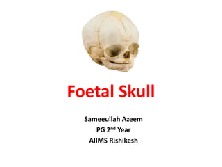
SlideShare a Scribd company logo
1 of 36
Foetal Skull
Sameeullah Azeem
PG 2nd Year
AIIMS Rishikesh
Contents
• Regions of foetal skull
• Bones
• Sutures
• Fontanelles
• MCQs
Introduction
• At birth, the calvaria is large in proportion to
other skeletal parts, reflecting cerebral
maturation.
• The facial region is relatively small and
constitutes only about one-eighth of the
neonatal skull, whereas it constitutes about
half of the adult skull.
Introduction
Adult Skull Fetal Skull
Adult and Fetal Skull
Fetal Skull
• Skull bones encase
and protect the
brain
• Major 3 Parts
Vault
Face
Base
Bones of the Vault
2 Frontal Bones
2 Parietal Bones
1 Occipital Bones
2 Temporal Bones
Vault
• Large, dome shaped part above an imaginary
line drawn between the orbital ridges and
nape of the neck
• Base
Base comprises bones that firmly united to
protect the vital centers in the medula
oblangata
Face
Comprises of 14 bones that are firmly united and are
non-compressible
2 Frontal Bones
• Form the forehead or
Sinciput
• The ossification center
of each bone is frontal
eminence
• Fuse into single bone by
8 years of age.
2-Parietal Bones
• Lie on either side
and occupy most
of the skull
• Parietal
eminence(Ossificat
ion Centre)
Occipital Bone
Temporal Bone
Fontaneles
• The fibrous membrane that
forms the calvaria remains
unossified at the six angles of
the parietal bones, producing
six fontanelles:
• Two single midline (anterior
and posterior) and
• Two lateral pairs
(sphenoidal/anterolateral and
mastoid/posterolateral).
Fontanelles
Anterior Fontanelle
• Largest and Rhomboid
• 4 cm AP
• 2.5 Transverse
• Formed between Sagittal,
Transverse and Frontal
Suture
• Closes at 12-18 months.
Clinical
• Palpation of fontanelle
is useful clinically.
• Sunken fontanelle
indicates dehydration
• Tense or bulging
anterior fontanelle
indicates
raised intracranial
pressure. E.g
Meningitis ,
Hydrocephalus
Posterior Fontanelle
• Triangular
• Between sagital and
lambdoid suture
• Closes at 6-8 weeks
• Delayed closure
suggests
Hyperthyrodism
Anteriolateral Fontanelle
• Sphenoidal Fontanelle
• Lies at sphenoidal angle
of Parietal Bone between
the sphenoid, parietal,
temporal, and frontal
bones
• Fuses at 6 Months
Posteriolateral Fontanelle
• Mastoid Fontanelle
• Bilateral soft
membranous gaps at the
junction of
the parietomastoid, occi
pitomastoid,
and lambdoid sutures
• Fuse at 6-18 months
• Persists as Asterion
Sutures
Metopic Suture
• Also known as median
frontal suture
• Vertical fibrous joint that
divides the two halves of the
frontal bone runs cross the
frontal bone from
the nasion to the bregma.
• Fusion 3-9 months
Metopic Synostosis
• Premature fusion of the
metopic suture
• Results
in Trigonocephaly.
Coronal Suture
• Cranial suture formed
between the two parietal
bones and the frontal
bone.
• Junction of the Anterior
Fontanelle
• Fuses at 24 years of age
Oxycephaly
• Craniosynostosis in which
top of the skull is pointed or
conical due to premature
closure of the coronal
suture.
• Most severe form of
craniosynostosis.
• Characterised by
• 8th cranial nerve lesion
• Optic nerve compression
• Mental deficiency
Plagiocephaly
• Flat Head Syndrome
• Asymmetric premature
closure of the coronal
suture/lambdoid suture.
• Widespread form is
characterized by a flat
spot on the back or one
side of the head caused
by remaining in a supine
position for prolonged
periods
Sagittal Suture
• Midline articulation that
joins the two parietal
bones
• Normal fusion at 22
years.
Scaphocephaly
• Most common form
of craniosynostosis
• Premature closure of
the sagittal suture
• Results in an impediment
to the lateral growth of
the skull while
anteroposterior growth
continues, producing a
narrow and elongated
skull.
Squamosal Sutures
• Arches backward from
the pterion and connects
the temporal squama
with the lower border of
the parietal bone
• May not completely
close until 60 years of
age.
Lambdoid Suture
• Junction between the
superior border of
the occipital bone and the
posterior borders of the
right and left parietal bones
• Fuses at approximately 26
years
Sphenofrontal Suture
• Between Frontal and Sphenoid Bone
• Anteriorly suture appears on the roof of the
bony orbit.
• Laterally as meeting point of inferior posterior
edges of frontal bone and anterior, superior
edge of the greater wing of the sphenoid bone
• Fuses at 15 years of age.
Questions
• 1) At what age the anterior fontanelle fuse?
a) 12-18 Months
b) 18-36 Months
c) 6-12 Months
d) 3-6 Months
Ans : A
• 2) A mother bought her 1 month old baby in the OPD
with the complaint of fever. The physician did the
physical examination and found raised intracranial
pressure. Which of the following fontanelles suggest
the same after proper palpation ?
a) Anterior
b) Posterior
c) Sphenoidal
d) Mastoid
Ans : A
3) Trigonocephaly results from which of the
following Synostosis?
a) Lambdoid
b) Sagital
c) Coronal
d) Mitopic
Ans: D
Bibilography
Grays Anatomy 41st Edition
Radiopedia

More Related Content

What's hot

What's hot (20)

applied anatomy of ankle and foot
applied anatomy of ankle and footapplied anatomy of ankle and foot
applied anatomy of ankle and foot
 
Forearm muscles
Forearm musclesForearm muscles
Forearm muscles
 
Flat Foot, Tibialis Posterior Tendon Dysfunction & Accessory Navicularis
Flat Foot, Tibialis Posterior Tendon Dysfunction & Accessory NavicularisFlat Foot, Tibialis Posterior Tendon Dysfunction & Accessory Navicularis
Flat Foot, Tibialis Posterior Tendon Dysfunction & Accessory Navicularis
 
Fascia
FasciaFascia
Fascia
 
Wrist ligaments
Wrist ligamentsWrist ligaments
Wrist ligaments
 
CTEV/ Clubfoot
CTEV/ ClubfootCTEV/ Clubfoot
CTEV/ Clubfoot
 
Embryology
EmbryologyEmbryology
Embryology
 
Embryology musculoskeletal system
Embryology musculoskeletal systemEmbryology musculoskeletal system
Embryology musculoskeletal system
 
Scfe seminar
Scfe seminarScfe seminar
Scfe seminar
 
Dr. kamal kant applied anatomy of hip
Dr. kamal kant   applied anatomy of hipDr. kamal kant   applied anatomy of hip
Dr. kamal kant applied anatomy of hip
 
arches of foot.pptx
arches of foot.pptxarches of foot.pptx
arches of foot.pptx
 
Connective tissue
Connective tissueConnective tissue
Connective tissue
 
Classification of bones and Anatomy of a long bone
Classification of bones and Anatomy of a long boneClassification of bones and Anatomy of a long bone
Classification of bones and Anatomy of a long bone
 
Ppt mscler system
Ppt mscler systemPpt mscler system
Ppt mscler system
 
8. hip joint
8. hip joint8. hip joint
8. hip joint
 
Knee Joint Anatomy
Knee Joint AnatomyKnee Joint Anatomy
Knee Joint Anatomy
 
3.5 cervical vertebra
3.5 cervical vertebra3.5 cervical vertebra
3.5 cervical vertebra
 
Brachial plexus
Brachial plexusBrachial plexus
Brachial plexus
 
The axilla
The axillaThe axilla
The axilla
 
Knee instability
Knee instabilityKnee instability
Knee instability
 

Similar to Foetal Skull Anatomy

fetalskullppt-160707124535-converted.pptx
fetalskullppt-160707124535-converted.pptxfetalskullppt-160707124535-converted.pptx
fetalskullppt-160707124535-converted.pptx
AkhilaLukose
 
OVERVIEW OF THE HEAD REGION.pptx.pdf
OVERVIEW OF THE HEAD REGION.pptx.pdfOVERVIEW OF THE HEAD REGION.pptx.pdf
OVERVIEW OF THE HEAD REGION.pptx.pdf
AbooddSandoqaa
 

Similar to Foetal Skull Anatomy (20)

SKULL SUTURES.pptx
SKULL SUTURES.pptxSKULL SUTURES.pptx
SKULL SUTURES.pptx
 
FETAL_SKULL .Its Anatomy and physiology.
FETAL_SKULL .Its Anatomy and physiology.FETAL_SKULL .Its Anatomy and physiology.
FETAL_SKULL .Its Anatomy and physiology.
 
Feotal skull presentation
Feotal skull presentationFeotal skull presentation
Feotal skull presentation
 
Axial skeletal
Axial skeletalAxial skeletal
Axial skeletal
 
3. cranio facial development
3. cranio facial development3. cranio facial development
3. cranio facial development
 
Fetal skull
Fetal skullFetal skull
Fetal skull
 
Development of skull embryology copy.pptx
Development of skull embryology copy.pptxDevelopment of skull embryology copy.pptx
Development of skull embryology copy.pptx
 
Basic Skull Anatomy
Basic Skull AnatomyBasic Skull Anatomy
Basic Skull Anatomy
 
FETAL SKULL.pptx
FETAL SKULL.pptxFETAL SKULL.pptx
FETAL SKULL.pptx
 
Osteology of skull and cervial vertebrae
Osteology of skull and cervial vertebraeOsteology of skull and cervial vertebrae
Osteology of skull and cervial vertebrae
 
Fetal skull ppt
Fetal skull pptFetal skull ppt
Fetal skull ppt
 
EMBRYOLOGY,GROWTH & DEVELOPMENT OF CRANIUM & CRANIAL BASE IRT ORTHODONTICS.pptx
EMBRYOLOGY,GROWTH & DEVELOPMENT OF CRANIUM & CRANIAL BASE IRT ORTHODONTICS.pptxEMBRYOLOGY,GROWTH & DEVELOPMENT OF CRANIUM & CRANIAL BASE IRT ORTHODONTICS.pptx
EMBRYOLOGY,GROWTH & DEVELOPMENT OF CRANIUM & CRANIAL BASE IRT ORTHODONTICS.pptx
 
fetalskullppt-160707124535-converted.pptx
fetalskullppt-160707124535-converted.pptxfetalskullppt-160707124535-converted.pptx
fetalskullppt-160707124535-converted.pptx
 
Anatomy of the head and neck
Anatomy of the head and neckAnatomy of the head and neck
Anatomy of the head and neck
 
FETAL SKULL PPT (1).pptx
FETAL SKULL PPT (1).pptxFETAL SKULL PPT (1).pptx
FETAL SKULL PPT (1).pptx
 
FETAL SKULL PPT .pptx
FETAL SKULL PPT .pptxFETAL SKULL PPT .pptx
FETAL SKULL PPT .pptx
 
Skull Development
Skull DevelopmentSkull Development
Skull Development
 
OVERVIEW OF THE HEAD REGION.pptx.pdf
OVERVIEW OF THE HEAD REGION.pptx.pdfOVERVIEW OF THE HEAD REGION.pptx.pdf
OVERVIEW OF THE HEAD REGION.pptx.pdf
 
Prenatal development of cranial base
Prenatal development of cranial basePrenatal development of cranial base
Prenatal development of cranial base
 
Growth and development of the mandible
Growth and development of the mandibleGrowth and development of the mandible
Growth and development of the mandible
 

Recently uploaded

Jual Obat Aborsi Di Dubai UAE Wa 0838-4800-7379 Obat Penggugur Kandungan Cytotec
Jual Obat Aborsi Di Dubai UAE Wa 0838-4800-7379 Obat Penggugur Kandungan CytotecJual Obat Aborsi Di Dubai UAE Wa 0838-4800-7379 Obat Penggugur Kandungan Cytotec
Jual Obat Aborsi Di Dubai UAE Wa 0838-4800-7379 Obat Penggugur Kandungan Cytotec
jualobat34
 
Physiologic Anatomy of Heart_AntiCopy.pdf
Physiologic Anatomy of Heart_AntiCopy.pdfPhysiologic Anatomy of Heart_AntiCopy.pdf
Physiologic Anatomy of Heart_AntiCopy.pdf
MedicoseAcademics
 
Difference Between Skeletal Smooth and Cardiac Muscles
Difference Between Skeletal Smooth and Cardiac MusclesDifference Between Skeletal Smooth and Cardiac Muscles
Difference Between Skeletal Smooth and Cardiac Muscles
MedicoseAcademics
 

Recently uploaded (20)

HISTORY, CONCEPT AND ITS IMPORTANCE IN DRUG DEVELOPMENT.pptx
HISTORY, CONCEPT AND ITS IMPORTANCE IN DRUG DEVELOPMENT.pptxHISTORY, CONCEPT AND ITS IMPORTANCE IN DRUG DEVELOPMENT.pptx
HISTORY, CONCEPT AND ITS IMPORTANCE IN DRUG DEVELOPMENT.pptx
 
Circulatory Shock, types and stages, compensatory mechanisms
Circulatory Shock, types and stages, compensatory mechanismsCirculatory Shock, types and stages, compensatory mechanisms
Circulatory Shock, types and stages, compensatory mechanisms
 
Drug development life cycle indepth overview.pptx
Drug development life cycle indepth overview.pptxDrug development life cycle indepth overview.pptx
Drug development life cycle indepth overview.pptx
 
Physicochemical properties (descriptors) in QSAR.pdf
Physicochemical properties (descriptors) in QSAR.pdfPhysicochemical properties (descriptors) in QSAR.pdf
Physicochemical properties (descriptors) in QSAR.pdf
 
VIP ℂall Girls Kothanur {{ Bangalore }} 6378878445 WhatsApp: Me 24/7 Hours Se...
VIP ℂall Girls Kothanur {{ Bangalore }} 6378878445 WhatsApp: Me 24/7 Hours Se...VIP ℂall Girls Kothanur {{ Bangalore }} 6378878445 WhatsApp: Me 24/7 Hours Se...
VIP ℂall Girls Kothanur {{ Bangalore }} 6378878445 WhatsApp: Me 24/7 Hours Se...
 
Top 10 Most Beautiful Chinese Pornstars List 2024
Top 10 Most Beautiful Chinese Pornstars List 2024Top 10 Most Beautiful Chinese Pornstars List 2024
Top 10 Most Beautiful Chinese Pornstars List 2024
 
See it and Catch it! Recognizing the Thought Traps that Negatively Impact How...
See it and Catch it! Recognizing the Thought Traps that Negatively Impact How...See it and Catch it! Recognizing the Thought Traps that Negatively Impact How...
See it and Catch it! Recognizing the Thought Traps that Negatively Impact How...
 
VIP ℂall Girls Arekere Bangalore 6378878445 WhatsApp: Me All Time Serviℂe Ava...
VIP ℂall Girls Arekere Bangalore 6378878445 WhatsApp: Me All Time Serviℂe Ava...VIP ℂall Girls Arekere Bangalore 6378878445 WhatsApp: Me All Time Serviℂe Ava...
VIP ℂall Girls Arekere Bangalore 6378878445 WhatsApp: Me All Time Serviℂe Ava...
 
Jual Obat Aborsi Di Dubai UAE Wa 0838-4800-7379 Obat Penggugur Kandungan Cytotec
Jual Obat Aborsi Di Dubai UAE Wa 0838-4800-7379 Obat Penggugur Kandungan CytotecJual Obat Aborsi Di Dubai UAE Wa 0838-4800-7379 Obat Penggugur Kandungan Cytotec
Jual Obat Aborsi Di Dubai UAE Wa 0838-4800-7379 Obat Penggugur Kandungan Cytotec
 
Physiologic Anatomy of Heart_AntiCopy.pdf
Physiologic Anatomy of Heart_AntiCopy.pdfPhysiologic Anatomy of Heart_AntiCopy.pdf
Physiologic Anatomy of Heart_AntiCopy.pdf
 
ANATOMY AND PHYSIOLOGY OF REPRODUCTIVE SYSTEM.pptx
ANATOMY AND PHYSIOLOGY OF REPRODUCTIVE SYSTEM.pptxANATOMY AND PHYSIOLOGY OF REPRODUCTIVE SYSTEM.pptx
ANATOMY AND PHYSIOLOGY OF REPRODUCTIVE SYSTEM.pptx
 
Test bank for critical care nursing a holistic approach 11th edition morton f...
Test bank for critical care nursing a holistic approach 11th edition morton f...Test bank for critical care nursing a holistic approach 11th edition morton f...
Test bank for critical care nursing a holistic approach 11th edition morton f...
 
Part I - Anticipatory Grief: Experiencing grief before the loss has happened
Part I - Anticipatory Grief: Experiencing grief before the loss has happenedPart I - Anticipatory Grief: Experiencing grief before the loss has happened
Part I - Anticipatory Grief: Experiencing grief before the loss has happened
 
Face and Muscles of facial expression.pptx
Face and Muscles of facial expression.pptxFace and Muscles of facial expression.pptx
Face and Muscles of facial expression.pptx
 
Difference Between Skeletal Smooth and Cardiac Muscles
Difference Between Skeletal Smooth and Cardiac MusclesDifference Between Skeletal Smooth and Cardiac Muscles
Difference Between Skeletal Smooth and Cardiac Muscles
 
Top 10 Most Beautiful Russian Pornstars List 2024
Top 10 Most Beautiful Russian Pornstars List 2024Top 10 Most Beautiful Russian Pornstars List 2024
Top 10 Most Beautiful Russian Pornstars List 2024
 
Intro to disinformation and public health
Intro to disinformation and public healthIntro to disinformation and public health
Intro to disinformation and public health
 
TEST BANK For Guyton and Hall Textbook of Medical Physiology, 14th Edition by...
TEST BANK For Guyton and Hall Textbook of Medical Physiology, 14th Edition by...TEST BANK For Guyton and Hall Textbook of Medical Physiology, 14th Edition by...
TEST BANK For Guyton and Hall Textbook of Medical Physiology, 14th Edition by...
 
MOTION MANAGEMANT IN LUNG SBRT BY DR KANHU CHARAN PATRO
MOTION MANAGEMANT IN LUNG SBRT BY DR KANHU CHARAN PATROMOTION MANAGEMANT IN LUNG SBRT BY DR KANHU CHARAN PATRO
MOTION MANAGEMANT IN LUNG SBRT BY DR KANHU CHARAN PATRO
 
The Clean Living Project Episode 23 - Journaling
The Clean Living Project Episode 23 - JournalingThe Clean Living Project Episode 23 - Journaling
The Clean Living Project Episode 23 - Journaling
 

Foetal Skull Anatomy

  • 1. Foetal Skull Sameeullah Azeem PG 2nd Year AIIMS Rishikesh
  • 2. Contents • Regions of foetal skull • Bones • Sutures • Fontanelles • MCQs
  • 3. Introduction • At birth, the calvaria is large in proportion to other skeletal parts, reflecting cerebral maturation. • The facial region is relatively small and constitutes only about one-eighth of the neonatal skull, whereas it constitutes about half of the adult skull.
  • 6. Fetal Skull • Skull bones encase and protect the brain • Major 3 Parts Vault Face Base
  • 7. Bones of the Vault 2 Frontal Bones 2 Parietal Bones 1 Occipital Bones 2 Temporal Bones
  • 8. Vault • Large, dome shaped part above an imaginary line drawn between the orbital ridges and nape of the neck • Base Base comprises bones that firmly united to protect the vital centers in the medula oblangata Face Comprises of 14 bones that are firmly united and are non-compressible
  • 9. 2 Frontal Bones • Form the forehead or Sinciput • The ossification center of each bone is frontal eminence • Fuse into single bone by 8 years of age.
  • 10. 2-Parietal Bones • Lie on either side and occupy most of the skull • Parietal eminence(Ossificat ion Centre)
  • 13. Fontaneles • The fibrous membrane that forms the calvaria remains unossified at the six angles of the parietal bones, producing six fontanelles: • Two single midline (anterior and posterior) and • Two lateral pairs (sphenoidal/anterolateral and mastoid/posterolateral).
  • 15. Anterior Fontanelle • Largest and Rhomboid • 4 cm AP • 2.5 Transverse • Formed between Sagittal, Transverse and Frontal Suture • Closes at 12-18 months.
  • 16. Clinical • Palpation of fontanelle is useful clinically. • Sunken fontanelle indicates dehydration • Tense or bulging anterior fontanelle indicates raised intracranial pressure. E.g Meningitis , Hydrocephalus
  • 17. Posterior Fontanelle • Triangular • Between sagital and lambdoid suture • Closes at 6-8 weeks • Delayed closure suggests Hyperthyrodism
  • 18. Anteriolateral Fontanelle • Sphenoidal Fontanelle • Lies at sphenoidal angle of Parietal Bone between the sphenoid, parietal, temporal, and frontal bones • Fuses at 6 Months
  • 19. Posteriolateral Fontanelle • Mastoid Fontanelle • Bilateral soft membranous gaps at the junction of the parietomastoid, occi pitomastoid, and lambdoid sutures • Fuse at 6-18 months • Persists as Asterion
  • 21. Metopic Suture • Also known as median frontal suture • Vertical fibrous joint that divides the two halves of the frontal bone runs cross the frontal bone from the nasion to the bregma. • Fusion 3-9 months
  • 22. Metopic Synostosis • Premature fusion of the metopic suture • Results in Trigonocephaly.
  • 23. Coronal Suture • Cranial suture formed between the two parietal bones and the frontal bone. • Junction of the Anterior Fontanelle • Fuses at 24 years of age
  • 24. Oxycephaly • Craniosynostosis in which top of the skull is pointed or conical due to premature closure of the coronal suture. • Most severe form of craniosynostosis. • Characterised by • 8th cranial nerve lesion • Optic nerve compression • Mental deficiency
  • 25. Plagiocephaly • Flat Head Syndrome • Asymmetric premature closure of the coronal suture/lambdoid suture. • Widespread form is characterized by a flat spot on the back or one side of the head caused by remaining in a supine position for prolonged periods
  • 26. Sagittal Suture • Midline articulation that joins the two parietal bones • Normal fusion at 22 years.
  • 27. Scaphocephaly • Most common form of craniosynostosis • Premature closure of the sagittal suture • Results in an impediment to the lateral growth of the skull while anteroposterior growth continues, producing a narrow and elongated skull.
  • 28. Squamosal Sutures • Arches backward from the pterion and connects the temporal squama with the lower border of the parietal bone • May not completely close until 60 years of age.
  • 29. Lambdoid Suture • Junction between the superior border of the occipital bone and the posterior borders of the right and left parietal bones • Fuses at approximately 26 years
  • 30. Sphenofrontal Suture • Between Frontal and Sphenoid Bone • Anteriorly suture appears on the roof of the bony orbit. • Laterally as meeting point of inferior posterior edges of frontal bone and anterior, superior edge of the greater wing of the sphenoid bone • Fuses at 15 years of age.
  • 31.
  • 33. • 1) At what age the anterior fontanelle fuse? a) 12-18 Months b) 18-36 Months c) 6-12 Months d) 3-6 Months Ans : A
  • 34. • 2) A mother bought her 1 month old baby in the OPD with the complaint of fever. The physician did the physical examination and found raised intracranial pressure. Which of the following fontanelles suggest the same after proper palpation ? a) Anterior b) Posterior c) Sphenoidal d) Mastoid Ans : A
  • 35. 3) Trigonocephaly results from which of the following Synostosis? a) Lambdoid b) Sagital c) Coronal d) Mitopic Ans: D
  • 36. Bibilography Grays Anatomy 41st Edition Radiopedia