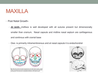
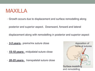
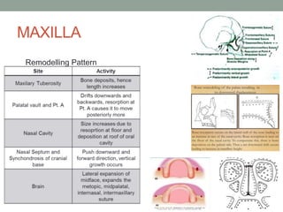
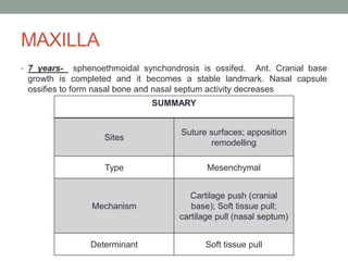
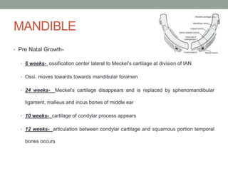
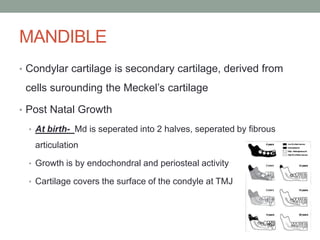
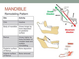
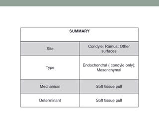
CRANIOFACIAL
DEVELOPMENT
DEPARTMENT OF ORTHODONTICS
FINAL YEAR BDS
CRANIAL VAULT
• Pre Natal Growth
• 7 weeks- brain is prominent and covered by dense membrane
• 8 weeks- bone blastema forms in middle layer of membrane.
Ossification starts with frontal bone squamous temporal
parietal squamous occipital
• 9 weeks- increase in bone size. Bone growth exceeds brain
growth
• 12 weeks- single thickness plate of flat bone with sutures in the
middle
8 weeks 12 weeks
CRANIAL VAULT
• Post Natal Growth
• At birth- cranial vault larger than the face
• All sutures present including metopic suture
• Fontanels are present
• Till 2 years- fontanels and sutures, including metopic suture closes
• 4 years- brain and cranial vault have achieved 80% of adult size,
suture grows
• 10 years- brain and cranial vault achieve 95% of adult size, suture
still growing
• Age 25, sagittal suture closed, 2- 3 years later coronal suture close
SUMMARY
Site Sutures (Major), Surfaces (Minor)
Type Mesenchymal
Mechanism Pressure to separate sutures
Determinant
Intracranial pressure ( brain
growth)
CRANIAL BASE
• Pre Natal Growth-
• At 6 weeks- Parachordal cartilages which surround the notochord
form the basioccipital bone
• Development is from posterior to anterior towards otic capsule.
Bones formed are: Petrous portion of temporal bone cartilages
of sphenoid bone nasal capsule ethmoid bone
inferior turbinate nasal septum
• 8 weeks- the cartilages join to form a single cartilage. This is
primary hyaline cartilage called basal plate extending from foramen
magnum to tip of nasal cavity
• 9-16 weeks- ossification centers form from
parachordal cartilages through the sphenoid
complex
• 36 wks- ossi. centers extend till ethmoid region
• Synchondrosis form within ossi. centers
CRANIAL BASE
• Post Natal Growth
• 3 synchondrosis
• Intersphenoid- fuses at time of birth
• Sphenoethmoidal – fuses at 7 years
• Spheno- occipital- fuses after puberty
Between birth and 7 years- anterior cranial base grows more than posterior
cranial base. Maximum growth occurs in first 2- 3 post natal years with
anterior more growth than posterior as well as cranial base angulations
SUMMARY
Site Synchondroses, Sutures (
laterally)
Center Synchondroses
Type Endochondral, Mesenchymal
(laterally)
Mechanism Interstitial growth at
synchondroses
Determinant Genetic ( at synchondroses),
cartilage pull (at lateral
sutures)
MAXILLA
• Pre Natal Growth-
• Formed by intramembranous ossification
• Centers of ossi. develop within the mesenchyme. They are covered by a periosteum which
becomes the source of osteoblastic cells
• Nasal capsular cartilage provide a signalling mechanism for development, growth and
maintenance of sutures
• 6 weeks- Palatal shelf elevation
• 7 weeks- palatal shelf ossi.
• Development direction is laterally and anteroposteriorly with expansion of the brain, cranial
cavity, oral cavity, and oronasal pharynx. Nasal capsular cartilage provides growth factors
MAXILLA
• Post Natal Growth-
• At birth- midface is well developed with all sutures present but dimensionally
smaller than cranium. Nasal capsule and midline nasal septum are cartilagenous
and continous with cranial base
• Ossi. is primarily intramembranous and at nasal capsule it is endochondral
MAXILLA
• Growth occurs due to displacement and surface remodelling along
posterior and superior aspect. Downward, forward and lateral
displacement along with remodelling in posterior and superior aspect
• 3-5 years- premx/mx suture close
• 15-18 years- midpalatal suture close
• 20-25 years- transpalatal suture close
Apposition of
bone at sutures
Surface modelling
and remodelling
MAXILLA
Remodelling Pattern
MAXILLA
• 7 years- sphenoethmoidal synchondrosis is ossifed. Ant. Cranial base
growth is completed and it becomes a stable landmark. Nasal capsule
ossifies to form nasal bone and nasal septum activity decreases
SUMMARY
Sites
Suture surfaces; apposition
remodelling
Type Mesenchymal
Mechanism
Cartilage push (cranial
base); Soft tissue pull;
cartilage pull (nasal septum)
Determinant Soft tissue pull
MANDIBLE
• Pre Natal Growth-
• 6 weeks- ossification center lateral to Meckel’s cartilage at division of IAN
• Ossi. moves towards towards mandibular foramen
• 24 weeks- Meckel’s cartilage disappears and is replaced by sphenomandibular
ligament, malleus and incus bones of middle ear
• 10 weeks- cartilage of condylar process appears
• 12 weeks- articulation between condylar cartilage and squamous portion temporal
bones occurs
MANDIBLE
• Condylar cartilage is secondary cartilage, derived from
cells surounding the Meckel’s cartilage
• Post Natal Growth
• At birth- Md is seperated into 2 halves, seperated by fibrous
articulation
• Growth is by endochondral and periosteal activity
• Cartilage covers the surface of the condyle at TMJ
MANDIBLE
Remodeling Pattern
Site Activity
Chin Inactive
Body of mandible Grows longer by
bone deposition
on posterior
surface
Ramus Grows higher by
bone formation at
condyle head and
surface
remodelling
Posterior surface
of Ramus
Bone deposition
Anterior surface
of Ramus
Bone removed
SUMMARY
Site
Condyle; Ramus; Other
surfaces
Type
Endochondral ( condyle only);
Mesenchymal
Mechanism Soft tissue pull
Determinant Soft tissue pull
FACIAL SOFT TISSUES
• Growth of Lips
• Trail behind jaw growth and undergo a growth spurt
• Short lip in mixed dentition years, lip seperation is called “ lip
incompetence”
• Lip thickness reaches maximum during adolescence then decreases
• Growth of Nose
• Nasal bone growth completes at age 10, after which cartilage and soft
tissue growth occurs. They undergo adolescent growth spurt
• Prominence of nose increases in adolescence esp. for boys
THEORIES OF GROWTH
• Three theories- 1) bone 2) cartilage 3) soft tissue matrix
• Difference is in location at which genetic control is
expressed
• Indirect genetic control is called “epigenetic”
• “Growth site”- location at which growth occurs
• “Growth center”- independent (genetically controlled)
growth occurs

3. cranio facial development

  • 1.
  • 2.
    CRANIAL VAULT • PreNatal Growth • 7 weeks- brain is prominent and covered by dense membrane • 8 weeks- bone blastema forms in middle layer of membrane. Ossification starts with frontal bone squamous temporal parietal squamous occipital • 9 weeks- increase in bone size. Bone growth exceeds brain growth • 12 weeks- single thickness plate of flat bone with sutures in the middle 8 weeks 12 weeks
  • 3.
    CRANIAL VAULT • PostNatal Growth • At birth- cranial vault larger than the face • All sutures present including metopic suture • Fontanels are present • Till 2 years- fontanels and sutures, including metopic suture closes • 4 years- brain and cranial vault have achieved 80% of adult size, suture grows • 10 years- brain and cranial vault achieve 95% of adult size, suture still growing • Age 25, sagittal suture closed, 2- 3 years later coronal suture close
  • 4.
    SUMMARY Site Sutures (Major),Surfaces (Minor) Type Mesenchymal Mechanism Pressure to separate sutures Determinant Intracranial pressure ( brain growth)
  • 5.
    CRANIAL BASE • PreNatal Growth- • At 6 weeks- Parachordal cartilages which surround the notochord form the basioccipital bone • Development is from posterior to anterior towards otic capsule. Bones formed are: Petrous portion of temporal bone cartilages of sphenoid bone nasal capsule ethmoid bone inferior turbinate nasal septum • 8 weeks- the cartilages join to form a single cartilage. This is primary hyaline cartilage called basal plate extending from foramen magnum to tip of nasal cavity • 9-16 weeks- ossification centers form from parachordal cartilages through the sphenoid complex • 36 wks- ossi. centers extend till ethmoid region • Synchondrosis form within ossi. centers
  • 6.
    CRANIAL BASE • PostNatal Growth • 3 synchondrosis • Intersphenoid- fuses at time of birth • Sphenoethmoidal – fuses at 7 years • Spheno- occipital- fuses after puberty Between birth and 7 years- anterior cranial base grows more than posterior cranial base. Maximum growth occurs in first 2- 3 post natal years with anterior more growth than posterior as well as cranial base angulations
  • 7.
    SUMMARY Site Synchondroses, Sutures( laterally) Center Synchondroses Type Endochondral, Mesenchymal (laterally) Mechanism Interstitial growth at synchondroses Determinant Genetic ( at synchondroses), cartilage pull (at lateral sutures)
  • 8.
    MAXILLA • Pre NatalGrowth- • Formed by intramembranous ossification • Centers of ossi. develop within the mesenchyme. They are covered by a periosteum which becomes the source of osteoblastic cells • Nasal capsular cartilage provide a signalling mechanism for development, growth and maintenance of sutures • 6 weeks- Palatal shelf elevation • 7 weeks- palatal shelf ossi. • Development direction is laterally and anteroposteriorly with expansion of the brain, cranial cavity, oral cavity, and oronasal pharynx. Nasal capsular cartilage provides growth factors
  • 9.
    MAXILLA • Post NatalGrowth- • At birth- midface is well developed with all sutures present but dimensionally smaller than cranium. Nasal capsule and midline nasal septum are cartilagenous and continous with cranial base • Ossi. is primarily intramembranous and at nasal capsule it is endochondral
  • 10.
    MAXILLA • Growth occursdue to displacement and surface remodelling along posterior and superior aspect. Downward, forward and lateral displacement along with remodelling in posterior and superior aspect • 3-5 years- premx/mx suture close • 15-18 years- midpalatal suture close • 20-25 years- transpalatal suture close Apposition of bone at sutures Surface modelling and remodelling
  • 11.
  • 12.
    MAXILLA • 7 years-sphenoethmoidal synchondrosis is ossifed. Ant. Cranial base growth is completed and it becomes a stable landmark. Nasal capsule ossifies to form nasal bone and nasal septum activity decreases SUMMARY Sites Suture surfaces; apposition remodelling Type Mesenchymal Mechanism Cartilage push (cranial base); Soft tissue pull; cartilage pull (nasal septum) Determinant Soft tissue pull
  • 13.
    MANDIBLE • Pre NatalGrowth- • 6 weeks- ossification center lateral to Meckel’s cartilage at division of IAN • Ossi. moves towards towards mandibular foramen • 24 weeks- Meckel’s cartilage disappears and is replaced by sphenomandibular ligament, malleus and incus bones of middle ear • 10 weeks- cartilage of condylar process appears • 12 weeks- articulation between condylar cartilage and squamous portion temporal bones occurs
  • 14.
    MANDIBLE • Condylar cartilageis secondary cartilage, derived from cells surounding the Meckel’s cartilage • Post Natal Growth • At birth- Md is seperated into 2 halves, seperated by fibrous articulation • Growth is by endochondral and periosteal activity • Cartilage covers the surface of the condyle at TMJ
  • 15.
    MANDIBLE Remodeling Pattern Site Activity ChinInactive Body of mandible Grows longer by bone deposition on posterior surface Ramus Grows higher by bone formation at condyle head and surface remodelling Posterior surface of Ramus Bone deposition Anterior surface of Ramus Bone removed
  • 16.
    SUMMARY Site Condyle; Ramus; Other surfaces Type Endochondral( condyle only); Mesenchymal Mechanism Soft tissue pull Determinant Soft tissue pull
  • 17.
    FACIAL SOFT TISSUES •Growth of Lips • Trail behind jaw growth and undergo a growth spurt • Short lip in mixed dentition years, lip seperation is called “ lip incompetence” • Lip thickness reaches maximum during adolescence then decreases • Growth of Nose • Nasal bone growth completes at age 10, after which cartilage and soft tissue growth occurs. They undergo adolescent growth spurt • Prominence of nose increases in adolescence esp. for boys
  • 18.
    THEORIES OF GROWTH •Three theories- 1) bone 2) cartilage 3) soft tissue matrix • Difference is in location at which genetic control is expressed • Indirect genetic control is called “epigenetic” • “Growth site”- location at which growth occurs • “Growth center”- independent (genetically controlled) growth occurs