1. Parts of the Nerve Cell and Their Functions
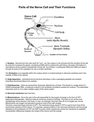
1. Nucleus - Derived from the Latin word for "nux", nut, the nucleus is the archivist and the architect of the cell.
As archivist it contains the genes, consisting of DNA which contains the cell history, the basic information to
manufacture all the proteins characteristic of that cell. As architect, it synthesizes RNA from DNA and ships it
through its pores to the cytoplasm for use in protein synthesis.
The.Nucleolus is an organelle within the nucleus which is involved actively in ribosome synthesis and in the
transfer of RNA to the cytosol.
2. Golgi Apparatus - membrane-bound structure that plays a role in packaging peptides and proteins
(including neurotransmitters) into vesicles.
3. Polyribosomes - there are several free ribosomes attached by a thread. The thread is a single strand of
mRNA (messenger RNA, a molecule involved in the synthesis of proteins outside the nucleus). The associated
ribosomes work on it to make multiple copies of the same protein.
4. Neuronal membrane (see next box)
5. Mitochondrium - this is the part of the cell responsible for the supply of energy in the form of ATP
(adenosine triphosphate). Neurons need an enormous amount of energy. The brain is one of the most
metabolically active tissues in the body. In man, for example, the brain uses 40 ml of oxygen per minute.
Mitochondria use oxygen and glucose to produce most of the cell's energy.
The brain consumes large amounts of ATP. The chemical energy stored in ATP is used to fuel most of the
biochemical reactions of the neuron. For example, special proteins in the neuronal membrane use the energy
released by the breakdown of ATP into ADP to pump certain substances across the membrane to establish
concentration differences between the inside of the neuron and the outside.
2. 6. Rough Endoplasmic Reticulum and Smooth Endoplasmic Reticulum (7) - A system of tubes for the
transportation of materials within the cytoplasm. It may have ribosomes (rough ER) or no ribosomes (smooth
ER). With ribosomes, the ER is important for protein synthesis.
2. Neuronal Membrane
The neuronal membrane serves as a barrier to
enclose the cytoplasm inside the neuron, and to
exclude certain substances that float in the fluid that
bathes the neuron.
The membrane with its mosaic of proteins is
responsible for many important functions:
• keeping certain ions and small molecules out
of the cell and letting others in,
• accumulating nutrients, and rejecting harmful
substances,
• catalyzing enzymatic reactions,
• establishing an electrical potential inside the
cell,
• conducting an impulse
• being sensitive to particular
neurotransmitters and modulators .
3. The membrane is made of lipids and proteins - fats and chains of aminoacids. The basic structure of this
membrane is a bilayer or sandwich of phospholipids, organized in such a way that the polar (charged) regions
face outward and the non polar regions face inward.
The external face of the membrane contains the receptors, small specialized molecular regions which provide
a kind of "attachment port" for other external molecules, in a scheme analogous to a a key and a keyhole. For
each external molecule there is a corresponding receptor. Whenever receptors become attached to a
molecule, some alterations of the membrane and in the interior of the cell ensue, such as the modification of
permeability to some ions.
3. Dendrites
These structures branch out in treelike fashion and serve as the main apparatus for receiving
signals from other nerve cells. They function as an "antennae" of the neuron and are covered
by thousands of synapses. The dendritic membrane under the synapse (the post-synaptic
membrane) has many specialized protein molecules called receptors that detect the
neurotransmitters in the synaptic cleft. A nerve cell can have many dendrites which branch
many times, their surface is irregular and covered in dendritic spines which are where the
synaptic input connections are made.
4. Axon
A
xon
Usually a long process which often projects to distant regions of the nervous system.
The axon is the main conducting unit of the neuron, capable of conveying electrical
signals along distances that range from as short as 0.1 mm to as long as 2 m. Many
axon split into several branches, thereby conveying information to different targets.
Many neurons do not have axons. In these so-called amacrine neurons, all the
neuronal processess are dendrites. Neurons with very short axons are also found.
The axons of many neurons are wrapped in a myelin sheat, which is composed of the
membranes of intersticial cells and is wrapped around the axons to form several
concentric layers. The myelin sheath is broken at various points by the nodes of
Ranvier, so that in cross section it looks like a string of sausages. The myelin
protects the axon, and prevents interference between axons as they pass along in
bundles, sometimes thousands at time.
The cells that wrap around peripheral nerve fibers - that is, nerve fibers outside of the
brain and spinal cord - are called Schwann cells (because they were first described
by Theodor Schwann). The cells that wrap around axons within the central nervous
system (brain and spinal cord) are called oligodendrocytes. The axon, with its
surrounded sheath, is called a nerve fiber. Between each pair of sucessive Schwann
cells is a gap of a node of Ranvier.
The Axon Hillock
The axon hillock is where the axon is joined to the cell. It is from here that the
electrical firing known as an action potential usually occurs.
4. 5. Nerve Ending (Presynaptic Terminals)
Synapses are the junctions formed with other nerve cells where the presynaptic terminal of
one cell comes into 'contact' with the postsynaptic membrane of another. It is at these
junctions that neurons are excited, inhibited, or modulated. There are two types of synapse,
electrical and chemical.
Electrical synapses occur where the presynaptic terminal is in electrical continuity with the
postsynaptic. Ions and small molecules passing through, thus connecting channels from
one cell to the next, so that electrical changes in one cell are transmitted almost
instantaneously to the next. Ions can generally flow both ways at these junctions i.e. they
tend to be bi-directional, although there are electrical junctions where the ions can only flow
one way, these are know as rectifying junctions. Rectifying junctions are used to
synchronise the firing of nerve cells.
Chemical synaptic junction is more complicated. The gap between the post- and
presynaptic terminals is larger, and the mode of transmission is not electrical, but carried by
neurotransmitters, neuroactive substances released at the presynaptic side of the junction.
There are two types of chemical junctions. Type I is an excitatory synapse, generally found
on dendrites, type II is an inhibitory synapse, generally found on cell bodies. Different
substances are released at these two types of synapse. The direction of flow of information
is usually one way at these junctions.
Each terminal button is connected to other neurons across a small gap called a synapse.
The physical and neurochemical characteristics of each synapse determines the strength
and polarity of the new input signal. This is where the brain is the most flexible, and the
most vulnerable. Changing the constitution of various neurotransmitter chemicals can
increase or decrease the amount of stimulation that the firing axon imparts on the
neighbouring dendrite. Altering the neurotransmitters can also change whether the
stimulation is excitatory or inhibitory.