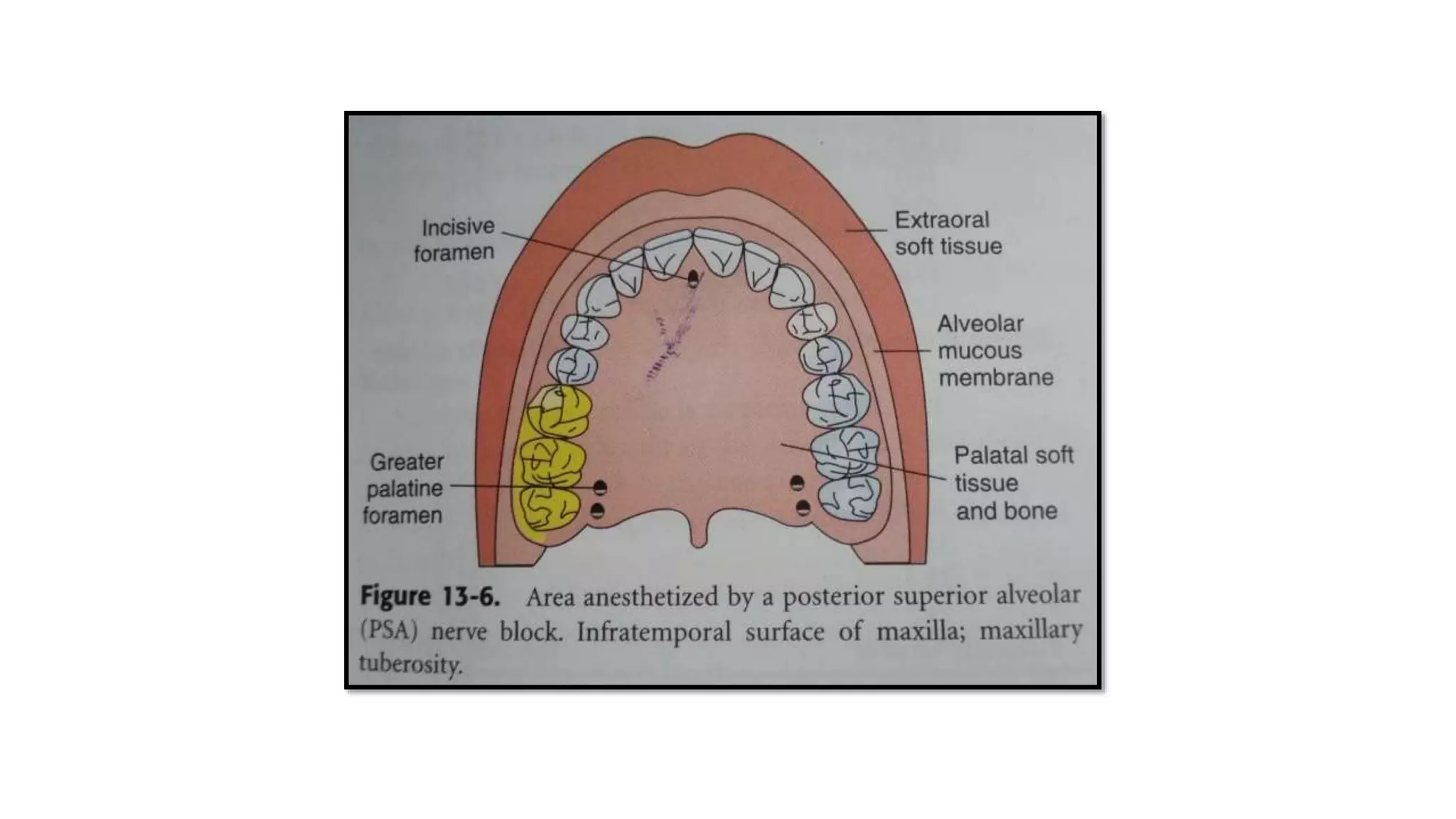
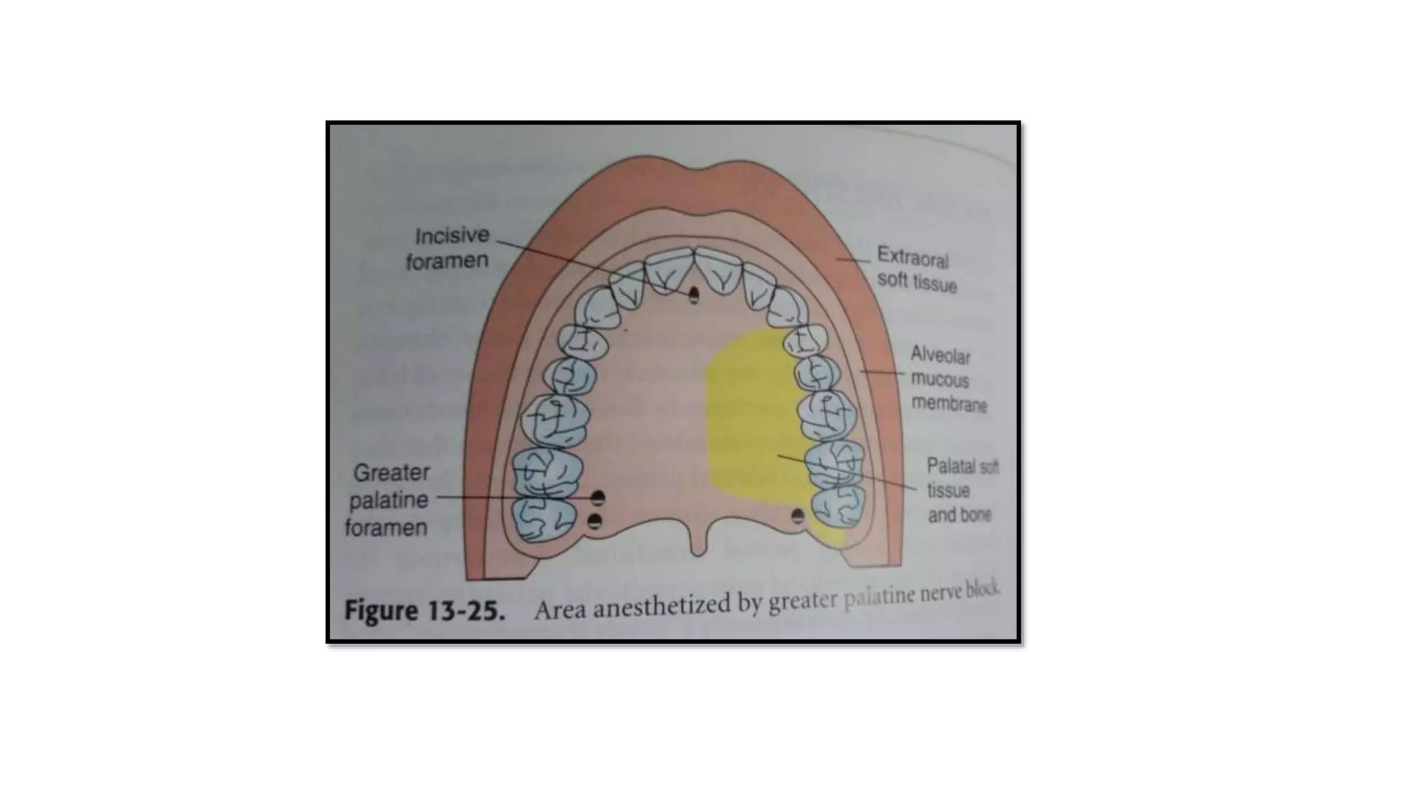
NERVE BLOCK
posterior superior alveolar nerve block
greater palatine nerve block
BY SAADIA ASHRAF
FINAL YEAR PART II
DEPARTMENT OF ORAL AND MAXILLOFACIAL SURGERY
CONTENTS
 INTRODUCTION
 POSTERIOR SUPERIOR ALVEOLAR NERVE BLOCK
 AREAS ANESTHETIZED
 INDICATIONS
 CONTRAINDICATIONS
 ADVANTAGES
 DISADVANTAGES
 TECHNIQUE
 FAILURE OF ANESTHESIA
 COMPLICATION
 GREATER PALATINE NERVE BLOCK
 AREAS ANESTHETIZED
 INDICATIONS
 CONTRAINDICATIONS
 ADVANTAGES
 DISADVANTAGES
 TECHNIQUE
 FAILURE OF ANESTHESIA
 COMPLICATION
 REFERENCE
INTRODUCTION
 The posterior superior alveolar (PSA) nerve descends from the main trunk of
maxillary division in the pterygopalatine fossa just before the maxillary division
enters the infraorbital canal.
 The greater palatine nerve descends through the pterygopalatine canal emerging
on the hard palate through the greater palatine foremen . The nerve course
anteriorly between the mucoperiosteum and the osseous hard palate , supplying
sensory innervation to the palatal soft tissues as far as anterior to first molar .
 The posterior superior alveolar nerve block is a most commonly used nerve block,
although it is a highly successful technique (>95%) •The PSA nerve block is
effective for the maxillary 3rd , 2nd and 1st molar except the mesio buccal root of
maxillary 1st molar (doesn’t anesthetize in 28%of patients), which is supplied by
middle superior alveolar nerve.
 Other common names: Tuberosity block and Zygomatic block
 Nerves anesthetized: Posterior superior alveolar nerves and its branches
 Area anesthetized: 1) Pulps of maxillary 3rd, 2nd and 1st molars (entire tooth =
72%; mesio buccal root of maxillary 1st molar not anesthetized = 28%) 2) Buccal
periodontium and bone overlying these teeth
 Contraindications: 1. When the risk of hemorrhage is too great (as with a
hemophiliac), in which case a supraperiosteal injection or PDL injection is
recommended
 Indications: 1.treatment involving two or more maxillary molars 2.When
supraperiosteal injection is contraindicated like infections and acute
3.When supraperiosteal injection has proved ineffective.
 Advantages: 1. Atraumatic 2. High success rate (>95%) 3. Minimum number of
injections, 1 injection compared with 3 infiltrations 4. Minimizes the total volume
of local anesthetic solution administered
 Disadvantages: 1. Risk of hematoma 2. Technique somewhat arbitrary 3. Second
injection necessary for treatment of the mesio buccal root of maxillary 1st molar
28% of patients
 Alternatives:
1) supraperiosteal or pdl injection for pulpal and root anesthesia
2) infiltration for buccal periodontium and hard tissues
3) maxillary nerve block
 TECHNIQUE:
 25 gauge short needle is recommended.
 Insert needle at the height of the mucobuccal fold above the maxillary 2nd molar.
 Target area is the PSA nerve which is posterior, superior and medial to the posterior border of
the maxilla
 Landmarks: mucobuccal fold, maxillary tuberosity and zygomatic process of maxilla
 Orientation of bevel : towards bone during the injection . If bone is accidently touched ,
sensation is less unpleasant
 PROCEDURE:
 A. assume a correct position
 For left PSA nerve block, right handed administrator should be at 10’o clock
position facing patient
 for right PSA nerve block , a right handed administrator should be at 8’o
position facing the patient.
 Prepare the tissues at the height of mucobuccal fold for penetration
1. dry with a sterile gauze
2. Apply a topical antiseptic
3. Apply a topical anesthetic for a minimum of 1 minute
 Orient the bevel of needle towards bone
 Partially open the patients mouth pulling the mandible to the side of injection
 Retract patients cheek with fingers.
 Pull the tissues at the injection site taut
 Insert the needle into the height mucobuccal fold over the second molar
 Advance the needle slowly in an upward , inward and backward direction in one movement
1. Upward superiorly at 45° angle to the occlusal plane
2. Inward medially towards the midline at a 45° angle to the occlusal palne
3. Backward posteriorly at a 45° angle to the long axis of second molar.
 Slowly advance needle through soft tissues
1. There should be no resistance and therefore no discomfort to the patient
2. If resistance is felt ,the angle of the needle in toward the midline is too
great.
a) withdraw the needle slightly ( but do not remove
it entirely from the tissues ) and bring syringe barrel
close to occlusal plane.
a) Readvance the needle .
 Advance needle to desired depth.
 Aspirate in two planes
1. Rotate the syringe barrel ( needle bevel ) one fourth turn & reaspirate
 If both aspirations are negative
1. Slowly over 30-60 seconds deposit 0.9-1.8ml of anesthetic solution.
2. Aspirate several additional times during drug administration.
3. The PSA injection is normally atraumatic because of the large tissue space available
to accommodate the anesthetic solution and the fact that bone is not touched
 Slowly withdraw the syringe
 Make the needle safe
 Wait minimally 3-5 minute before commencing the dental procedure
 Signs and symptoms:
1. Subjective : usually none
2. objective. Use of electrical pulp testing with no response from tooth with maximal
EPT output.
3. Absence of pain during treatment
 Safety features:
1. Slow injection
2. No anatomic safety features to prevent over insertion of the needle; therefore
careful observation is necessary
 Precautions: The depth of needle penetration should be correct: over insertion,
increases the risk of hematoma and too shallow might still provide adequate
Anesthesia
 Failure of Anesthesia:
1. Needle too lateral. To correct: redirect the tip medially
2. 2. Needle not high enough. To correct: redirect the needle tip superiorly
3. 3. Needle too far posterior. To correct: withdraw the needle to the proper depth
 Complications:
1. Hematoma: commonly produced by inserting the needle too far posteriorly into
pterygoid plexus of veins. In addition the maxillary artery may be perforated .use of a
short needle minimize the risk of pterygoid plexus puncture
2. A visible intraoral hematoma develops within several minutes usually noted in the buccal
tissues of mandibular region
a) Theres no early accessible intra oral area to which pressure can be applied to stop the
hemorrhage
b) Bleeding continuous until the pressure of extravascular blood is equal or greater than that of
intravascular blood
3. Mandibular anesthesia: The mandibular division of the 5th cranial nerve is
located lateral to the PSA nerve. Deposition of LA lateral to the desired location
may produce varying degrees of mandibular anesthesia.
GREATER PALATINE NERVE BLOCK
 The greater palatine nerve block is useful for dental procedures involving palatal
soft tissues distal to canine. Minimum volumes of solution (0.45-0.6ml) provide
profound hard and soft tissue anesthesia.
 Other common names : Anterior Palatine Nerve Block
 Nerve anesthetized : Greater Palatine
 Areas anesthetized : posterior portion of the hard palate and its overlying soft
tissues , anteriorly as far as the first premolar and medially to the midline.
 INDICATIONS :
 When palatal soft tissue anesthesia is necessary for restorative therapy on more
than 2 teeth
1. For pain control during periodontal or oral surgical procedures involving palatal
soft and hard tissues
 CONTRAINDICATIONS
1. Inflammation and infection at the injection site
2. Smaller area of therapy
 ADVANTAGES
1. Minimizes needle penetration and volume of solution
2. Minimize patient discomfort
 DISADVANTAGES
1. No homeostasis except in the immediate area of injection
2. Potentially traumatic
 ALTERNATIVES
1. Local infiltration into specific region
2. Maxillary nerve block
 TECHNIQUE
 A 27G short needle is recommended
 Area of insertion: soft TISSUE anterior to greater palatine foremen
 Target area: greater palatine nerve as it passes anteriorly between soft tissues and
bone of the hard palate
 Landmarks: greater palatine foramen and junction of the maxillary alveolar process
and palatine bone
 Path of insertion:advnce the syringe from the opposite side of the mouth at right
angle to the target area
 Orientation of bevel: towards the palatal soft tissues
 PROCEDURE
 Assume the correct position
1. For a right greater palatine nerve block , a right handed administrator should sit
facing the patient at 7or 8 o clock position
2. For a left greater palatine nerve block , a right handed administrator should sit facing
the patient at 11'o clock position
 Ask the patient who is in a supine position to do the following
1. Open wide
2. Extend the neck
3. Turn the head to the left or right
 Locate the greater palatine foremen
1. Place a cotton swab at the junction of the maxillary alveolar process and the
hard palate.
2. Start in the region of the maxillary first molar and palpate posteriorly by
pressing firmly into tissues with the swab.
3. The swab falls into the depression created by the greater palatine foramen
4. The foramen is most frequently located distal to the maxillary second molar , but it may be
located anterior or posterior to its usual location
 Prepare the tissue at the injection site , just 1 – 2mm anterior to the greater palatine foramen.
1. dry with a sterile gauze
2. Apply a topical antiseptic
3. Apply a topical anesthetic for a minimum of 1 minute
 After 2 minutes of local anesthetic application move the swab posteriorly so it is directly over
the greater palatine foramen.
1. Apply considerable pressure at the area of the foramen with the swab in the left hand
2. Note the ischemic at the injection site.
3. Apply pressure for a minimum of 30 seconds
 Direct the syringe in the mouth from the opposite side with the needle
approaching the injection site at a right angle.
 Place the bevel of the needle gently against the previously blanched soft tissue
at the injection site
 With the bevel lying against the tissue
1. Apply enough pressure to bow the needle slightly
2. Deposit a small volume of anesthetic
 Straighten the needle and permit the bevel to penetrate the mucosa
1. Continue to deposit small volumes of anesthetic through out the procedure.
2. Ischemia spreads into adjacent tissues as the anesthetic is deposited .
 Slowly advance the needle until palatine bone is gently contacted
1. The depth of penetration is usually about 5mm
2. Continue to deposit small volume of anesthetic
 Aspirate in two planes
 If negative slowly deposit not more than one fourth to one third of a catridge.
 Withdraw the syringe
 Make the needle safe
 Wait 2-3 minutes before commencing the procedure.
 SIGNS AND SYMPTOMS
1. Subjective : numbness in the posterior portion of the palate
2. Objective : no pain during dental therapy
 Safety features
1. Contact with bone
2. Aspiration
 Precautions : do not enter the greater palatine canal
 FAILURE OF ANESTHESIA
1. If local anesthetic is deposited too far anterior to the foremen , adequate soft
tissue anesthesia may not occur in the palatal tissues posterior to the site of
injection
2. Anesthesia on the palate in the area of the maxillary first premolar may prove
inadequate because of overlapping fibers from nasopalatine nerve.
 To correct : local infiltration may be necessary as a supplement in the area of inadequate
anesthesia
 COMPLICATIONS
a) Ischemia and necrosis of soft tissue when highly concentrated vasoconstricting
solution used for homeostasis over a prolonged period.
a) Norepinephrine should never be used for homeostasis on the palatal soft tissues.
b) Hematoma is possible
c) Some patients may be uncomfortable if their soft palate becomes anesthetized .
REFERENCE
 Handbook of local anesthesia
- Stanley F Malamed

Nerv block - PSA & GREATER PALATINE NERVE BLOCK

  • 1.
    NERVE BLOCK posterior superioralveolar nerve block greater palatine nerve block BY SAADIA ASHRAF FINAL YEAR PART II DEPARTMENT OF ORAL AND MAXILLOFACIAL SURGERY
  • 2.
    CONTENTS  INTRODUCTION  POSTERIORSUPERIOR ALVEOLAR NERVE BLOCK  AREAS ANESTHETIZED  INDICATIONS  CONTRAINDICATIONS  ADVANTAGES  DISADVANTAGES  TECHNIQUE  FAILURE OF ANESTHESIA  COMPLICATION
  • 3.
     GREATER PALATINENERVE BLOCK  AREAS ANESTHETIZED  INDICATIONS  CONTRAINDICATIONS  ADVANTAGES  DISADVANTAGES  TECHNIQUE  FAILURE OF ANESTHESIA  COMPLICATION  REFERENCE
  • 4.
    INTRODUCTION  The posteriorsuperior alveolar (PSA) nerve descends from the main trunk of maxillary division in the pterygopalatine fossa just before the maxillary division enters the infraorbital canal.  The greater palatine nerve descends through the pterygopalatine canal emerging on the hard palate through the greater palatine foremen . The nerve course anteriorly between the mucoperiosteum and the osseous hard palate , supplying sensory innervation to the palatal soft tissues as far as anterior to first molar .
  • 5.
     The posteriorsuperior alveolar nerve block is a most commonly used nerve block, although it is a highly successful technique (>95%) •The PSA nerve block is effective for the maxillary 3rd , 2nd and 1st molar except the mesio buccal root of maxillary 1st molar (doesn’t anesthetize in 28%of patients), which is supplied by middle superior alveolar nerve.  Other common names: Tuberosity block and Zygomatic block  Nerves anesthetized: Posterior superior alveolar nerves and its branches  Area anesthetized: 1) Pulps of maxillary 3rd, 2nd and 1st molars (entire tooth = 72%; mesio buccal root of maxillary 1st molar not anesthetized = 28%) 2) Buccal periodontium and bone overlying these teeth
  • 7.
     Contraindications: 1.When the risk of hemorrhage is too great (as with a hemophiliac), in which case a supraperiosteal injection or PDL injection is recommended  Indications: 1.treatment involving two or more maxillary molars 2.When supraperiosteal injection is contraindicated like infections and acute 3.When supraperiosteal injection has proved ineffective.  Advantages: 1. Atraumatic 2. High success rate (>95%) 3. Minimum number of injections, 1 injection compared with 3 infiltrations 4. Minimizes the total volume of local anesthetic solution administered
  • 8.
     Disadvantages: 1.Risk of hematoma 2. Technique somewhat arbitrary 3. Second injection necessary for treatment of the mesio buccal root of maxillary 1st molar 28% of patients  Alternatives: 1) supraperiosteal or pdl injection for pulpal and root anesthesia 2) infiltration for buccal periodontium and hard tissues 3) maxillary nerve block
  • 9.
     TECHNIQUE:  25gauge short needle is recommended.  Insert needle at the height of the mucobuccal fold above the maxillary 2nd molar.  Target area is the PSA nerve which is posterior, superior and medial to the posterior border of the maxilla  Landmarks: mucobuccal fold, maxillary tuberosity and zygomatic process of maxilla  Orientation of bevel : towards bone during the injection . If bone is accidently touched , sensation is less unpleasant
  • 10.
     PROCEDURE:  A.assume a correct position  For left PSA nerve block, right handed administrator should be at 10’o clock position facing patient  for right PSA nerve block , a right handed administrator should be at 8’o position facing the patient.  Prepare the tissues at the height of mucobuccal fold for penetration 1. dry with a sterile gauze 2. Apply a topical antiseptic 3. Apply a topical anesthetic for a minimum of 1 minute
  • 11.
     Orient thebevel of needle towards bone  Partially open the patients mouth pulling the mandible to the side of injection  Retract patients cheek with fingers.  Pull the tissues at the injection site taut  Insert the needle into the height mucobuccal fold over the second molar  Advance the needle slowly in an upward , inward and backward direction in one movement 1. Upward superiorly at 45° angle to the occlusal plane 2. Inward medially towards the midline at a 45° angle to the occlusal palne 3. Backward posteriorly at a 45° angle to the long axis of second molar.
  • 12.
     Slowly advanceneedle through soft tissues 1. There should be no resistance and therefore no discomfort to the patient 2. If resistance is felt ,the angle of the needle in toward the midline is too great. a) withdraw the needle slightly ( but do not remove it entirely from the tissues ) and bring syringe barrel close to occlusal plane. a) Readvance the needle .  Advance needle to desired depth.
  • 13.
     Aspirate intwo planes 1. Rotate the syringe barrel ( needle bevel ) one fourth turn & reaspirate  If both aspirations are negative 1. Slowly over 30-60 seconds deposit 0.9-1.8ml of anesthetic solution. 2. Aspirate several additional times during drug administration. 3. The PSA injection is normally atraumatic because of the large tissue space available to accommodate the anesthetic solution and the fact that bone is not touched  Slowly withdraw the syringe  Make the needle safe
  • 14.
     Wait minimally3-5 minute before commencing the dental procedure  Signs and symptoms: 1. Subjective : usually none 2. objective. Use of electrical pulp testing with no response from tooth with maximal EPT output. 3. Absence of pain during treatment  Safety features: 1. Slow injection 2. No anatomic safety features to prevent over insertion of the needle; therefore careful observation is necessary
  • 15.
     Precautions: Thedepth of needle penetration should be correct: over insertion, increases the risk of hematoma and too shallow might still provide adequate Anesthesia  Failure of Anesthesia: 1. Needle too lateral. To correct: redirect the tip medially 2. 2. Needle not high enough. To correct: redirect the needle tip superiorly 3. 3. Needle too far posterior. To correct: withdraw the needle to the proper depth
  • 16.
     Complications: 1. Hematoma:commonly produced by inserting the needle too far posteriorly into pterygoid plexus of veins. In addition the maxillary artery may be perforated .use of a short needle minimize the risk of pterygoid plexus puncture 2. A visible intraoral hematoma develops within several minutes usually noted in the buccal tissues of mandibular region a) Theres no early accessible intra oral area to which pressure can be applied to stop the hemorrhage b) Bleeding continuous until the pressure of extravascular blood is equal or greater than that of intravascular blood
  • 17.
    3. Mandibular anesthesia:The mandibular division of the 5th cranial nerve is located lateral to the PSA nerve. Deposition of LA lateral to the desired location may produce varying degrees of mandibular anesthesia.
  • 18.
    GREATER PALATINE NERVEBLOCK  The greater palatine nerve block is useful for dental procedures involving palatal soft tissues distal to canine. Minimum volumes of solution (0.45-0.6ml) provide profound hard and soft tissue anesthesia.  Other common names : Anterior Palatine Nerve Block  Nerve anesthetized : Greater Palatine  Areas anesthetized : posterior portion of the hard palate and its overlying soft tissues , anteriorly as far as the first premolar and medially to the midline.
  • 20.
     INDICATIONS : When palatal soft tissue anesthesia is necessary for restorative therapy on more than 2 teeth 1. For pain control during periodontal or oral surgical procedures involving palatal soft and hard tissues  CONTRAINDICATIONS 1. Inflammation and infection at the injection site 2. Smaller area of therapy
  • 21.
     ADVANTAGES 1. Minimizesneedle penetration and volume of solution 2. Minimize patient discomfort  DISADVANTAGES 1. No homeostasis except in the immediate area of injection 2. Potentially traumatic
  • 22.
     ALTERNATIVES 1. Localinfiltration into specific region 2. Maxillary nerve block  TECHNIQUE  A 27G short needle is recommended  Area of insertion: soft TISSUE anterior to greater palatine foremen  Target area: greater palatine nerve as it passes anteriorly between soft tissues and bone of the hard palate  Landmarks: greater palatine foramen and junction of the maxillary alveolar process and palatine bone
  • 23.
     Path ofinsertion:advnce the syringe from the opposite side of the mouth at right angle to the target area  Orientation of bevel: towards the palatal soft tissues  PROCEDURE  Assume the correct position 1. For a right greater palatine nerve block , a right handed administrator should sit facing the patient at 7or 8 o clock position 2. For a left greater palatine nerve block , a right handed administrator should sit facing the patient at 11'o clock position
  • 24.
     Ask thepatient who is in a supine position to do the following 1. Open wide 2. Extend the neck 3. Turn the head to the left or right  Locate the greater palatine foremen 1. Place a cotton swab at the junction of the maxillary alveolar process and the hard palate. 2. Start in the region of the maxillary first molar and palpate posteriorly by pressing firmly into tissues with the swab. 3. The swab falls into the depression created by the greater palatine foramen
  • 25.
    4. The foramenis most frequently located distal to the maxillary second molar , but it may be located anterior or posterior to its usual location  Prepare the tissue at the injection site , just 1 – 2mm anterior to the greater palatine foramen. 1. dry with a sterile gauze 2. Apply a topical antiseptic 3. Apply a topical anesthetic for a minimum of 1 minute  After 2 minutes of local anesthetic application move the swab posteriorly so it is directly over the greater palatine foramen. 1. Apply considerable pressure at the area of the foramen with the swab in the left hand 2. Note the ischemic at the injection site. 3. Apply pressure for a minimum of 30 seconds
  • 26.
     Direct thesyringe in the mouth from the opposite side with the needle approaching the injection site at a right angle.  Place the bevel of the needle gently against the previously blanched soft tissue at the injection site  With the bevel lying against the tissue 1. Apply enough pressure to bow the needle slightly 2. Deposit a small volume of anesthetic  Straighten the needle and permit the bevel to penetrate the mucosa
  • 27.
    1. Continue todeposit small volumes of anesthetic through out the procedure. 2. Ischemia spreads into adjacent tissues as the anesthetic is deposited .  Slowly advance the needle until palatine bone is gently contacted 1. The depth of penetration is usually about 5mm 2. Continue to deposit small volume of anesthetic  Aspirate in two planes  If negative slowly deposit not more than one fourth to one third of a catridge.
  • 28.
     Withdraw thesyringe  Make the needle safe  Wait 2-3 minutes before commencing the procedure.  SIGNS AND SYMPTOMS 1. Subjective : numbness in the posterior portion of the palate 2. Objective : no pain during dental therapy  Safety features 1. Contact with bone 2. Aspiration
  • 29.
     Precautions :do not enter the greater palatine canal  FAILURE OF ANESTHESIA 1. If local anesthetic is deposited too far anterior to the foremen , adequate soft tissue anesthesia may not occur in the palatal tissues posterior to the site of injection 2. Anesthesia on the palate in the area of the maxillary first premolar may prove inadequate because of overlapping fibers from nasopalatine nerve.  To correct : local infiltration may be necessary as a supplement in the area of inadequate anesthesia
  • 30.
     COMPLICATIONS a) Ischemiaand necrosis of soft tissue when highly concentrated vasoconstricting solution used for homeostasis over a prolonged period. a) Norepinephrine should never be used for homeostasis on the palatal soft tissues. b) Hematoma is possible c) Some patients may be uncomfortable if their soft palate becomes anesthetized .
  • 31.
    REFERENCE  Handbook oflocal anesthesia - Stanley F Malamed