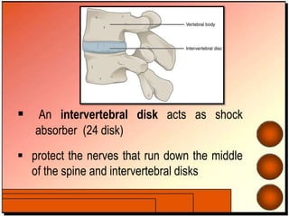
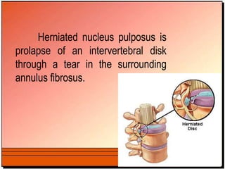
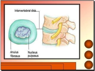
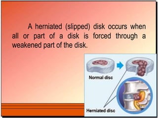
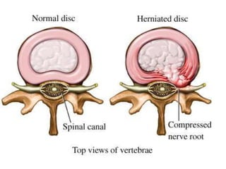
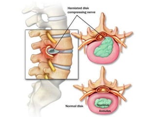
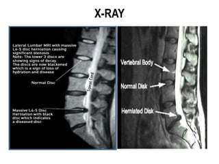
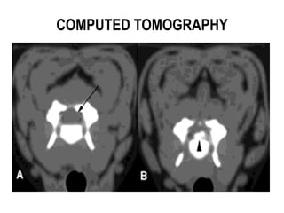
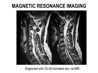
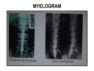
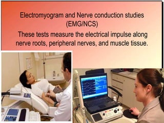
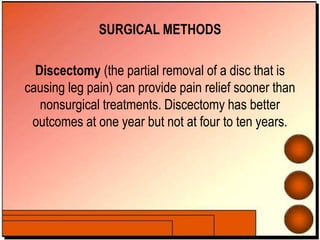
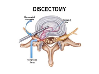
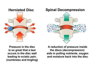
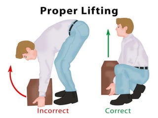
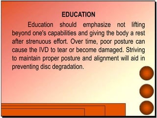
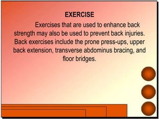
Prepared by: Roxanne Mae Birador SN
 An intervertebral disk acts as shock
absorber (24 disk)
 protect the nerves that run down the middle
of the spine and intervertebral disks
Herniated nucleus pulposus is
prolapse of an intervertebral disk
through a tear in the surrounding
annulus fibrosus.
A herniated (slipped) disk occurs when
all or part of a disk is forced through a
weakened part of the disk.
Alternative Names
Lumbar radiculopathy
Cervical radiculopathy
Herniated intervertebral disk/ disc
Prolapsed intervertebral disk/ disc
Slipped disk/ disc
Ruptured disk/ disc
deterioration and loss of function in the
cells of a tissue or organ
slipping forward
process of pushing out
abnormal separation
ETIOLOGY
Most disc herniations occur when a person is in
their 30’s or 40’s
After age 50 or 60, osteoarthritic degeneration
(spondylosis) or spinal stenosis are more likely
causes of low back pain or leg pain.
Then the disc does press on a nerve, symptoms
may include:
Pain that travels through the buttock and
down a leg to the ankle or foot because of
pressure on the sciatic nerve. Low back pain
(LUMBAGO) may accompany the leg pain.
Tingling ("pins-and-needles“ sensation) or
numbness in one leg that can begin in the buttock or
behind the knee and extend to the thigh, ankle, or
foot.
 Weakness in certain muscles in one or both legs.
 Pain in the front of the thigh.
 Severe deep muscle pain and muscle spasms.
Weakness in both legs and the loss of
bladder and/or bowel control are symptoms of
a specific and severe type of nerve root
compression called CAUDA EQUINA
SYNDROME.
If the herniated disc is in the lumbar region
the patient may also experience SCIATICA due
to irritation of one of the nerve roots of the sciatic
nerve.
-The lower back (lumbar area) of the spine
is the most common area affected by a
slipped disk.
-The neck (cervical area) disks are the
second most commonly affected area.
-The upper-to-mid-back (thoracic area)
disks are rarely involved.
 4.8% males and 2.5% females older than 35
experience sciatica during their lifetime.
 Of all individuals, 60% to 80% experience back
pain during their lifetime.
 In 14%, pain lasts more than 2 weeks.
 Generally, males have a slightly higher incidence
than females.
DIAGNOSIS
Physical examination - Straight Leg Raise
The straight leg raise, also called Lasègue's
sign, Lasègue test or Lazarević's sign, is a test done
during the physical examination to determine whether
a patient with low back pain has an underlying
herniated disk, often located at L5 (fifth lumbar spinal
nerve).
TECHNIQUE
With the patient lying down on his or her back on
an examination table or exam floor, the examiner
lifts the patient's leg while the knee is straight.
A variation is to lift the leg while the patient is
sitting. However, this reduces the sensitivity of the
test.
In order to make this test more specific, the ankle
can be dorsiflexed and the cervical spine flexed.
This increases the stretching of the nerve root and
dura.
Lasègue's sign was named after Charles
Lasègue (1816-1883).In 1864 Lasègue described
the signs of developing low back pain while
straightening the knee when the leg has already
been lifted.
CHARLES LASÈGUE
In 1880, Serbian doctor Laza Lazarević
described the straight leg raise test as it is used
today, so the sign is often named Lazarević's sign in
Serbia and some other countries.
LAZA LAZAREVIĆ
IMAGING
 X-ray
 Computed tomography
 Magnetic resonance imaging
 Myelogram
X-RAY
COMPUTED TOMOGRAPHY
MAGNETIC RESONANCE IMAGING
Diagnosed with C5-C6 herniated disc via MRI
MYELOGRAM
Electromyogram and Nerve conduction studies
(EMG/NCS)
These tests measure the electrical impulse along
nerve roots, peripheral nerves, and muscle tissue.
This will indicate whether there is ongoing
nerve damage, if the nerves are in a state of healing
from a past injury, or whether there is another site of
nerve compression. EMG/NCS studies are typically
used to pinpoint the sources of nerve dysfunction
distal to the spine.
The presence and severity of myelopathy
(known as (acute) spinal cord injury) can be
evaluated by means of Transcranial Magnetic
Stimulation (TMS).
A neurophysiological method that allows the
measurement of the time required for a neural
impulse to cross the pyramidal tracts, starting from
the cerebral cortex and ending at the anterior horn
cells of the cervical, thoracic or lumbar spinal cord.
This measurement is called Central Conduction
Time (CCT).
MEDICATIONS
 Acetaminophen (paracetamol)
 NSAIDs
 Muscle relaxants
 If the pain is still not managed adequately,
short term use of opioids such as morphine
may be useful
Antidepressants may be effective for treating
chronic pain associated with symptoms of
depression, but they have a risk of side effects.
Antiseizure drugs gabapentin and
carbamazepine are sometimes used for chronic
low back pain and may relieve sciatic pain, there
is insufficient evidence to support their use.
NON-SURGICAL METHODS
 Education on proper body mechanics
 Physical therapy, to address mechanical
factors, and may include modalities to
temporarily relieve pain (i.e. traction, electrical
stimulation, massage)
 Non-steroidal anti-inflammatory drugs
(NSAIDs)
 Weight control
 Spinal manipulation: Moderate quality evidence
suggests that spinal manipulation is more
effective than placebo for the treatment of acute
(less than 3 months duration) lumbar disc
herniation and acute sciatica.
Contraindication: Spinal manipulation is
contraindicated for disc herniations when there are
progressive neurological deficits such as with cauda
equina syndrome.
TRACTION BELT
PELVIC GIRDLE TRACTION
SURGICAL METHODS
Discectomy (the partial removal of a disc that is
causing leg pain) can provide pain relief sooner than
nonsurgical treatments. Discectomy has better
outcomes at one year but not at four to ten years.
DISCECTOMY
LUMBAR DISCECTOMY
NECK DISCECTOMY
The presence of cauda equina syndrome (in
which there is incontinence, weakness and genital
numbness) is considered a medical emergency
requiring immediate attention and possibly Surgical
Decompression.
PREVENTION
There are various causes for back injuries,
prevention must be comprehensive. Back injuries
are predominant in manual labor so the majority low
back pain prevention methods have been applied
primarily toward biomechanics. Prevention must
come from multiple sources such as education,
proper body mechanics, and physical fitness.
EDUCATION
Education should emphasize not lifting
beyond one's capabilities and giving the body a rest
after strenuous effort. Over time, poor posture can
cause the IVD to tear or become damaged. Striving
to maintain proper posture and alignment will aid in
preventing disc degradation.
EXERCISE
Exercises that are used to enhance back
strength may also be used to prevent back injuries.
Back exercises include the prone press-ups, upper
back extension, transverse abdominus bracing, and
floor bridges.
Other preventative measures are to lose
weight and to not work oneself past fatigue. Signs of
fatigue include shaking, poor coordination, muscle
burning and loss of the transverse abdominal brace.
Heavy lifting should be done with the legs
performing the work, and not the back.
Swimming is a common tool used in strength
training. The usage of lumbar sacral support belts
may restrict movement at the spine and support the
back during lifting.
Herniated Nucleus Pulposus

Herniated Nucleus Pulposus

  • 1.
    Prepared by: RoxanneMae Birador SN
  • 2.
     An intervertebraldisk acts as shock absorber (24 disk)  protect the nerves that run down the middle of the spine and intervertebral disks
  • 3.
    Herniated nucleus pulposusis prolapse of an intervertebral disk through a tear in the surrounding annulus fibrosus.
  • 5.
    A herniated (slipped)disk occurs when all or part of a disk is forced through a weakened part of the disk.
  • 6.
    Alternative Names Lumbar radiculopathy Cervicalradiculopathy Herniated intervertebral disk/ disc Prolapsed intervertebral disk/ disc Slipped disk/ disc Ruptured disk/ disc
  • 7.
    deterioration and lossof function in the cells of a tissue or organ slipping forward process of pushing out abnormal separation
  • 9.
    ETIOLOGY Most disc herniationsoccur when a person is in their 30’s or 40’s
  • 10.
    After age 50or 60, osteoarthritic degeneration (spondylosis) or spinal stenosis are more likely causes of low back pain or leg pain.
  • 13.
    Then the discdoes press on a nerve, symptoms may include: Pain that travels through the buttock and down a leg to the ankle or foot because of pressure on the sciatic nerve. Low back pain (LUMBAGO) may accompany the leg pain.
  • 16.
    Tingling ("pins-and-needles“ sensation)or numbness in one leg that can begin in the buttock or behind the knee and extend to the thigh, ankle, or foot.
  • 17.
     Weakness incertain muscles in one or both legs.  Pain in the front of the thigh.  Severe deep muscle pain and muscle spasms.
  • 18.
    Weakness in bothlegs and the loss of bladder and/or bowel control are symptoms of a specific and severe type of nerve root compression called CAUDA EQUINA SYNDROME.
  • 19.
    If the herniateddisc is in the lumbar region the patient may also experience SCIATICA due to irritation of one of the nerve roots of the sciatic nerve.
  • 22.
    -The lower back(lumbar area) of the spine is the most common area affected by a slipped disk. -The neck (cervical area) disks are the second most commonly affected area. -The upper-to-mid-back (thoracic area) disks are rarely involved.
  • 23.
     4.8% malesand 2.5% females older than 35 experience sciatica during their lifetime.  Of all individuals, 60% to 80% experience back pain during their lifetime.  In 14%, pain lasts more than 2 weeks.  Generally, males have a slightly higher incidence than females.
  • 24.
  • 25.
    The straight legraise, also called Lasègue's sign, Lasègue test or Lazarević's sign, is a test done during the physical examination to determine whether a patient with low back pain has an underlying herniated disk, often located at L5 (fifth lumbar spinal nerve).
  • 26.
    TECHNIQUE With the patientlying down on his or her back on an examination table or exam floor, the examiner lifts the patient's leg while the knee is straight. A variation is to lift the leg while the patient is sitting. However, this reduces the sensitivity of the test.
  • 27.
    In order tomake this test more specific, the ankle can be dorsiflexed and the cervical spine flexed. This increases the stretching of the nerve root and dura.
  • 28.
    Lasègue's sign wasnamed after Charles Lasègue (1816-1883).In 1864 Lasègue described the signs of developing low back pain while straightening the knee when the leg has already been lifted.
  • 29.
  • 30.
    In 1880, Serbiandoctor Laza Lazarević described the straight leg raise test as it is used today, so the sign is often named Lazarević's sign in Serbia and some other countries.
  • 31.
  • 32.
    IMAGING  X-ray  Computedtomography  Magnetic resonance imaging  Myelogram
  • 33.
  • 34.
  • 35.
    MAGNETIC RESONANCE IMAGING Diagnosedwith C5-C6 herniated disc via MRI
  • 36.
  • 37.
    Electromyogram and Nerveconduction studies (EMG/NCS) These tests measure the electrical impulse along nerve roots, peripheral nerves, and muscle tissue.
  • 38.
    This will indicatewhether there is ongoing nerve damage, if the nerves are in a state of healing from a past injury, or whether there is another site of nerve compression. EMG/NCS studies are typically used to pinpoint the sources of nerve dysfunction distal to the spine.
  • 39.
    The presence andseverity of myelopathy (known as (acute) spinal cord injury) can be evaluated by means of Transcranial Magnetic Stimulation (TMS).
  • 40.
    A neurophysiological methodthat allows the measurement of the time required for a neural impulse to cross the pyramidal tracts, starting from the cerebral cortex and ending at the anterior horn cells of the cervical, thoracic or lumbar spinal cord. This measurement is called Central Conduction Time (CCT).
  • 41.
    MEDICATIONS  Acetaminophen (paracetamol) NSAIDs  Muscle relaxants  If the pain is still not managed adequately, short term use of opioids such as morphine may be useful
  • 42.
    Antidepressants may beeffective for treating chronic pain associated with symptoms of depression, but they have a risk of side effects.
  • 43.
    Antiseizure drugs gabapentinand carbamazepine are sometimes used for chronic low back pain and may relieve sciatic pain, there is insufficient evidence to support their use.
  • 44.
    NON-SURGICAL METHODS  Educationon proper body mechanics  Physical therapy, to address mechanical factors, and may include modalities to temporarily relieve pain (i.e. traction, electrical stimulation, massage)  Non-steroidal anti-inflammatory drugs (NSAIDs)
  • 45.
     Weight control Spinal manipulation: Moderate quality evidence suggests that spinal manipulation is more effective than placebo for the treatment of acute (less than 3 months duration) lumbar disc herniation and acute sciatica.
  • 47.
    Contraindication: Spinal manipulationis contraindicated for disc herniations when there are progressive neurological deficits such as with cauda equina syndrome.
  • 48.
  • 49.
  • 50.
    SURGICAL METHODS Discectomy (thepartial removal of a disc that is causing leg pain) can provide pain relief sooner than nonsurgical treatments. Discectomy has better outcomes at one year but not at four to ten years.
  • 51.
  • 52.
  • 53.
  • 54.
    The presence ofcauda equina syndrome (in which there is incontinence, weakness and genital numbness) is considered a medical emergency requiring immediate attention and possibly Surgical Decompression.
  • 56.
    PREVENTION There are variouscauses for back injuries, prevention must be comprehensive. Back injuries are predominant in manual labor so the majority low back pain prevention methods have been applied primarily toward biomechanics. Prevention must come from multiple sources such as education, proper body mechanics, and physical fitness.
  • 58.
    EDUCATION Education should emphasizenot lifting beyond one's capabilities and giving the body a rest after strenuous effort. Over time, poor posture can cause the IVD to tear or become damaged. Striving to maintain proper posture and alignment will aid in preventing disc degradation.
  • 59.
    EXERCISE Exercises that areused to enhance back strength may also be used to prevent back injuries. Back exercises include the prone press-ups, upper back extension, transverse abdominus bracing, and floor bridges.
  • 60.
    Other preventative measuresare to lose weight and to not work oneself past fatigue. Signs of fatigue include shaking, poor coordination, muscle burning and loss of the transverse abdominal brace. Heavy lifting should be done with the legs performing the work, and not the back.
  • 61.
    Swimming is acommon tool used in strength training. The usage of lumbar sacral support belts may restrict movement at the spine and support the back during lifting.