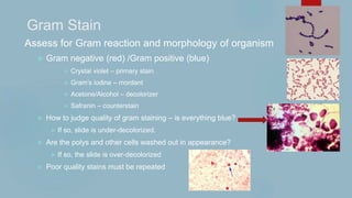
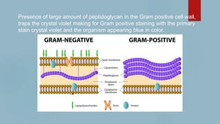
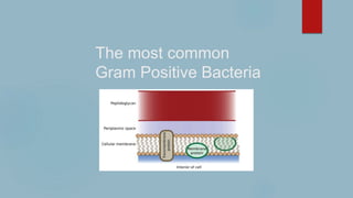
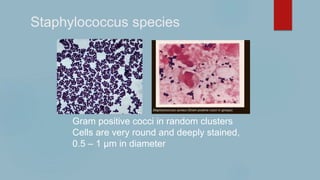
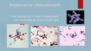
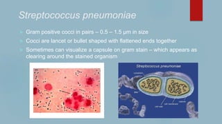
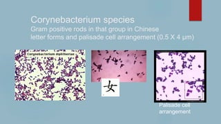
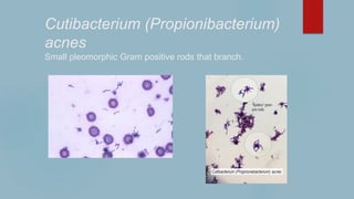
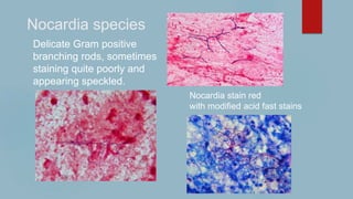
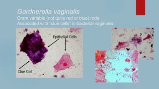
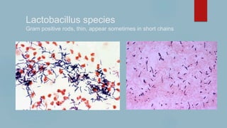
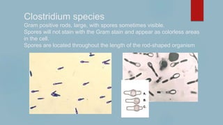
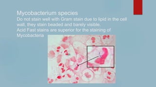
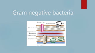
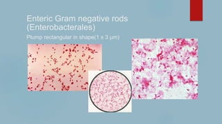
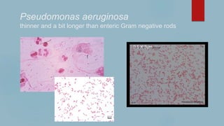
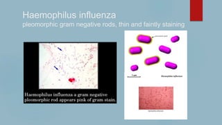
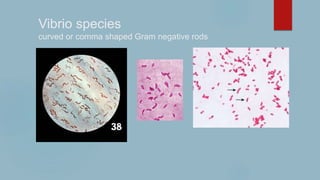
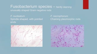
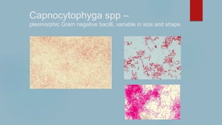
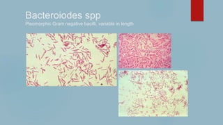
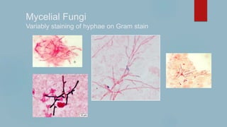
Advertisement
Advertisement

More Related Content

Advertisement
Advertisement

Gram Stains 2021

  1. Gram stains 2021 Margie A. Morgan, PhD. D(ABMM)
  2. Gram Stain Assess for Gram reaction and morphology of organism  Gram negative (red) /Gram positive (blue)  Crystal violet – primary stain  Gram’s iodine – mordant  Acetone/Alcohol – decolorizer  Safranin – counterstain  How to judge quality of gram staining – is everything blue?  If so, slide is under-decolorized.  Are the polys and other cells washed out in appearance?  If so, the slide is over-decolorized  Poor quality stains must be repeated
  3. Prepare a thin smear of material onto the glass slide. Air dry and then heat fix specimen on slide prior to staining. Stain approximately one minute per step and rinse with sterile water between each step. Air dry before scope reading.
  4. Presence of large amount of peptidoglycan in the Gram positive cell wall, traps the crystal violet making for Gram positive staining with the primary stain crystal violet and the organism appearing blue in color.
  5. Cellular Material – Gram stains are not able to differentiate many cell types. Best for identifying polymorphonuclear leukocytes (PMNs), mononuclear cells, and epithelial cells. For other cell types, differential stains must be used, such as Wright-Giemsa)
  6. Artifacts in Gram stains Safranin crystals Crystal Violet crystals When reading a Gram stain, check for the uniformity of the cell borders of suspected organisms, there should be repeating/consistent size and shape of the cells that is supportive of a micro-organism. Artifacts such as stain crystals are usually not consistent in shape or size.
  7. The most common Gram Positive Bacteria
  8. Staphylococcus species Gram positive cocci in random clusters Cells are very round and deeply stained, 0.5 – 1 µm in diameter
  9. Streptococcus / Beta hemolytic  Gram positive cocci in chains of variable length  Cocci are 0.5 wide by 2.0 µm in length (oval)
  10. Streptococcus pneumoniae  Gram positive cocci in pairs – 0.5 – 1.5 µm in size  Cocci are lancet or bullet shaped with flattened ends together  Sometimes can visualize a capsule on gram stain – which appears as clearing around the stained organism
  11. Viridans Streptococcus  Gram positive cocci in short chains and pairs  Chains are usually not as long as the Beta hemolytic Streptococcus, size is 0.5 µm – 1.5 µm.
  12. Enterococcus species  Gram positive cocci in short chains and pairs  Can be difficult to differentiate from viridans Streptococcus, size is also size is 0.5 µm – 1.5 µm.
  13. Bacillus species Gram positive rods – large (1.0 X 4.0 µm) Box car shaped, sometimes spores are visible. Spores will not stain with gentian violet. Can be over-decolorized and appear Gram negative on Gram stain Over-decolorized Spores
  14. Listeria monocytogenes Gram positive short rods (0.5 X 1.5 µm) Do not appear in chains and they do not branch.
  15. Corynebacterium species Gram positive rods in that group in Chinese letter forms and palisade cell arrangement (0.5 X 4 µm) Palisade cell arrangement
  16. Cutibacterium (Propionibacterium) acnes Small pleomorphic Gram positive rods that branch.
  17. Nocardia species Delicate Gram positive branching rods, sometimes staining quite poorly and appearing speckled. Nocardia stain red with modified acid fast stains
  18. Actinomyces species Gram positive rods with antler like branching, can be seen in sulfur granules in tissue Sulfur Granule Formation
  19. Gardnerella vaginalis Gram variable (not quite red or blue) rods Associated with “clue cells” in bacterial vaginosis
  20. Lactobacillus species Gram positive rods, thin, appear sometimes in short chains
  21. Clostridium species Gram positive rods, large, with spores sometimes visible. Spores will not stain with the Gram stain and appear as colorless areas in the cell. Spores are located throughout the length of the rod-shaped organism
  22. Mycobacterium species Do not stain well with Gram stain due to lipid in the cell wall, they stain beaded and barely visible. Acid Fast stains are superior for the staining of Mycobacteria
  23. Gram negative bacteria
  24. Enteric Gram negative rods (Enterobacterales) Plump rectangular in shape(1 x 3 µm)
  25. Pseudomonas aeruginosa thinner and a bit longer than enteric Gram negative rods 0.5 X 3µm
  26. Haemophilus influenza pleomorphic gram negative rods, thin and faintly staining
  27. Vibrio species curved or comma shaped Gram negative rods
  28. Fusobacterium species – faintly staining unusually shaped Gram negative rods F. nucleatum F. necrophorum Spindle shaped, with pointed Chaining pleomorphic rods ends
  29. Campylobacter sea gull shaped, faintly staining Gram negative rods
  30. Capnocytophyga spp – pleomorphic Gram negative bacilli, variable in size and shape
  31. Bacteroiodes spp Pleomorphic Gram negative bacilli, variable in length
  32. Yersinia pestis Safety pin like with bipolar staining ---
  33. (0.5 X 1.5 µm)
  34. Neisseria species (meningitidis and gonorrhoeae) Gram negative diplococci Kidney bean shaped in pairs
  35. Yeast – Candida species  Large Gram positive oval globules (@ 8 um) that produce a smaller attached cell known as a bud  Sometimes pseudo-hyphae can be seen which are extensions of the yeast cells into long hyphae like forms
  36. Cryptococcus neoformans/gatti Due to the polysaccharide capsule of C. neoformans/gatti the yeast appears somewhat like a round blob on Gram stain  An India ink prep can be used to visualize the polysaccharide capsule
  37. Mycelial Fungi Variably staining of hyphae on Gram stain
Advertisement