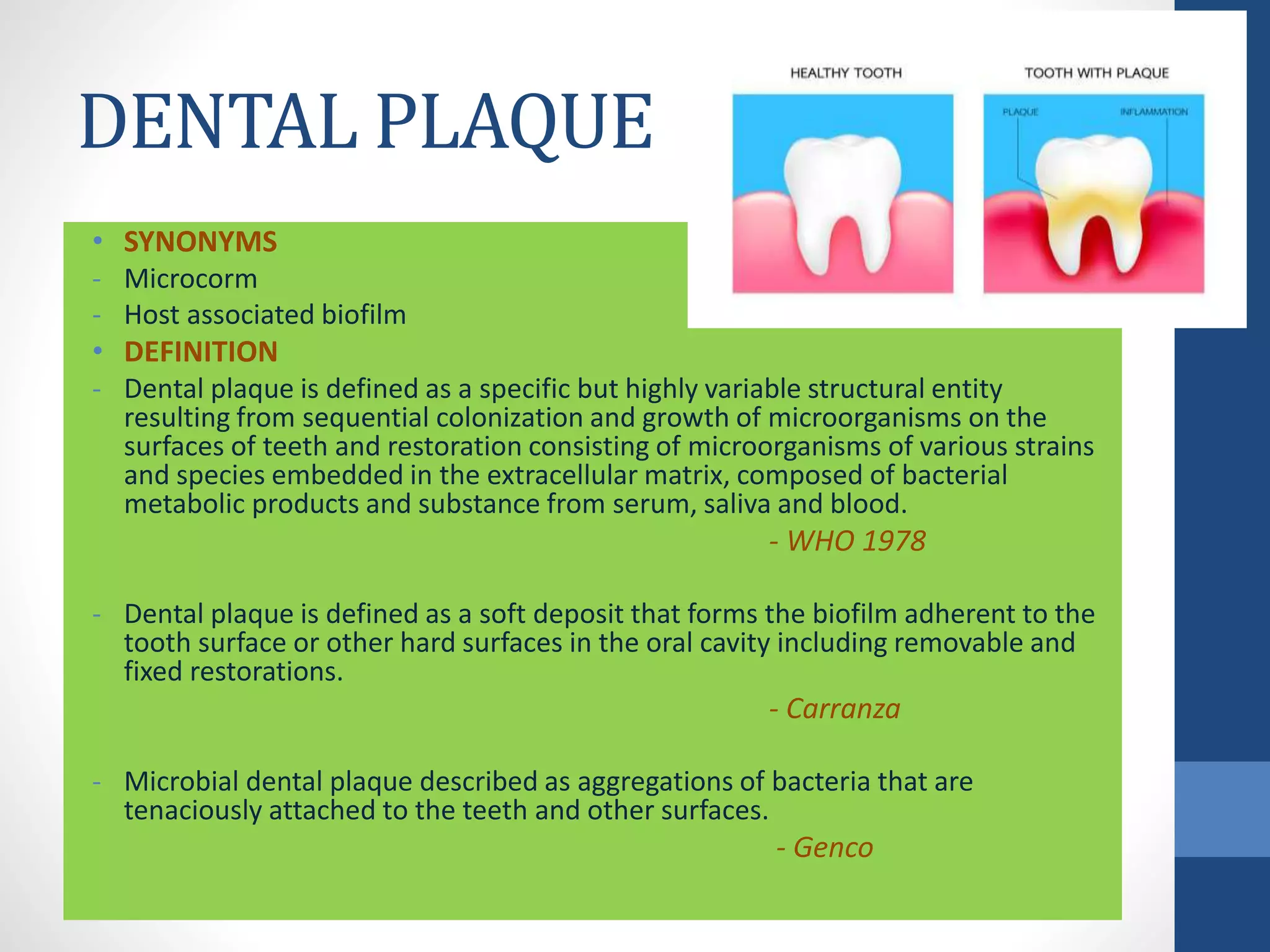
DENTAL PLAQUE
GUIDED BY: PRESENTED BY:
DR. PARUL ANEJA (READER) DR. MARIYAM MOMIN
II YEAR PG
DEPARTMENT OF PERIODONTOLOGY & ORAL IMPLANTOLOGY.
CONTENTS
• Biofilms
• Quorum sensing
• Horizontal gene transfer
• Tooth accumulated deposits
• Materia alba
• Dental pellicle
• Dental stains
• Dental plaque
• Classification of dental
plaque
• Composition of plaque
• Plaque formation
• Subgingival microbial
complexes
• Metabolism of dental
plaque bacteria
• Structure of dental
plaque
• Clinical assessment of
plaque
• Plaque hypothesis
BIOFILMS
• Biofilm is defined as matrix enclosed bacterial population
adherent to each other and/or to surface or interfaces -
Costerton
QUORUM SENSING
HORIZONTAL GENE
TRANSFER
General properties of biofilm &
microbial communities
S.No. General property Dental plaque example
1 Open architecture Presence of channel or pores
2 Broader habitat range Obligate anaerobes in an overtly aerobic environment
3 More efficient metabolism Complete catabolism of complete host macromolecule
(mucins) by consortium of food webs
4 Enhanced virulence Pathogenic synergism in abscess & periodontal disease
5 Protection from host defense
desiccation
Production of extracellular matrix to form a functional
matrix
6 Neutralization of inhibitors Beta lactamases production by neighboring cells to
protect sensitive organism
7 Novel gene expression Synthesis of novel proteins upon attachment
8 Co-coordinated gene response Production of cell to cell signaling molecules
9 Cell to cell signaling Production of CSP, AI2 etc. cross talk with host epithelial
cells
10 Spatial &environmental
heterogenticity
pH & oxygen gradients; co-adhesion.
TOOTH ACCUMULATED
DEPOSITS
• DEVELOPMENTAL ORIGIN
1. Dental cuticle
2. Coronal cementum
3. Sub-surface enamel matrix
4. Reduced enamel epithelium
• ACQUIRED COATINGS
1. Salivary pellicle
2. Bacterial coating – plaque &
materia alba
3. Sub-surface pellicle
4. Surface stains
MATERIA ALBA
• Materia alba refers to the soft accumulation of bacteria &
tissue cells that lack the organized structure of dental
plaque and are easily displaced with a water spray –
Carranza
• COMPOSITION
- Food debris
- Dead tissue elements
- Purulent material
- Mixture of salivary proteins & lipids
• CONSISTENCY : soft & sticky
• SIGNIFICANCE : ideal growth medium for bacteria which can
induce gingival inflammation.
DENTAL PELLICLE
• SYNONYM – Acquired pellicle
• DEFINITION
- Pellicle is defined as tooth or mucosal adherent salivary protein -
PLR
• TYPES
1. Unstained pellicle
2. Stained pellicle
3. Sub-surface pellicle
• COMPOSITION
1. Glycoproteins
2. Phosphoproteins
3. Proline rich proteins
4. Histidine rich proteins
5. Enzymes - amylase
• FORMATION
Components of
GCF & saliva
(+ve)
Hydroxyapatite
on the enamel
surface
(-ve)
Pellicle
+
Difference between Food
debris, Materia alba & Plaque
S.No. Feature Food debris Materia alba Plaque
1 Structure No structure Amorphous Definite
structure
2 Adherence None Loose Close
3 Effect of rinsing Dislodge
readily
Dislodge by
forceful
rinsing
Do not
dislodge
Food
debris
Materia
alba
Plaque
DENTAL STAINS
• They are the pigmented deposits on the tooth surface.
Green stains:
- Gingival 3rd of facial
surface of maxillary
anterior teeth.
- Penicillium & aspergillus
- Common in children
(boys).
Black stains:
- Thin black line on the facial
& lingual surfaces of teeth
near gingival margin in
people with good oral
hygiene
- Actinomyces species
- Common in women.
Brown stains:
- Buccal surface of maxillary molars
& lingual surface of mandibular
incisors.
- Presence of tanin
- Thin & translucent Acquired
pigmented pellicle which is free
from bacteria.
- Common in people with poor oral
hygiene.
Metallic stains:
- Bismuth pigmentation : black line
- Arsenic pigmenation: black line
- Mercury pigmentation: black line
- Lead pigmentation: bluish red,
deep blue or grey (burtonian
line)
- Silver pigmentation : Violet
marginal line
Tobacco stains:
- Lingual and palatal surfaces
- Common in pits, fissures &
other enamel irregularities
- Tobacco stain is directly
proprtional to no. of
cigarettes smoked per day
Chlorhexidine stains:
- Frequent use of
chlorhexidine leads to dark
yellow to brownish stain
- Artificial as well as natural
teeth are affected
- At higher concentrations
stains are more severe.
Orange stains:
- Facial & lingual surfaces
of anterior teeth
- Serratia marcescens &
Flavobacterium
lutescens
- Rare stain
DENTAL PLAQUE
• SYNONYMS
- Microcorm
- Host associated biofilm
• DEFINITION
- Dental plaque is defined as a specific but highly variable structural entity
resulting from sequential colonization and growth of microorganisms on the
surfaces of teeth and restoration consisting of microorganisms of various strains
and species embedded in the extracellular matrix, composed of bacterial
metabolic products and substance from serum, saliva and blood.
- WHO 1978
- Dental plaque is defined as a soft deposit that forms the biofilm adherent to the
tooth surface or other hard surfaces in the oral cavity including removable and
fixed restorations.
- Carranza
- Microbial dental plaque described as aggregations of bacteria that are
tenaciously attached to the teeth and other surfaces.
- Genco
PLAQUE RETENTIVE AREAS
• NATURAL AREAS / FACTORS
 Supra-gingival
1. Carious lesions
2. Exposed cementum
3. Supra-gingival calculus
 Sub-gingival
1. sub-gingival calculus
2. Cavitated caries lesions
3. Furcation involvement
4. Cemento-enamel junction
5. Rough, unplaned cementum
6. Root grooved & enamel projections
7. Deep & narrow infrabony pockets.
• IATROGENIC FACTORS
1. Orthodontic bands
2. Overhanging restorative margins
3. Overcontoured & inadequate crown
margins
4. Portions of removable prosthesis that
impinge on gingiva.
CLASSIFICATION OF PLAQUE
Dental
plaque
Supra-
gingival
Coronal Marginal
Sub-gingival
Attached
Tooth
associated
Tissue
associated
Peri-implant
plaque
Unattached
Differencebetweensupra-gingival& sub-
gingivalplaque
Differencebetweentoothassociatedandtissue
associatedsubgingivalplaque
S.N
o.
Tooth associated
subgingival plaque
Tissue associated
subgingival plaque
1 Gram +ve bacteria
predominates
Gram +ve and gram –ve
bacteria
2 Does not extend to
junctional epithelium
Extends to junctional
epithelium
3 May penetrate
cementum
May penetrate
epithelium &
connective tissue
4 Microorganisms:
S.sanguinis, S. mitis,
A. naeslundii, A.
viscosus,
Eubacterium
Microorganisms:
Staphylococcus
intermedius, S.oralis,
P.micros, P. gingivalis, P.
intermedia,
F.nucleatum, T.
forsythia.
5 Plays a role in calculus
formation & root
caries
Plays a role in gingivitis
& periodontitis.
Differencebetweenearly plaque& matured
plaque
COMPOSITION OF DENTAL
PLAQUE
PLAQUE FORMATION
Steps in plaque formation:
1. Formation of pellicle on the tooth surface
2. Initial adhesion/ attachment of bacteria
3. Colonization/ plaque maturation.
SUBGINGIVAL MICROBIAL
COMPLEXES
METABOLISM OF DENTAL
PLAQUE BACTERIA
STRUCTURE OF PLAQUE
Supragingival plaque Subgingival plaque
Corncob formation Test tube-brush formation
FACTORS AFFECTING SUPRAGINGIVAL
PLAQUE FORMATION
MICROBIAL SHIFT DURING
PERIODONTAL DISEASES
S. No. Shift from Shift to
1 Gram positive Gram negative
2 Cocci Rods & spirochetes
3 Non-motile Motile
4 Facultative anaerobes Obligate anaerobes
5 Fermenting Proteolytic species
CLINICAL ASSESSMENT OF
DENTAL PLAQUE
• Direct vision
• Use of
explorers
• Disclosing
solutions
• Clinical
records
Criteria for identificationof
periodontopathogens
Koch’s postulates (1870s)
• It must be routinely
isolated from diseased
individuals.
• It must be grown in pure
culture in the laboratory.
• It must produce a similar
disease when inoculated
into a susceptible host.
• It must be recovered from
lesions in a diseased
laboratory animal.
Limitations in periodontal
microbiology
• The inability to culture all
the organisms
associated with the
disease.
• The difficulties inherent
in defining and culturing
sites of the active
disease.
• Lack of good animal
model system for the
study of periodontitis.
SOCRANSKY’S POSTULATES
• Must be associated with the disease, as evident by
increase in the number of organisms at diseased
sites.
• Must be eliminated or decreased in sites that
demonstrate clinical resolution of disease with the
treatment.
• Must demonstrate a host response, in the form of
an alteration in the host cellular or humoral immune
response.
• Must be capable of causing disease in experimental
animal models.
• Must demonstrate virulence factors responsible for
enabling the microorganism to cause destruction of
the periodontal tissues.
CLINICAL SIGNIFICANCE OF
PLAQUE
PLAQUE IN INITIATION OF PERIODONTAL DISEASES
Noxious products
release from plaque
bacteria
Release of gingipains
from P. gingivalis
Destruction of host
collagen
Direct destruction of
host soft tissue and
hard tissues.
Release of endotoxins
from plaque bacteria
Stimulation of host
immune and
inflammatory systems
Stimulation of IL &
TNF
Destruction of host
tissue.
Direct effect of plaque Indirect effect of plaque
Role of bacteria in periodontal diseases
/ etiological significance of dental plaque
• Plaque vs gingivitis
• Plaque vs chronic periodontitis
• Plaque vs LAP
• Plaque Vs necrotizing periodontal diseases
• Abscesses of periodontium
• Periimplantitis
MICROBIAL FLORA IN PERIODONTAL
HEALTH & DISEASES
PERIODONTAL HEALTH
• S. sanguis
• S. mitis
• Veillonella
• Streptococcus
• Capnocytophaga
GINGIVITIS
• Gram +ve species
- S. mitis
- S. oralis
- S. sanguis
- A. viscosus
- Peptostreptococcus micros
• Gram –ve species
- F. nucleatum
- P. intermedia
- Hemophilus
- Capnocytophaga
- Campylobacter species
PREGNANCY ASSOCIATED
GINGIVITIS
Prevotella intermedia
CHRONIC
PERIODONTITIS
• P. gingivalis
• B. forsythus
• P. intermedia
• C. rectus
• E. corrodens
• F. nucleatum
• A. actinomycetum
comitans
REFRACTORY
PERIODONTITIS
• A. actinomycetum
comitans
• P. gingivalis
• P. intermedia
• Bacteroides forsythus
NECROTIZING ULCERATIVE
PERIODONTITIS
- Spirochetes
- P. intermedia
Abscesses of the
periodontium
- P. intermedia
- P. gingivalis
- Bacteroides forsythus
- Peptostreptococcus micros
- Fusobacterium nucleatum
LOCALIZED
AGGRESSIVE
PERIODONTITIS
• A. actinomycetum
comitans
• C. rectus
• P. gingivalis
• F. nucleatum
• Capnocytophaga
• E. corrodens
• Bacteroides capillus
GENERALIZED
AGGRESSIVE
PERIODONTITIS
• P. gingivalis
• A. actinomycetum
comitans
• P. intermedia
• E. corrodens
• Capnocytophaga
• Nisseria
PLAQUE HYPOTHESIS
ECOLOGICAL PLQUE
HYPOTHESIS
References
• Newman & Carranza’s Clinical Periodontology-(13th edition)
• Textbook of Periodontics – (Shalu Bathla 2nd edition)
• Essentials of Periodontology – (Sahitya Reddy S)
Dental plaque

Dental plaque

  • 1.
    DENTAL PLAQUE GUIDED BY:PRESENTED BY: DR. PARUL ANEJA (READER) DR. MARIYAM MOMIN II YEAR PG DEPARTMENT OF PERIODONTOLOGY & ORAL IMPLANTOLOGY.
  • 2.
    CONTENTS • Biofilms • Quorumsensing • Horizontal gene transfer • Tooth accumulated deposits • Materia alba • Dental pellicle • Dental stains • Dental plaque • Classification of dental plaque • Composition of plaque • Plaque formation • Subgingival microbial complexes • Metabolism of dental plaque bacteria • Structure of dental plaque • Clinical assessment of plaque • Plaque hypothesis
  • 3.
    BIOFILMS • Biofilm isdefined as matrix enclosed bacterial population adherent to each other and/or to surface or interfaces - Costerton
  • 4.
  • 5.
  • 6.
    General properties ofbiofilm & microbial communities S.No. General property Dental plaque example 1 Open architecture Presence of channel or pores 2 Broader habitat range Obligate anaerobes in an overtly aerobic environment 3 More efficient metabolism Complete catabolism of complete host macromolecule (mucins) by consortium of food webs 4 Enhanced virulence Pathogenic synergism in abscess & periodontal disease 5 Protection from host defense desiccation Production of extracellular matrix to form a functional matrix 6 Neutralization of inhibitors Beta lactamases production by neighboring cells to protect sensitive organism 7 Novel gene expression Synthesis of novel proteins upon attachment 8 Co-coordinated gene response Production of cell to cell signaling molecules 9 Cell to cell signaling Production of CSP, AI2 etc. cross talk with host epithelial cells 10 Spatial &environmental heterogenticity pH & oxygen gradients; co-adhesion.
  • 7.
    TOOTH ACCUMULATED DEPOSITS • DEVELOPMENTALORIGIN 1. Dental cuticle 2. Coronal cementum 3. Sub-surface enamel matrix 4. Reduced enamel epithelium • ACQUIRED COATINGS 1. Salivary pellicle 2. Bacterial coating – plaque & materia alba 3. Sub-surface pellicle 4. Surface stains
  • 8.
    MATERIA ALBA • Materiaalba refers to the soft accumulation of bacteria & tissue cells that lack the organized structure of dental plaque and are easily displaced with a water spray – Carranza • COMPOSITION - Food debris - Dead tissue elements - Purulent material - Mixture of salivary proteins & lipids • CONSISTENCY : soft & sticky • SIGNIFICANCE : ideal growth medium for bacteria which can induce gingival inflammation.
  • 9.
    DENTAL PELLICLE • SYNONYM– Acquired pellicle • DEFINITION - Pellicle is defined as tooth or mucosal adherent salivary protein - PLR • TYPES 1. Unstained pellicle 2. Stained pellicle 3. Sub-surface pellicle • COMPOSITION 1. Glycoproteins 2. Phosphoproteins 3. Proline rich proteins 4. Histidine rich proteins 5. Enzymes - amylase • FORMATION Components of GCF & saliva (+ve) Hydroxyapatite on the enamel surface (-ve) Pellicle +
  • 10.
    Difference between Food debris,Materia alba & Plaque S.No. Feature Food debris Materia alba Plaque 1 Structure No structure Amorphous Definite structure 2 Adherence None Loose Close 3 Effect of rinsing Dislodge readily Dislodge by forceful rinsing Do not dislodge Food debris Materia alba Plaque
  • 11.
    DENTAL STAINS • Theyare the pigmented deposits on the tooth surface. Green stains: - Gingival 3rd of facial surface of maxillary anterior teeth. - Penicillium & aspergillus - Common in children (boys). Black stains: - Thin black line on the facial & lingual surfaces of teeth near gingival margin in people with good oral hygiene - Actinomyces species - Common in women. Brown stains: - Buccal surface of maxillary molars & lingual surface of mandibular incisors. - Presence of tanin - Thin & translucent Acquired pigmented pellicle which is free from bacteria. - Common in people with poor oral hygiene. Metallic stains: - Bismuth pigmentation : black line - Arsenic pigmenation: black line - Mercury pigmentation: black line - Lead pigmentation: bluish red, deep blue or grey (burtonian line) - Silver pigmentation : Violet marginal line Tobacco stains: - Lingual and palatal surfaces - Common in pits, fissures & other enamel irregularities - Tobacco stain is directly proprtional to no. of cigarettes smoked per day Chlorhexidine stains: - Frequent use of chlorhexidine leads to dark yellow to brownish stain - Artificial as well as natural teeth are affected - At higher concentrations stains are more severe. Orange stains: - Facial & lingual surfaces of anterior teeth - Serratia marcescens & Flavobacterium lutescens - Rare stain
  • 12.
    DENTAL PLAQUE • SYNONYMS -Microcorm - Host associated biofilm • DEFINITION - Dental plaque is defined as a specific but highly variable structural entity resulting from sequential colonization and growth of microorganisms on the surfaces of teeth and restoration consisting of microorganisms of various strains and species embedded in the extracellular matrix, composed of bacterial metabolic products and substance from serum, saliva and blood. - WHO 1978 - Dental plaque is defined as a soft deposit that forms the biofilm adherent to the tooth surface or other hard surfaces in the oral cavity including removable and fixed restorations. - Carranza - Microbial dental plaque described as aggregations of bacteria that are tenaciously attached to the teeth and other surfaces. - Genco
  • 13.
    PLAQUE RETENTIVE AREAS •NATURAL AREAS / FACTORS  Supra-gingival 1. Carious lesions 2. Exposed cementum 3. Supra-gingival calculus  Sub-gingival 1. sub-gingival calculus 2. Cavitated caries lesions 3. Furcation involvement 4. Cemento-enamel junction 5. Rough, unplaned cementum 6. Root grooved & enamel projections 7. Deep & narrow infrabony pockets. • IATROGENIC FACTORS 1. Orthodontic bands 2. Overhanging restorative margins 3. Overcontoured & inadequate crown margins 4. Portions of removable prosthesis that impinge on gingiva.
  • 14.
    CLASSIFICATION OF PLAQUE Dental plaque Supra- gingival CoronalMarginal Sub-gingival Attached Tooth associated Tissue associated Peri-implant plaque Unattached
  • 15.
  • 16.
    Differencebetweentoothassociatedandtissue associatedsubgingivalplaque S.N o. Tooth associated subgingival plaque Tissueassociated subgingival plaque 1 Gram +ve bacteria predominates Gram +ve and gram –ve bacteria 2 Does not extend to junctional epithelium Extends to junctional epithelium 3 May penetrate cementum May penetrate epithelium & connective tissue 4 Microorganisms: S.sanguinis, S. mitis, A. naeslundii, A. viscosus, Eubacterium Microorganisms: Staphylococcus intermedius, S.oralis, P.micros, P. gingivalis, P. intermedia, F.nucleatum, T. forsythia. 5 Plays a role in calculus formation & root caries Plays a role in gingivitis & periodontitis.
  • 17.
  • 18.
  • 19.
    PLAQUE FORMATION Steps inplaque formation: 1. Formation of pellicle on the tooth surface 2. Initial adhesion/ attachment of bacteria 3. Colonization/ plaque maturation.
  • 21.
  • 22.
  • 23.
    STRUCTURE OF PLAQUE Supragingivalplaque Subgingival plaque Corncob formation Test tube-brush formation
  • 24.
  • 25.
    MICROBIAL SHIFT DURING PERIODONTALDISEASES S. No. Shift from Shift to 1 Gram positive Gram negative 2 Cocci Rods & spirochetes 3 Non-motile Motile 4 Facultative anaerobes Obligate anaerobes 5 Fermenting Proteolytic species
  • 26.
    CLINICAL ASSESSMENT OF DENTALPLAQUE • Direct vision • Use of explorers • Disclosing solutions • Clinical records
  • 27.
    Criteria for identificationof periodontopathogens Koch’spostulates (1870s) • It must be routinely isolated from diseased individuals. • It must be grown in pure culture in the laboratory. • It must produce a similar disease when inoculated into a susceptible host. • It must be recovered from lesions in a diseased laboratory animal. Limitations in periodontal microbiology • The inability to culture all the organisms associated with the disease. • The difficulties inherent in defining and culturing sites of the active disease. • Lack of good animal model system for the study of periodontitis.
  • 28.
    SOCRANSKY’S POSTULATES • Mustbe associated with the disease, as evident by increase in the number of organisms at diseased sites. • Must be eliminated or decreased in sites that demonstrate clinical resolution of disease with the treatment. • Must demonstrate a host response, in the form of an alteration in the host cellular or humoral immune response. • Must be capable of causing disease in experimental animal models. • Must demonstrate virulence factors responsible for enabling the microorganism to cause destruction of the periodontal tissues.
  • 29.
    CLINICAL SIGNIFICANCE OF PLAQUE PLAQUEIN INITIATION OF PERIODONTAL DISEASES Noxious products release from plaque bacteria Release of gingipains from P. gingivalis Destruction of host collagen Direct destruction of host soft tissue and hard tissues. Release of endotoxins from plaque bacteria Stimulation of host immune and inflammatory systems Stimulation of IL & TNF Destruction of host tissue. Direct effect of plaque Indirect effect of plaque
  • 30.
    Role of bacteriain periodontal diseases / etiological significance of dental plaque • Plaque vs gingivitis • Plaque vs chronic periodontitis • Plaque vs LAP • Plaque Vs necrotizing periodontal diseases • Abscesses of periodontium • Periimplantitis
  • 31.
    MICROBIAL FLORA INPERIODONTAL HEALTH & DISEASES PERIODONTAL HEALTH • S. sanguis • S. mitis • Veillonella • Streptococcus • Capnocytophaga GINGIVITIS • Gram +ve species - S. mitis - S. oralis - S. sanguis - A. viscosus - Peptostreptococcus micros • Gram –ve species - F. nucleatum - P. intermedia - Hemophilus - Capnocytophaga - Campylobacter species PREGNANCY ASSOCIATED GINGIVITIS Prevotella intermedia
  • 32.
    CHRONIC PERIODONTITIS • P. gingivalis •B. forsythus • P. intermedia • C. rectus • E. corrodens • F. nucleatum • A. actinomycetum comitans REFRACTORY PERIODONTITIS • A. actinomycetum comitans • P. gingivalis • P. intermedia • Bacteroides forsythus NECROTIZING ULCERATIVE PERIODONTITIS - Spirochetes - P. intermedia Abscesses of the periodontium - P. intermedia - P. gingivalis - Bacteroides forsythus - Peptostreptococcus micros - Fusobacterium nucleatum
  • 33.
    LOCALIZED AGGRESSIVE PERIODONTITIS • A. actinomycetum comitans •C. rectus • P. gingivalis • F. nucleatum • Capnocytophaga • E. corrodens • Bacteroides capillus GENERALIZED AGGRESSIVE PERIODONTITIS • P. gingivalis • A. actinomycetum comitans • P. intermedia • E. corrodens • Capnocytophaga • Nisseria
  • 34.
  • 35.
  • 36.
    References • Newman &Carranza’s Clinical Periodontology-(13th edition) • Textbook of Periodontics – (Shalu Bathla 2nd edition) • Essentials of Periodontology – (Sahitya Reddy S)