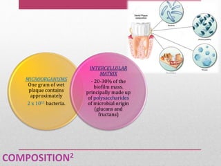
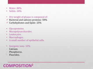
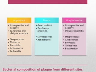
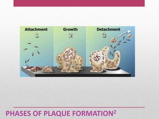
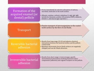
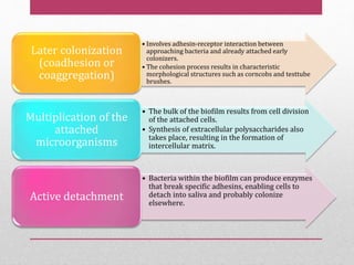
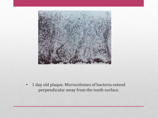
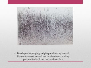
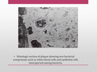
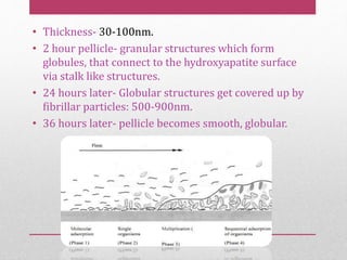
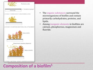
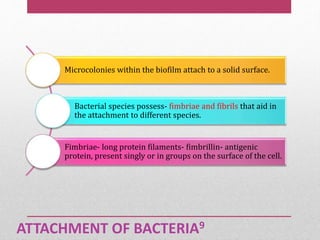
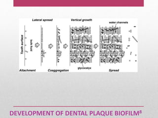
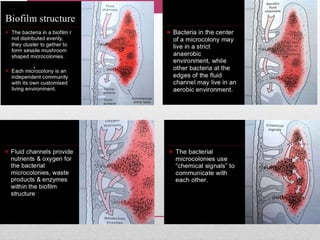
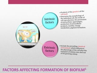
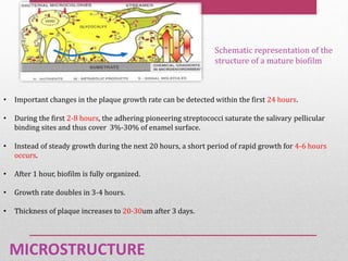
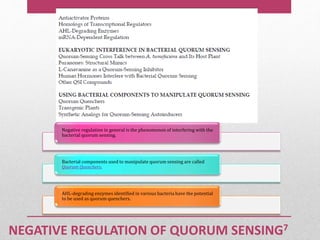
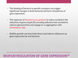
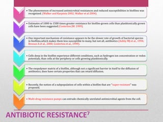
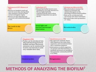
DENTAL
PLAQUE
Presented by:
Dr. Sucheta Prabhu
II MDS
Department of Pedodontics and Preventive
Dentistry
Table of contents• Introduction
• Definition
• History
• Terminology
• Detection of dental plaque
• Classification
• Timeline of plaque development
• Composition of plaque
• Phases of plaque formation
• Dental plaque as a biofilm
• Plaque hypothesis
• Microbial interactions
• Biofilms and infectious disease
• Management protocol
INTRODUCTION1
• Bacteria found in the saliva can be observed as planktonic
bacteria (i.e. single floating bacteria in a liquid phase).
• However bacteria found on the surface of hard structures
such as teeth, restorations, prostheses and implants form
an adherent gelatinous film called dental plaque (Fine
1988).
• Dental plaque, an adherent, bacterial biofilm that forms on
all hard and soft tissue, is the principal aetiologic agent in
caries and periodontal diseases (Fine 1988).
DEFINITION
Dental plaque is defined as a specific but highly variable structural
entity resulting from sequential colonization and growth of
microorganisms on the surface of teeth and restoration consisting of
microorganisms of various strains and species embedded in the
extracellular matrix, composed of bacterial metabolic products and
substance from serum, saliva and blood.
- WHO(1978)
DEFINITION
• Dental plaque is defined clinically as a structured, resilient,
yellowish grayish substance that adheres tenaciously to
intraoral hard surfaces, including removable or fixed
restorations.
- Bowen(1976)
• Dental plaque is a general term for complex microbial
community that develops on the tooth surface, embedded in a
matrix of polymers of bacterial and salivary origin.
- Marsh
• Dental plaque can be defined as the soft deposits that form the
biofilm adhering to the tooth surface or other hard surfaces in
the oral cavity, including removable and fixed restorations.
- Carranza
• Plaque can be defined as a complex microbial
community, with greater than 1010 bacteria per
milligram.
- Socransky (1998)
• It is a soft amorphous granular deposits which
accumulate on surface of teeth, dental restoration and
dental calculus.
- Glickman
DEFINITION
HISTORY
• J Leon Williams (1897)- described “dental plaque”.
• G. V. Black (1899)- coined the term “Gelatinous dental
plaque”.
• Waerhaung (1950)- described the importance of
bacterial plaque in the etiology of dental disease.
TERMINOLOGY11
• Materia Alba- refers to soft accumulations of bacteria, food
matter, and tissue cells that lack the organized structure of
dental plaque and are easily displaced with a water spray.
• Calculus- is a hard deposit that forms by mineralization of
dental plaque and is generally covered by a layer of
unmineralized plaque.
• Biofilm: it is defined as a microbially derived sessile
community characterized by cells that are irreversibly
attached to a substratum or interface or to each other, are
embedded in a matrix of extracellular polymeric substances
that they have produced, and exhibit an altered phenotype
with respect to growth rate and gene transcription. (Donla
and Costerton 2002)
• Acquired pellicle- may be defined as a homogenous,
membranous, acellular film that covers the tooth surface and
frequently form on the interface between the tooth, the dental
plaque and calculus.
TERMINOLOGY
DETECTION
• Direct vision:
 Plaque is whitish yellow.
 Identification of the dental plaque is difficult because of the color similarity between
the tooth surface and dental plaque.
 It can be readily seen on the teeth after 1-2 days of uninterrupted plaque formation
with no oral hygiene measures.
 Thin plaque- translucent- not visible.
 Stained plaque- may be acquired.(eg tobacco)
 Thick plaque- tooth appears dull and dirty.
DETECTION
• By instrumentation:
 By using an explorer or a periodontal probe.
 This is done by running the explorer or probe along the
gingival third of the tooth.
 When plaque is present it will adhere to the tip of the
explorer.
 Slippery due to coating of soft slimy layer.
 When calcification has started, appears slightly rough.
• Disclosing agents:
 Dental plaque has the ability to retain a large number of
dye substances because of the polarity difference between
the components of the plaque and the dyes.
 Disclosing dyes work by changing the color of dental
plaque so that it contrasts with the tooth surface.
 The first chemical reported to stain plaque was iodine but
now a days many dyes have been used as disclosing agents.
DETECTION
CLASSIFICATION12
Essentials of clinical periodontology and periodontics.Shantipriya reddy. 4th ed.
Sr. no Feature Supragingival
plaque
Subgingival
plaque
1 Matrix 50% matrix Little or no
matrix
2 Flora Mostly gram
positive
Mostly gram
negative
3 Motile bacteria Few Common
4 Anaerobic/ aerobic Aerobic unless
thick
Highly aerobic
areas present
5 Metabolism Predominantly
carbohydrates
Predominantly
proteins
Differences in supragingival and subgingival plaque
TIMELINE11
At birth
• sterile
Hours
• Facultative, aerobic bacteria
Second
day
• anaerobic bacteria
2 weeks
• mature microbiota
Weaning
(> 2years)
• 400 different types of bacteria
Niche of plaque accumulation11:
• Intraoral, supragingival hard surfaces (teeth, implant,
restorations, prosthesis).
• Subgingival regions- Periodontal pocket (hard: root
cementum, soft: pocket epithelium).
• Buccal, palatal and floor of the mouth epithelium.
• Dorsum of the tongue.
• Tonsils.
COMPOSITION2
MICROORGANISMS
One gram of wet
plaque contains
approximately
2 x 10¹¹ bacteria.
INTERCELLULAR
MATRIX
- 20-30% of the
biofilm mass.
principally made up
of polysaccharides
of microbial origin
(glucans and
fructans)
COMPOSITION2
• Water: 80%
• Solids- 20%
• Dry weight of plaque is composed of:
 Bacterial and salivary proteins- 50%
 Carbohydrates and lipids- 25%
• Glycoproteins.
• Mucopolysaccharides.
• Leukocytes.
• Macrophages.
• A small number of epithelial cells.
• Inorganic ions- 10%
- Calcium.
- Phosphorus.
- Fluorides.
Bacterial composition of plaque from different sites.
Approximal
• Gram positive and
negative;
• Facultative and
obligate anaerobs.
• Streptococcus
• Neisseria
• Prevotella
• Actinomyces
• Veillonella
Fissure
• Gram positive;
• Facultative
anaerobs.
• Streptococcus
• Actinomyces
Gingival crevice
• Gram positive and
negative;
• Obligate anaerobs.
• Streptococcus
• Actinomyces
• Prevotella
• Treponema
• Eubacterium
PHASES OF PLAQUE FORMATION2
• Forms immediately by selective adsorption of salivary,
microbial, molecules to the tooth surface.
• Albumin, amylase, carbonic anhydrase II, sIgA, IgG, IgM,
lactoferrin, lysozyme, proline-rich proteins (PRP), statherin,
histatin 1, and mucous gly-coprotein 1.
Formation of the
acquired enamel (or
dental) pellicle
• Passive transport of microorganisms to the coated
tooth surface by the flow of oral fluids.
Transport
• Results from long-range (10-20 nm) physico-chemical
interactions between the bacterial surface and the pellicle-
coated tooth.
• Repulsive electrostatic forces (both surfaces are negatively
charged) and van der Waals attraction.
Reversible bacterial
adhesion
• Results from short-range (<1nm) stronger, specific
stereochemical interactions involving bacterial surface
components (adhesins) and cognate receptors on the pellicle.Irreversible bacterial
adhesion
• Involves adhesin-receptor interaction between
approaching bacteria and already attached early
colonizers.
• The cohesion process results in characteristic
morphological structures such as corncobs and testtube
brushes.
Later colonization
(coadhesion or
coaggregation)
• The bulk of the biofilm results from cell division
of the attached cells.
• Synthesis of extracellular polysaccharides also
takes place, resulting in the formation of
intercellular matrix.
Multiplication of the
attached
microorganisms
• Bacteria within the biofilm can produce enzymes
that break specific adhesins, enabling cells to
detach into saliva and probably colonize
elsewhere.
Active detachment
• 1 day old plaque. Microcolonies of bacteria extend
perpendicular away from the tooth surface.
• Developed supragingival plaque showing overall
filamentous nature and microcolonies extending
perpendicular from the tooth surface
• Histologic section of plaque showing non bacterial
components such as white blood cells and epithelial cells
interspersed among bacteria.
Corn cob appearance Test tube brush appearance
DENTAL PLAQUE AS A BIOFILM5
• Costerton et al stated that biofilm consists of single cells and
microcolonies, all embedded in a highly hydrated,
predominantly anionic exopolymer matrix.
• The new definition of a biofilm is a microbially derived
community characterized by cells that are irreversibly
attached to a substratum or interface or to each other, are
embedded in a matrix of extra-cellular polysaccharides that
they have produced and exhibit an altered phenotype with
respect to growth rate and gene transcription.
HISTORICAL PERSPECTIVE11
• 17th century- Anton Von Leeuwenhoek- saw microbial
aggregates on scrapings of plaque from his teeth.
• Bill Costerton- coined the term “Biofilm”.(1978).
• Donlan and Costerton- most salient description of a
biofilm. (2002)
• The basic structural units of a biofilm are the colonies or cell clusters formed by
the surface adherent bacterial cells. Colonies are discrete units of densely packed
bacterial cell aggregates.
• A glycocalyx matrix made up of extra-cellular polymeric substances surrounds the
microcolonies and anchors the bacterial cell to the substrate.
• Probably 85% volume of the biofilm structure is made up of matrix material,
while 15% is made up of cells.
• A viable, fully hydrated biofilm- “tower-” or “mushroom” shaped structures
adherent to a substrate. The water channels- primitive circulatory system in a
biofilm→ intersect the structure of biofilm to establish connections between the
microcolonies.
• Presence of water channels facilitates efficient exchange of materials between
bacterial cells and bulk fluid, which in turn helps to coordinate functions in a
biofilm community.
Ultrastructure of biofilm5
• Thickness- 30-100nm.
• 2 hour pellicle- granular structures which form
globules, that connect to the hydroxyapatite surface
via stalk like structures.
• 24 hours later- Globular structures get covered up by
fibrillar particles: 500-900nm.
• 36 hours later- pellicle becomes smooth, globular.
Composition of a biofilm5
• The organic substances surround the
microorganisms of biofilm and contain
primarily carbohydrates, proteins, and
lipids.
• Among inorganic elements in biofilms are
calcium, phosphorous, magnesium and
fluoride.
ATTACHMENT OF BACTERIA9
Microcolonies within the biofilm attach to a solid surface.
Bacterial species possess- fimbriae and fibrils that aid in
the attachment to different species.
Fimbriae- long protein filaments- fimbrillin- antigenic
protein, present singly or in groups on the surface of the cell.
DEVELOPMENT OF DENTAL PLAQUE BIOFILM8
EARLY COLONIZERS, LATE COLONIZERS8
FACTORS AFFECTING FORMATION OF BIOFILM5
MICROSTRUCTURE
Schematic representation of the
structure of a mature biofilm
• Important changes in the plaque growth rate can be detected within the first 24 hours.
• During the first 2-8 hours, the adhering pioneering streptococci saturate the salivary pellicular
binding sites and thus cover 3%-30% of enamel surface.
• Instead of steady growth during the next 20 hours, a short period of rapid growth for 4-6 hours
occurs.
• After 1 hour, biofilm is fully organized.
• Growth rate doubles in 3-4 hours.
• Thickness of plaque increases to 20-30um after 3 days.
CHARACTERISTICS OF A BIOFILM5
Biofilm structure protects the residing bacteria from
environmental threats.
Structure of biofilm permits trapping of nutrients and
metabolic cooperativity between resident cells of same species
and/or different species.
Biofilm structures display organized internal compartmentalization,
which allows bacterial species with different growth
requirements to survive in each compartment.
Bacterial cells in a biofilm community may communicate and
exchange genetic materials to acquire new traits.
SIGNIFICANCE OF BIOFILMS7
PLAQUE HYPOTHESIS4
• Traditionally, there have been two hypotheses: the non-
specific and specific plaque hypotheses (NSPH and SPH), first
delineated by Loesche .
• The NSPH assumes that the entire plaque flora elaborate
noxious products that, if exceeding the host detoxification
threshold, result in slow tissue destruction.
• Consequently, the hypothesis relies upon mechanical
debridement of dental biofilm from the tooth surfaces for
treatment and prevention.
SPECIFIC PLAQUE HYPOTHESES11
• The SPH, on the other hand, states that only plaque with
certain pathogens and/or a relative increase in levels of
given indigenous plaque organisms causes infections.
• It therefore entails that treatment should be aimed at
diagnosis and then elimination of causative organisms,
involving an anti-microbial component.
THE ECOLOGICAL PLAQUE HYPOTHESIS (EPH)3
• According to this, species in the dental biofilm, including
opportunistic pathogens in low numbers, represent a
stable homeostatic microbial community maintained by a
number of synergistic and antagonistic interactions and
negative feedback mechanisms.
• The EPH implies that disease can be prevented not only
by targeting pathogens, but also by an ecological
approach that interferes with environmental stresses that
can break microbial homeostasis of dental biofilms.
• Caries is a result of changes in the environment due to acid production from the fermentation
of dietary carbohydrates, which selects for acidogenic and acid-tolerating species such as
mutans streptococci and lactobacilli.
THE "ECOLOGICAL PLAQUE HYPOTHESIS" AND THE PREVENTION OF DENTAL CARIES6
• Disease- prevented not only by- targeting the putative pathogens directly, interfering with
the key environmental factors- deleterious ecological shifts in the composition of the plaque
biofilms.
MICROBIAL COMPLEXES
MICROBIAL INTERACTIONS2
• Microorganisms within dental biofilm→ spatially arranged in close
proximity to each other→ facilitates interactions among them.
• Interactions can be synergistic or antagonistic.
• Beneficial interactions are necessary for nutrition acquisition.
1. Bacteria within the biofilm have overlapping patterns of
enzymes→ enables complete degradation of complex host
molecules. Eg. Mucin→ liberating an array of nutrients.
2. Biofilm bacteria→ involved in formation of food chains and
webs→ catabolize dietary sugars and other nutrients.
3. Degradation of extracellular polysaccharides.
MICROBIAL INTERACTIONS2
4) The other group of beneficial interactions is important for
persistence of involved species under environmental stresses such
as aeration, pH fluctuations, antimicrobials and host defenses.
• Antagonistic interactions also play an important role in determining
the composition of dental biofilm, and maintaining its homeostasis. A
classical example of such interactions is the production of
bacteriocins or bacteriocin-like substances.
• Antagonism is also manifested by production of other inhibitory
factors such as organic acids, hydrogen peroxide, and diacetyl.
QUORUM SENSING2
Bacteria→ communicate with each other within the biofilm using
signal molecules (autoinducers). When such signaling is activated
in response to cell density, it is called quorum sensing.
Quorum sensing→ mediated by competence stimulating peptides
(CSPs) & a group of homoserine lactones in Gram +ve and Gram-
ve bacteria, respectively.
• Molecules are specific→ serve intraspecies communication
purposes.
Recently, another communication system, called autoinducer
system 2 (AI-2)→ interspecies communication.
Signaling molecules are recognized by two-component signal
transduction systems→ involved in control of gene expression→
altered phenotype such as increased competence for natural
transformation and enhanced ability to form biofilms.
QUORUM SENSING7
KEY PLAYERS IN A QUORUM-SENSING NETWORK7
NEGATIVE REGULATION OF QUORUM SENSING7
Negative regulation in general is the phenomenon of interfering with the
bacterial quorum sensing.
Bacterial components used to manipulate quorum sensing are called
Quorum Quenchers.
AHL-degrading enzymes identified in various bacteria have the potential
to be used as quorum quenchers.
BIOFILM REGULATION OF GENE EXPRESSION10
• The binding of bacteria to specific receptors can trigger
significant changes in both bacterial and host cell patterns of
gene expression.
• The exposure of Streptococcus gordonii to saliva resulted in the
induction of genes (sspA/B) encoding adhesins that can bind to
salivary glycoproteins and engage in co aggregation with
Actinomyces spp.
• Biofilm growth can have both direct and indirect influences on
gene expression by oral bacteria.
ANTIBIOTIC RESISTANCE7
• The phenomenon of increased antimicrobial resistances and reduced susceptibilities in biofilms was
recognized. (Walker and Karpinia 2002; Walker et al 2004).
• Estimates of 1000 to 1500 times greater resistance for biofilm-grown cells than planktonically grown
cells have been suggested (Costerton JW. 1999).
• One important mechanism of resistance appears to be the slower rate of growth of bacterial species
in biofilms,which makes them less susceptible to many, but not all, antibiotics (Ashby MJ et al., 1994;
Brooun A et al., 2000; Costerton et al., 1999).
• Cells deep in the biofilm experience different conditions, such as hydrogen ion concentration or redox
potentials, than cells at the periphery or cells growing planktonically.
• The exopolymer matrix of a biofilm, although not a significant barrier in itself to the diffusion of
antibiotics, does have certain properties that can retard diffusion.
• Recently, the notion of a subpopulation of cells within a biofilm that are ‘‘super-resistant’’ was
proposed.
• Multi-drug resistance pumps can extrude chemically unrelated antimicrobial agents from the cell.
METHODS OF ANALYZING THE BIOFILM7
•Robinson et al 1997; Watson et al
2004.
•Devices are bonded to teeth and
worn for seven days, during which
time volunteers carried out their
normal oral hygiene regime.
•Devices are then debonded and
recovered, with undisturbed plaque
in situ.
The Leeds in situ
device
•Sutherland, 1977.
•Showed that biofilm bacteria were
enveloped in very large amounts of a
fibrous, highly hydrated,
exopolysaccharide matrix whose
chemical composition was species
specific.
Direct light and
electron microscopic
observation
•Christiane von Ohle, et al, 2010.
•Microelectrodes to measure the
influence of nutrients and
antimicrobial agents on the
physiology of human dental biofilms.
•Data can be corroborated with
microscopy and culture techniques.
•Microelectrodes with tip diameters
of < 10um.
Microelectrodes
• Haugland, 1992.
• Probes can be introduced into
fully hydrated living bacterial
biofilms and their fluorescent
emissions can be monitored for
location and intensity to yield
very valuable direct data.
Chemical probes
•Handelsman J,et al, 1998.
•The approach randomly shears
DNA sequences into many short
sequences, and reconstructs them
into a consensus sequence.
•Information on which organisms
are present and what functions or
metabolic processes are possible in
that particular community (Gill SR
et al 2006).
Metagenomics
BIOFILMS AND INFECTIOUS DISEASE.11
• Biofilms are involved in a wide variety of microbial infections like
dental caries, periodontal disease, otitis media, musculoskeletal
infections, necrotizing fascitis, cystic fibrosis, peri-implantitis.
• Salient features of these infections are persistence and chronicity.
PLAQUE IN CHILDREN
• In Children as in adults the cause of gingivitis is plaque, local
conditions and poor oral hygiene favor its accumulation.
• In preschool children the gingival response to bacterial plaque
has been found to be markedly less than that in adults.
• Dental plaque appears to form more rapidly in children age 8 to
12 years than in adults.
MANAGEMENT PROTOCOL11
• Detection and assessment:
• Revealed by staining with dyes (disclosing agents)- basic fuschin/
erythrosine.
• Two tone dyes- FDC Red no. 3 and FDC Green no. 3, stains
immature and mature plaque respectively.
• Laser Confocal microscopy-latest method to detect plaque.
CRITERIA FOR ASSESSMENT OF A BIOFILM11
• No visible biofilm.
0
• Thin biofilm only on anterior teeth.
1
• Easily removed thin biofilm distributed on
anterior and posterior tooth.
2
• Firmly adhered thick biofilm only on anterior or
posterior teeth.
3
• Firmly adhered thick biofilm on anterior teeth and thin
biofilm on posterior teeth, or firmly adhered thick biofilm
on posterior teeth and thin biofilm on anterior teeth.4
• Firmly adhered thick biofilm on anterior and
posterior teeth.
5
TREATMENT AND CONTROL OF BIOFILM FORMATION7
Physical removal by a professional and the individual remains
the most effective means of control.
Nanoparticles could be a new delivery mechanism for
antimicrobial agents or vaccines that could disrupt biofilms.
The bioelectric effect, in which electric fields are used to
enhance the efficacy of biocides and antibiotics in killing
biofilm bacteria.
Photodynamic therapy- non-toxic photosensitizers can be
preferentially localized in certain tissues and activated by light
of the appropriate wavelength to generate singlet oxygen and
free radicals that are cytotoxic to cells of the target tissue.
Efflux pump inhibitors reduce biofilm formation, and in
combination they could abolish biofilm formation completely.
CONCLUSION
• Biofilms are difficult to locate, remove and penetrate and
have an adaptive ability or to create their own environment.
• The need of the hour is to supplement the conventional
treatment strategies like scaling, root planing, surgery with
chemical plaque control agents, antimicrobials to fight the
menace of biofilms.
• Conventional mechanical plaque control methods like
efficient tooth brushing and interdental cleaning aids are
still the easiest, cheapest, and the most convenient weapon
against oral biofilms.
• Role of the dentist is to remove the biofilms and educate and
motivate the patients to do so.
REFERENCES
1. Bernimoulin J P. Recent concepts in plaque formation. J Clin Periodontol 2003;30:
7–9.
2. Al-Hebshi N, Skaug N. Current concepts on dental plaque and the microbiology of
periodontitis and dental caries - a concise review. Pakistan Oral & Dent. Jr 2006;
26:137-44.
3. Marsh PD, Bradshaw DJ. Physiological approaches to the control of oral biofilms.
Adv Dent Res. 1997 Apr;11:176-185.
4. Loesche WJ. The antimicrobial treatment of periodontal disease: changing the
treatment paradigm. Crit Rev Oral Biol Med. 1999;10:245-275.
5. Kanaparthy A, Kanaparthy R. Biofilms-The Unforgiving Film in Dentistry (Clinical
Endodontic Biofilms). Dentistry 2:145. doi:10.4172/2161-1122.1000145.
6. Marsh et al. Dental plaque as a biofilm and a microbial community - implications
for health and disease. BMC Oral Health 2006; 6(Suppl 1):S14. doi:10.1186/1472-
6831-6-S1-S14.
7. Venkataramaiah P, Biradar B. Plaque Biofilm. Gingival Diseases – Their
Aetiology, Prevention and Treatment. www.intechopen.com.
8. Lee S. Dental plaque biofilm. Dental conference 1. Columbia University.
9. Saini R et al. Dental Plaque: A complex biofilm. Pravara Med Rev 2015;7: 9-
14.
10. Marsh P. Plaque as a Biofilm. Caries Res 2004;38:204–11.
11. Carranza’s clinical periodontology. 11th ed. 309-60.
12. Essentials of clinical periodontology and periodontics.Shantipriya reddy.
4th ed.

Dental plaque

  • 1.
    DENTAL PLAQUE Presented by: Dr. SuchetaPrabhu II MDS Department of Pedodontics and Preventive Dentistry
  • 2.
    Table of contents•Introduction • Definition • History • Terminology • Detection of dental plaque • Classification • Timeline of plaque development • Composition of plaque • Phases of plaque formation • Dental plaque as a biofilm • Plaque hypothesis • Microbial interactions • Biofilms and infectious disease • Management protocol
  • 3.
    INTRODUCTION1 • Bacteria foundin the saliva can be observed as planktonic bacteria (i.e. single floating bacteria in a liquid phase). • However bacteria found on the surface of hard structures such as teeth, restorations, prostheses and implants form an adherent gelatinous film called dental plaque (Fine 1988). • Dental plaque, an adherent, bacterial biofilm that forms on all hard and soft tissue, is the principal aetiologic agent in caries and periodontal diseases (Fine 1988).
  • 4.
    DEFINITION Dental plaque isdefined as a specific but highly variable structural entity resulting from sequential colonization and growth of microorganisms on the surface of teeth and restoration consisting of microorganisms of various strains and species embedded in the extracellular matrix, composed of bacterial metabolic products and substance from serum, saliva and blood. - WHO(1978)
  • 5.
    DEFINITION • Dental plaqueis defined clinically as a structured, resilient, yellowish grayish substance that adheres tenaciously to intraoral hard surfaces, including removable or fixed restorations. - Bowen(1976) • Dental plaque is a general term for complex microbial community that develops on the tooth surface, embedded in a matrix of polymers of bacterial and salivary origin. - Marsh • Dental plaque can be defined as the soft deposits that form the biofilm adhering to the tooth surface or other hard surfaces in the oral cavity, including removable and fixed restorations. - Carranza
  • 6.
    • Plaque canbe defined as a complex microbial community, with greater than 1010 bacteria per milligram. - Socransky (1998) • It is a soft amorphous granular deposits which accumulate on surface of teeth, dental restoration and dental calculus. - Glickman DEFINITION
  • 7.
    HISTORY • J LeonWilliams (1897)- described “dental plaque”. • G. V. Black (1899)- coined the term “Gelatinous dental plaque”. • Waerhaung (1950)- described the importance of bacterial plaque in the etiology of dental disease.
  • 8.
    TERMINOLOGY11 • Materia Alba-refers to soft accumulations of bacteria, food matter, and tissue cells that lack the organized structure of dental plaque and are easily displaced with a water spray. • Calculus- is a hard deposit that forms by mineralization of dental plaque and is generally covered by a layer of unmineralized plaque.
  • 9.
    • Biofilm: itis defined as a microbially derived sessile community characterized by cells that are irreversibly attached to a substratum or interface or to each other, are embedded in a matrix of extracellular polymeric substances that they have produced, and exhibit an altered phenotype with respect to growth rate and gene transcription. (Donla and Costerton 2002) • Acquired pellicle- may be defined as a homogenous, membranous, acellular film that covers the tooth surface and frequently form on the interface between the tooth, the dental plaque and calculus. TERMINOLOGY
  • 10.
    DETECTION • Direct vision: Plaque is whitish yellow.  Identification of the dental plaque is difficult because of the color similarity between the tooth surface and dental plaque.  It can be readily seen on the teeth after 1-2 days of uninterrupted plaque formation with no oral hygiene measures.  Thin plaque- translucent- not visible.  Stained plaque- may be acquired.(eg tobacco)  Thick plaque- tooth appears dull and dirty.
  • 11.
    DETECTION • By instrumentation: By using an explorer or a periodontal probe.  This is done by running the explorer or probe along the gingival third of the tooth.  When plaque is present it will adhere to the tip of the explorer.  Slippery due to coating of soft slimy layer.  When calcification has started, appears slightly rough.
  • 12.
    • Disclosing agents: Dental plaque has the ability to retain a large number of dye substances because of the polarity difference between the components of the plaque and the dyes.  Disclosing dyes work by changing the color of dental plaque so that it contrasts with the tooth surface.  The first chemical reported to stain plaque was iodine but now a days many dyes have been used as disclosing agents. DETECTION
  • 13.
  • 14.
    Essentials of clinicalperiodontology and periodontics.Shantipriya reddy. 4th ed. Sr. no Feature Supragingival plaque Subgingival plaque 1 Matrix 50% matrix Little or no matrix 2 Flora Mostly gram positive Mostly gram negative 3 Motile bacteria Few Common 4 Anaerobic/ aerobic Aerobic unless thick Highly aerobic areas present 5 Metabolism Predominantly carbohydrates Predominantly proteins Differences in supragingival and subgingival plaque
  • 15.
    TIMELINE11 At birth • sterile Hours •Facultative, aerobic bacteria Second day • anaerobic bacteria 2 weeks • mature microbiota Weaning (> 2years) • 400 different types of bacteria
  • 16.
    Niche of plaqueaccumulation11: • Intraoral, supragingival hard surfaces (teeth, implant, restorations, prosthesis). • Subgingival regions- Periodontal pocket (hard: root cementum, soft: pocket epithelium). • Buccal, palatal and floor of the mouth epithelium. • Dorsum of the tongue. • Tonsils.
  • 17.
    COMPOSITION2 MICROORGANISMS One gram ofwet plaque contains approximately 2 x 10¹¹ bacteria. INTERCELLULAR MATRIX - 20-30% of the biofilm mass. principally made up of polysaccharides of microbial origin (glucans and fructans)
  • 18.
    COMPOSITION2 • Water: 80% •Solids- 20% • Dry weight of plaque is composed of:  Bacterial and salivary proteins- 50%  Carbohydrates and lipids- 25% • Glycoproteins. • Mucopolysaccharides. • Leukocytes. • Macrophages. • A small number of epithelial cells. • Inorganic ions- 10% - Calcium. - Phosphorus. - Fluorides.
  • 19.
    Bacterial composition ofplaque from different sites. Approximal • Gram positive and negative; • Facultative and obligate anaerobs. • Streptococcus • Neisseria • Prevotella • Actinomyces • Veillonella Fissure • Gram positive; • Facultative anaerobs. • Streptococcus • Actinomyces Gingival crevice • Gram positive and negative; • Obligate anaerobs. • Streptococcus • Actinomyces • Prevotella • Treponema • Eubacterium
  • 20.
    PHASES OF PLAQUEFORMATION2
  • 21.
    • Forms immediatelyby selective adsorption of salivary, microbial, molecules to the tooth surface. • Albumin, amylase, carbonic anhydrase II, sIgA, IgG, IgM, lactoferrin, lysozyme, proline-rich proteins (PRP), statherin, histatin 1, and mucous gly-coprotein 1. Formation of the acquired enamel (or dental) pellicle • Passive transport of microorganisms to the coated tooth surface by the flow of oral fluids. Transport • Results from long-range (10-20 nm) physico-chemical interactions between the bacterial surface and the pellicle- coated tooth. • Repulsive electrostatic forces (both surfaces are negatively charged) and van der Waals attraction. Reversible bacterial adhesion • Results from short-range (<1nm) stronger, specific stereochemical interactions involving bacterial surface components (adhesins) and cognate receptors on the pellicle.Irreversible bacterial adhesion
  • 22.
    • Involves adhesin-receptorinteraction between approaching bacteria and already attached early colonizers. • The cohesion process results in characteristic morphological structures such as corncobs and testtube brushes. Later colonization (coadhesion or coaggregation) • The bulk of the biofilm results from cell division of the attached cells. • Synthesis of extracellular polysaccharides also takes place, resulting in the formation of intercellular matrix. Multiplication of the attached microorganisms • Bacteria within the biofilm can produce enzymes that break specific adhesins, enabling cells to detach into saliva and probably colonize elsewhere. Active detachment
  • 23.
    • 1 dayold plaque. Microcolonies of bacteria extend perpendicular away from the tooth surface.
  • 24.
    • Developed supragingivalplaque showing overall filamentous nature and microcolonies extending perpendicular from the tooth surface
  • 25.
    • Histologic sectionof plaque showing non bacterial components such as white blood cells and epithelial cells interspersed among bacteria.
  • 26.
    Corn cob appearanceTest tube brush appearance
  • 27.
    DENTAL PLAQUE ASA BIOFILM5 • Costerton et al stated that biofilm consists of single cells and microcolonies, all embedded in a highly hydrated, predominantly anionic exopolymer matrix. • The new definition of a biofilm is a microbially derived community characterized by cells that are irreversibly attached to a substratum or interface or to each other, are embedded in a matrix of extra-cellular polysaccharides that they have produced and exhibit an altered phenotype with respect to growth rate and gene transcription.
  • 28.
    HISTORICAL PERSPECTIVE11 • 17thcentury- Anton Von Leeuwenhoek- saw microbial aggregates on scrapings of plaque from his teeth. • Bill Costerton- coined the term “Biofilm”.(1978). • Donlan and Costerton- most salient description of a biofilm. (2002)
  • 29.
    • The basicstructural units of a biofilm are the colonies or cell clusters formed by the surface adherent bacterial cells. Colonies are discrete units of densely packed bacterial cell aggregates. • A glycocalyx matrix made up of extra-cellular polymeric substances surrounds the microcolonies and anchors the bacterial cell to the substrate. • Probably 85% volume of the biofilm structure is made up of matrix material, while 15% is made up of cells. • A viable, fully hydrated biofilm- “tower-” or “mushroom” shaped structures adherent to a substrate. The water channels- primitive circulatory system in a biofilm→ intersect the structure of biofilm to establish connections between the microcolonies. • Presence of water channels facilitates efficient exchange of materials between bacterial cells and bulk fluid, which in turn helps to coordinate functions in a biofilm community. Ultrastructure of biofilm5
  • 30.
    • Thickness- 30-100nm. •2 hour pellicle- granular structures which form globules, that connect to the hydroxyapatite surface via stalk like structures. • 24 hours later- Globular structures get covered up by fibrillar particles: 500-900nm. • 36 hours later- pellicle becomes smooth, globular.
  • 31.
    Composition of abiofilm5 • The organic substances surround the microorganisms of biofilm and contain primarily carbohydrates, proteins, and lipids. • Among inorganic elements in biofilms are calcium, phosphorous, magnesium and fluoride.
  • 32.
    ATTACHMENT OF BACTERIA9 Microcolonieswithin the biofilm attach to a solid surface. Bacterial species possess- fimbriae and fibrils that aid in the attachment to different species. Fimbriae- long protein filaments- fimbrillin- antigenic protein, present singly or in groups on the surface of the cell.
  • 33.
    DEVELOPMENT OF DENTALPLAQUE BIOFILM8
  • 35.
  • 36.
  • 37.
    MICROSTRUCTURE Schematic representation ofthe structure of a mature biofilm • Important changes in the plaque growth rate can be detected within the first 24 hours. • During the first 2-8 hours, the adhering pioneering streptococci saturate the salivary pellicular binding sites and thus cover 3%-30% of enamel surface. • Instead of steady growth during the next 20 hours, a short period of rapid growth for 4-6 hours occurs. • After 1 hour, biofilm is fully organized. • Growth rate doubles in 3-4 hours. • Thickness of plaque increases to 20-30um after 3 days.
  • 38.
    CHARACTERISTICS OF ABIOFILM5 Biofilm structure protects the residing bacteria from environmental threats. Structure of biofilm permits trapping of nutrients and metabolic cooperativity between resident cells of same species and/or different species. Biofilm structures display organized internal compartmentalization, which allows bacterial species with different growth requirements to survive in each compartment. Bacterial cells in a biofilm community may communicate and exchange genetic materials to acquire new traits.
  • 39.
  • 40.
    PLAQUE HYPOTHESIS4 • Traditionally,there have been two hypotheses: the non- specific and specific plaque hypotheses (NSPH and SPH), first delineated by Loesche . • The NSPH assumes that the entire plaque flora elaborate noxious products that, if exceeding the host detoxification threshold, result in slow tissue destruction. • Consequently, the hypothesis relies upon mechanical debridement of dental biofilm from the tooth surfaces for treatment and prevention.
  • 41.
    SPECIFIC PLAQUE HYPOTHESES11 •The SPH, on the other hand, states that only plaque with certain pathogens and/or a relative increase in levels of given indigenous plaque organisms causes infections. • It therefore entails that treatment should be aimed at diagnosis and then elimination of causative organisms, involving an anti-microbial component.
  • 42.
    THE ECOLOGICAL PLAQUEHYPOTHESIS (EPH)3 • According to this, species in the dental biofilm, including opportunistic pathogens in low numbers, represent a stable homeostatic microbial community maintained by a number of synergistic and antagonistic interactions and negative feedback mechanisms. • The EPH implies that disease can be prevented not only by targeting pathogens, but also by an ecological approach that interferes with environmental stresses that can break microbial homeostasis of dental biofilms.
  • 43.
    • Caries isa result of changes in the environment due to acid production from the fermentation of dietary carbohydrates, which selects for acidogenic and acid-tolerating species such as mutans streptococci and lactobacilli. THE "ECOLOGICAL PLAQUE HYPOTHESIS" AND THE PREVENTION OF DENTAL CARIES6 • Disease- prevented not only by- targeting the putative pathogens directly, interfering with the key environmental factors- deleterious ecological shifts in the composition of the plaque biofilms.
  • 44.
  • 45.
    MICROBIAL INTERACTIONS2 • Microorganismswithin dental biofilm→ spatially arranged in close proximity to each other→ facilitates interactions among them. • Interactions can be synergistic or antagonistic. • Beneficial interactions are necessary for nutrition acquisition. 1. Bacteria within the biofilm have overlapping patterns of enzymes→ enables complete degradation of complex host molecules. Eg. Mucin→ liberating an array of nutrients. 2. Biofilm bacteria→ involved in formation of food chains and webs→ catabolize dietary sugars and other nutrients. 3. Degradation of extracellular polysaccharides.
  • 46.
    MICROBIAL INTERACTIONS2 4) Theother group of beneficial interactions is important for persistence of involved species under environmental stresses such as aeration, pH fluctuations, antimicrobials and host defenses. • Antagonistic interactions also play an important role in determining the composition of dental biofilm, and maintaining its homeostasis. A classical example of such interactions is the production of bacteriocins or bacteriocin-like substances. • Antagonism is also manifested by production of other inhibitory factors such as organic acids, hydrogen peroxide, and diacetyl.
  • 47.
    QUORUM SENSING2 Bacteria→ communicatewith each other within the biofilm using signal molecules (autoinducers). When such signaling is activated in response to cell density, it is called quorum sensing. Quorum sensing→ mediated by competence stimulating peptides (CSPs) & a group of homoserine lactones in Gram +ve and Gram- ve bacteria, respectively. • Molecules are specific→ serve intraspecies communication purposes. Recently, another communication system, called autoinducer system 2 (AI-2)→ interspecies communication. Signaling molecules are recognized by two-component signal transduction systems→ involved in control of gene expression→ altered phenotype such as increased competence for natural transformation and enhanced ability to form biofilms.
  • 48.
  • 49.
    KEY PLAYERS INA QUORUM-SENSING NETWORK7
  • 50.
    NEGATIVE REGULATION OFQUORUM SENSING7 Negative regulation in general is the phenomenon of interfering with the bacterial quorum sensing. Bacterial components used to manipulate quorum sensing are called Quorum Quenchers. AHL-degrading enzymes identified in various bacteria have the potential to be used as quorum quenchers.
  • 51.
    BIOFILM REGULATION OFGENE EXPRESSION10 • The binding of bacteria to specific receptors can trigger significant changes in both bacterial and host cell patterns of gene expression. • The exposure of Streptococcus gordonii to saliva resulted in the induction of genes (sspA/B) encoding adhesins that can bind to salivary glycoproteins and engage in co aggregation with Actinomyces spp. • Biofilm growth can have both direct and indirect influences on gene expression by oral bacteria.
  • 52.
    ANTIBIOTIC RESISTANCE7 • Thephenomenon of increased antimicrobial resistances and reduced susceptibilities in biofilms was recognized. (Walker and Karpinia 2002; Walker et al 2004). • Estimates of 1000 to 1500 times greater resistance for biofilm-grown cells than planktonically grown cells have been suggested (Costerton JW. 1999). • One important mechanism of resistance appears to be the slower rate of growth of bacterial species in biofilms,which makes them less susceptible to many, but not all, antibiotics (Ashby MJ et al., 1994; Brooun A et al., 2000; Costerton et al., 1999). • Cells deep in the biofilm experience different conditions, such as hydrogen ion concentration or redox potentials, than cells at the periphery or cells growing planktonically. • The exopolymer matrix of a biofilm, although not a significant barrier in itself to the diffusion of antibiotics, does have certain properties that can retard diffusion. • Recently, the notion of a subpopulation of cells within a biofilm that are ‘‘super-resistant’’ was proposed. • Multi-drug resistance pumps can extrude chemically unrelated antimicrobial agents from the cell.
  • 53.
    METHODS OF ANALYZINGTHE BIOFILM7 •Robinson et al 1997; Watson et al 2004. •Devices are bonded to teeth and worn for seven days, during which time volunteers carried out their normal oral hygiene regime. •Devices are then debonded and recovered, with undisturbed plaque in situ. The Leeds in situ device •Sutherland, 1977. •Showed that biofilm bacteria were enveloped in very large amounts of a fibrous, highly hydrated, exopolysaccharide matrix whose chemical composition was species specific. Direct light and electron microscopic observation •Christiane von Ohle, et al, 2010. •Microelectrodes to measure the influence of nutrients and antimicrobial agents on the physiology of human dental biofilms. •Data can be corroborated with microscopy and culture techniques. •Microelectrodes with tip diameters of < 10um. Microelectrodes • Haugland, 1992. • Probes can be introduced into fully hydrated living bacterial biofilms and their fluorescent emissions can be monitored for location and intensity to yield very valuable direct data. Chemical probes •Handelsman J,et al, 1998. •The approach randomly shears DNA sequences into many short sequences, and reconstructs them into a consensus sequence. •Information on which organisms are present and what functions or metabolic processes are possible in that particular community (Gill SR et al 2006). Metagenomics
  • 54.
    BIOFILMS AND INFECTIOUSDISEASE.11 • Biofilms are involved in a wide variety of microbial infections like dental caries, periodontal disease, otitis media, musculoskeletal infections, necrotizing fascitis, cystic fibrosis, peri-implantitis. • Salient features of these infections are persistence and chronicity.
  • 55.
    PLAQUE IN CHILDREN •In Children as in adults the cause of gingivitis is plaque, local conditions and poor oral hygiene favor its accumulation. • In preschool children the gingival response to bacterial plaque has been found to be markedly less than that in adults. • Dental plaque appears to form more rapidly in children age 8 to 12 years than in adults.
  • 56.
    MANAGEMENT PROTOCOL11 • Detectionand assessment: • Revealed by staining with dyes (disclosing agents)- basic fuschin/ erythrosine. • Two tone dyes- FDC Red no. 3 and FDC Green no. 3, stains immature and mature plaque respectively. • Laser Confocal microscopy-latest method to detect plaque.
  • 57.
    CRITERIA FOR ASSESSMENTOF A BIOFILM11 • No visible biofilm. 0 • Thin biofilm only on anterior teeth. 1 • Easily removed thin biofilm distributed on anterior and posterior tooth. 2 • Firmly adhered thick biofilm only on anterior or posterior teeth. 3 • Firmly adhered thick biofilm on anterior teeth and thin biofilm on posterior teeth, or firmly adhered thick biofilm on posterior teeth and thin biofilm on anterior teeth.4 • Firmly adhered thick biofilm on anterior and posterior teeth. 5
  • 58.
    TREATMENT AND CONTROLOF BIOFILM FORMATION7 Physical removal by a professional and the individual remains the most effective means of control. Nanoparticles could be a new delivery mechanism for antimicrobial agents or vaccines that could disrupt biofilms. The bioelectric effect, in which electric fields are used to enhance the efficacy of biocides and antibiotics in killing biofilm bacteria. Photodynamic therapy- non-toxic photosensitizers can be preferentially localized in certain tissues and activated by light of the appropriate wavelength to generate singlet oxygen and free radicals that are cytotoxic to cells of the target tissue. Efflux pump inhibitors reduce biofilm formation, and in combination they could abolish biofilm formation completely.
  • 59.
    CONCLUSION • Biofilms aredifficult to locate, remove and penetrate and have an adaptive ability or to create their own environment. • The need of the hour is to supplement the conventional treatment strategies like scaling, root planing, surgery with chemical plaque control agents, antimicrobials to fight the menace of biofilms. • Conventional mechanical plaque control methods like efficient tooth brushing and interdental cleaning aids are still the easiest, cheapest, and the most convenient weapon against oral biofilms. • Role of the dentist is to remove the biofilms and educate and motivate the patients to do so.
  • 60.
    REFERENCES 1. Bernimoulin JP. Recent concepts in plaque formation. J Clin Periodontol 2003;30: 7–9. 2. Al-Hebshi N, Skaug N. Current concepts on dental plaque and the microbiology of periodontitis and dental caries - a concise review. Pakistan Oral & Dent. Jr 2006; 26:137-44. 3. Marsh PD, Bradshaw DJ. Physiological approaches to the control of oral biofilms. Adv Dent Res. 1997 Apr;11:176-185. 4. Loesche WJ. The antimicrobial treatment of periodontal disease: changing the treatment paradigm. Crit Rev Oral Biol Med. 1999;10:245-275. 5. Kanaparthy A, Kanaparthy R. Biofilms-The Unforgiving Film in Dentistry (Clinical Endodontic Biofilms). Dentistry 2:145. doi:10.4172/2161-1122.1000145. 6. Marsh et al. Dental plaque as a biofilm and a microbial community - implications for health and disease. BMC Oral Health 2006; 6(Suppl 1):S14. doi:10.1186/1472- 6831-6-S1-S14.
  • 61.
    7. Venkataramaiah P,Biradar B. Plaque Biofilm. Gingival Diseases – Their Aetiology, Prevention and Treatment. www.intechopen.com. 8. Lee S. Dental plaque biofilm. Dental conference 1. Columbia University. 9. Saini R et al. Dental Plaque: A complex biofilm. Pravara Med Rev 2015;7: 9- 14. 10. Marsh P. Plaque as a Biofilm. Caries Res 2004;38:204–11. 11. Carranza’s clinical periodontology. 11th ed. 309-60. 12. Essentials of clinical periodontology and periodontics.Shantipriya reddy. 4th ed.