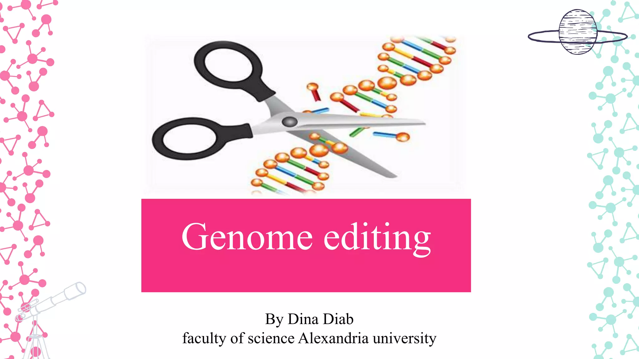
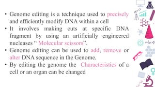
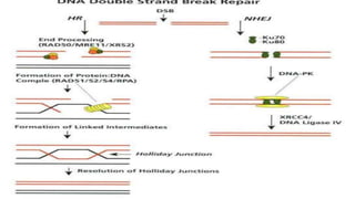
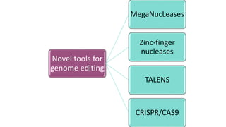
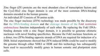
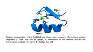
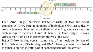
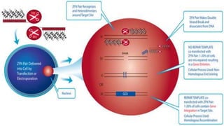
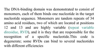
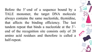
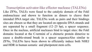
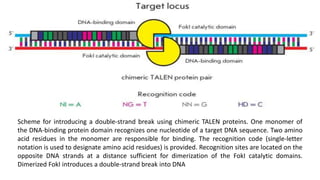
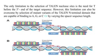
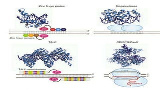
Genome editing is a technique used to precisely modify DNA within a cell. It involves using artificially engineered nucleases called "molecular scissors" to cut DNA at specific locations. This creates breaks that can then be repaired through natural cellular processes, allowing the genome to be altered. Early methods like homologous recombination were inefficient. New tools like zinc finger nucleases, TALENs, and the CRISPR/Cas9 system allow genome editing to be targeted to specific DNA sequences with greater accuracy and efficiency. These programmable nucleases make targeted cuts in the genome that can then be repaired through mechanisms like non-homologous end joining or homology-directed repair. CRISPR/Cas9 has become particularly
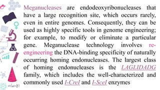
3. • Genome editing is a technique used to precisely
and efficiently modify DNA within a cell
• It involves making cuts at specific DNA
fragment by using an artificially engineered
nucleases “ Molecular scissors”.
• Genome editing can be used to add, remove or
alter DNA sequence in the Genome.
• By editing the genome the Characteristics of a
cell or an organ can be changed
4. The nucleases create a double-stranded
DNA Breaks (DSBs) at desired
locations in the genome and harness
the cell endogenous Mechanisms to
repair the induced break by natural
processes of homologous
recombinations (HR) and non-
homologous end joining (NHEJ).
5. Use a DNA templet to restore the
DSBs the outcome of this kind of
repair is precised
NHEJ vs HR
Rejoins the broken ends and is
often accompanied by loss/ gain
of some nucleotides, thus the
outcome of NHEJ is variable
8. Homologous recombination
The earliest method scientists used to edit genomes in living
cells was homologous recombination. which is the
exchange (recombination) of genetic information between
two similar (homologous) strands of DNA. Scientists began
developing this technique in the late 1970s following
observations that yeast, like other organisms, can carry out
homologous recombination naturally.
9. To perform homologous recombination in the
laboratory, one must generate and isolate DNA
fragments bearing genome sequences similar to the
portion of the genome that is to be edited. These
isolated fragments can be injected into individual
cells or taken up by cells using special chemicals.
Once inside a cell, these DNA fragments can then
recombine with the cell’s DNA to replace the
targeted portion of the genome.
10. Limitations
• Extremely inefficient in most cell
types. This technique can have as low
as a one-in-a-million probability of
successful editing.
• It is inaccurate and has a high rate of
error when the injected DNA
fragments insert into an unintended
part of the genome, causing what are
known as off-target edits.
14. Meganucleases are endodeoxyribonucleases that
have a large recognition site, which occurs rarely,
even in entire genomes. Consequently, they can be
used as highly specific tools in genome engineering;
for example, to modify or eliminate a particular
gene. Meganuclease technology involves re-
engineering the DNA-binding specificity of naturally
occurring homing endonucleases. The largest class
of homing endonucleases is the LAGLIDADG
family, which includes the well-characterized and
commonly used I-CreI and I-SceI enzymes
15. Through a combination of rational design and selection,
these homing endonucleases can be re-engineered to
target novel sequences. While many studies show promise
for the use of meganucleases in genome editing, the
DNA-binding and cleavage domains of homing
endonucleases are difficult to separate, and the relative
difficulty of engineering proteins with novel specificities
has traditionally limited the use of this platform.
16. To address this limitation, chimeric proteins comprising fusions of
meganucleases, ZFs, and TALEs have been engineered to generate
novel monomeric enzymes that take advantage of the binding
affinity of ZFs and TALEs and the cleavage specificity of
meganucleases.
One potential advantage associated with meganuclease technology
is that meganucleases are the smallest class of engineered nucleases,
making them potentially amenable to all standard gene delivery
methods. In fact, multiple meganuclease monomers could be readily
packaged into single viral vectors to simultaneously create multiple
DSBs.
17. Additionally,that DSB-formation by these
enzymes results in a 3’ overhang, which
may be more recombinogenic for HDR
than the 5’ overhang generated by FokI
cleavage.
19. Zinc finger (ZF) proteins are the most abundant class of transcription factors and
the Cys2-His2 zinc finger domain is one of the most common DNA-binding
domains encoded in the human genome.
An individual ZF Consists of 30 amino acids
The zinc finger nuclease (ZFN) technology was made possible by the discovery
that the DNA-binding domain and the cleavage domain of the FokI restriction
endonuclease function independently of each other. By replacing the FokI DNA-
binding domain with a zinc finger domain, it is possible to generate chimeric
nucleases with novel binding specificities. Because the FokI nuclease functions as
a dimer, two ZFNs binding opposite strands of DNA are required for induction of a
DSB. Initial experiments showed that ZFN-induced DSBs could be used to modify
the genome through either NHEJ or HDR and this technology has subsequently
been used to successfully modify genes in human somatic and pluripotent stem
cells.
20. Cartoon representation of the Cys2His2 zinc finger motif, consisting of an α helix and an
antiparallel β sheet. The zinc ion (green) is coordinated by two histidine residues and
two cysteine residues. The (Zn2+) stabilize the fold.
21. Each Zinc Finger Nuclease (ZFN) consists of two functional
domains: A) DNA-binding domains of individual ZFNs that typically
contain between three and six individual zinc finger repeats and can
each recognize between 9 and 18 basepairs. Each Finger makes
contact with 3 or 4 bp in the major groove of the DNA.
B.) A DNA-cleaving domain comprised of the nuclease domain of
Fok I. When the DNA-binding and DNA-cleaving domains are fused
together, a highly-specific pair of ‘genomic scissors’ are created.
22.
23. Disadvantages
Complexity and
high cost of protein
domains
construction for
each particular
genome locus
The probability of
inaccurate cleavage
of target DNA due
to single nucleotide
substitutions
Highly toxic
25. The history of this system’s development is
associated with the study of bacteria of the
Xanthomonas genus. These bacteria are pathogens
of crop plants, such as rice, pepper, and tomato; and
they cause significant economic damage to
agriculture, which was the motivate for their
thorough study. The bacteria were found to secrete
effector proteins (transcription activator-like
effectors, TALEs) to the cytoplasm of plant cells,
which affect processes in the plant cell and increase
its susceptibility to the pathogen
26. Further investigation of the effector protein action
mechanisms revealed that they are capable of DNA
binding and activating the expression of their target genes
via mimicking the eukaryotic transcription factors.
TALE proteins are composed of a central domain
responsible for DNA binding, a nuclear localization signal,
and a domain that activates the target gene transcription
The capability of these proteins to bind to DNA was first
described in 2007
27. The DNA-binding domain was demonstrated to consist of
monomers, each of them binds one nucleotide in the target
nucleotide sequence. Monomers are tandem repeats of 34
amino acid residues, two of which are located at positions
12 and 13 and are highly variable (repeat variable
diresidue, RVD), and it is they that are responsible for the
recognition of a specific nucleotide.This code is
degenerate; some RVDs can bind to several nucleotides
with different efficiencies
28. Before the 5’-end of a sequence bound by a
TALE monomer, the target DNA molecule
always contains the same nucleotide, thymidine,
that affects the binding efficiency. The last
tandem repeat that binds a nucleotide at the 3’-
end of the recognition site consists only of 20
amino acid residues and therefore is called a
half-repeat.
29. Transcription activator-like effector nuclease (TALENs)
Like ZFNs, TALEs were fused to the catalytic domain of the FokI
endonuclease and shown to function as dimers to cleave their
intended DNA target site. TALENs work as pairs and their bindings
sites are chosen so that they are located on opposite DNA strands and
are separated by a small fragment (12–25 bp), a spacer sequence.
Once in the nucleus, artificial nucleases bind to target sites: the FokI
domains located at the C-termini of a chimeric protein dimerize to
cause a double-strand break in a spacer sequenceAlso similar to
ZFNs, TALENs have been shown to efficiently induce both NHEJ
and HDR in human somatic and pluripotent stem cells.
30. Scheme for introducing a double-strand break using chimeric TALEN proteins. One monomer of
the DNA-binding protein domain recognizes one nucleotide of a target DNA sequence. Two amino
acid residues in the monomer are responsible for binding. The recognition code (single-letter
notation is used to designate amino acid residues) is provided. Recognition sites are located on the
opposite DNA strands at a distance sufficient for dimerization of the FokI catalytic domains.
Dimerized FokI introduces a double-strand break into DNA
31. The only limitation to the selection of TALEN nuclease sites is the need for T
before the 5’- end of the target sequence. However, this limitation can also be
overcome by selection of mutant variants of the TALEN N-terminal domain that
are capable of binding to A, G, or C Or by varying the spacer sequence length.
32. Additionally, the large size and repetitive
nature of TALE arrays presents a hurdle for
in vivo delivery of these proteins. As
opposed to a 30 amino acid zinc finger,
which binds three bases of DNA, TALENs
require 34 amino acids to specify a single
base pair and this size difference can
prohibit delivery of both TALEN monomers
in a single viral vector with limited
packaging capacity
33.
34. CRISPRs were first discovered in archaea (and later in
bacteria) by Francisco Mojica, a scientist at the University
of Alicante in Spain. He proposed that CRISPRs serve as
part of the bacterial immune system, defending against
invading viruses. They consist of repeating sequences of
genetic code, interrupted by “spacer” sequences – remnants
of genetic code from past invaders.
The system serves as a genetic memory that helps the cell
detect and destroy invaders when they return. Mojica’s
theory was experimentally demonstrated in 2007 by a team
of scientists led by Philippe Horvath.
In January 2013, the Zhang lab published the first method
to engineer CRISPR to edit the genome in mouse and
human cells.
35. Francisco Mojica
•Discovery of CRISPR and its function
•1993 – 2005
Sylvain Moineau
•December, 2010
•Moineau and colleagues demonstrated that CRISPR-Cas9 creates double-stranded breaks
in target DNA at precise position
Charpentier and Jennifer Doudna
•June, 2012
•Reported that the crRNA and the tracrRNA could be fused together to create a single,
synthetic guide, further simplifying the system.
Feng Zhang
January, 2013
CRISPR-Cas9 harnessed for genome editing
36.
37. Decades of work investigating CRISPR systems in various microbial
species has elucidated a mechanism by which short sequences of
invading nucleic acids are incorporated into CRISPR loci.They are
then transcribed and processed into CRISPR RNAs (crRNAs) which,
together with a trans-activating crRNAs (tracrRNAs), complex with
CRISPR-associated (Cas) proteins to dictate specificity of DNA
cleavage by Cas nucleases through Watson-Crick base pairing
between nucleic acids.
Doudna, Charpentier and colleagues showed through in vitro DNA
cleavage experiments that this system could be reduced to two
components by fusion of the crRNA and tracrRNA into a single
guide RNA (gRNA) 20 bases.
38. Furthermore, they showed that re-targeting of the Cas9/gRNA
complex to new sites could be accomplished by altering the
sequence of a short portion of the gRNA.
Thereafter, a series of publications demonstrated that the
CRISPR/Cas9 system could be engineered for efficient genetic
modification in mammalian cells. Collectively these studies have
propelled the CRISPR/Cas9 technology into the spotlight of the
genome-editing field.
The only sequence limitation of the CRISPR/Cas system derives
from the necessity of a protospacer-adjacent motif (PAM) located
immediately 3’ to the target site.
39. The PAM sequence is specific to the species of Cas9. For
example, the PAM sequence 5’-NGG-3’ is necessary for
binding and cleavage of DNA by the commonly used
Cas9 from Streptococcus pyogenes.
PAM with a more complex consensus sequence, will
overcome these drawbacks. For example, type II
crISPr/cas of N. meningitidis recognizes the PAM with
the 5’-NNNNGATT-3’ consensus, which certainly limits
the choice of a target but may increase the specificity.The
PAM sequence is specific to the species of Cas9.
40.
41. CRISPR-Cas systems
CRISPR-Cas systems can be divided in to two classes:
•Class 1, which uses several Cas proteins along with guide RNA ex:
type 1(CRISPR/cas3) & type 3 (CRISPR/CAS10)
•Class 2 system, which uses a single large Cas protein along with
guide RNA ex type 2 (CRISPR/cas9) & (CRISPR/ Cpf1).
Several variations of class 1 and class 2 have been identified, but all
of them use endonucleases of Cas family as effectors. However, the
CRISPR-Cpf1 system uses a large protein called Cpf1 (1,300 amino
acids) as the effector. Cpf1 is present in Prevotella and Francisella
bacteria and forms the basis of their acquired immune response.
42. pf1 Acts as molecular scissors and could cut DNA in human cells. Also, they
found that Cpf1 had avery different mechanism of action to Cas9:
1.Different target recognition sequences
The CRISPR-Cpf1 system recognize target DNA sequences using a short T-rich
protospacer-adjacent motif (PAM), which precedes the region of interest. On the
other hand, CRIPR-Cas 9 system recognizes a G-rich PAM region which follows
the target DNA sequence.
2.Different types of double-strand breaks
Cas9 endonuclease cuts both DNA strands at the same place, generating ‘blunt
ends’ while the Cpf1 endonuclease cuts DNA strands at different places,
generating a ‘staggered end’ where one strand is longer than the other. This type of
DNA end is also called ‘sticky end’.
43. Sticky ends are easier to work with as they will only pair with a
complimentary sticky end whereas blunt ends can often undergo
mutations upon rejoining.
3. Smaller guide RNA
The guide RNA in CRISPR-Cas9end nsists of a target-specific
CRISPR RNA (crRNA) and an auxiliary trans-activating crRNA
(tracrRNA), whereas CRISPR-Cpf1 arrays do not have this
additional tracrRNA. Thus, the Cpf1 system needs only one
RNA molecule to cut DNA instead of two.
This allows CRSIPR-Cpf1 to be much smaller than CRISPR,
making it easier for the enzyme to enter cells and tissues.
44.
45. As specificity is dictated by DNA complementarity (without
the need for multistep protein engineering), the CRISPR/Cas
technology has entered the picture as the faster, more
straightforward and affordable way for genome-editing in
comparison to traditional ZFN and TALENs approaches.
Furthermore, systems based on Cas9 have shown the
propensity to target heterochromatin sequences, targeting
DNase-inaccessible locations and cleaving at highly
methylated regions; this fact (in addition to the flexibility
offered by multiple variants of Cas9 for selection of targets)
lends this approach phenomenal versatility for genome
engineering.
46. Additionally, because the Cas9 protein is not directly coupled to
the gRNA, this system is highly amenable to multiplexing through
the concurrent use of multiple gRNAs to induce DSBs at several
loci.
47.
48. DELIVERY OF GENOME-EDITING TOOLS
transfection of plasmid DNA carrying nuclease and gRNA expression cassettes. This method
is not ideal for most gene and cell therapies due to low efficiency of transfection of primary
cells, DNA-related cytotoxicity, the presence of bacterial DNA sequences in plasmid
backbones, and the possibility of random integration of plasmid fragments into the genome.
Electroporation of mRNA encoding the nucleases and gRNAs generated through in vitro
transcription has become a preferred method for ex vivo gene editing of primary cells
relevant to gene therapy, such as T cells and hematopoietic stem cells (HSCs).
For many applications, viral vectors are still the optimal vehicle to maximize the efficiency of
delivery while minimizing cytotoxicity.
49. Uses of Genome editing techniques
• edit the genome of any organism. it is against the law to use
genome editing in human embryos that will be allowed to
develop beyond 14 days.
Genome editing can be used:
1. For research: Genome editing can be used to change the
DNA in cells or organisms to understand their biology and
how they work.
2. To treat disease: Genome editing has been used to modify
human blood cells that are then put back into the body to
treat conditions including leukaemia? And AIDS
50. 3. For biotechnology?: Genome editing has been used in agriculture
to genetically modify crops to improve their yields and resistance to
disease and drought, as well as to genetically modify cattle that
don’t have horns.