3. Mechanism of injury in head traumaMechanism of injury in head trauma
► Direct trauma by compression orDirect trauma by compression or
crushing.crushing.
► Acceleration-Deceleration InjuriesAcceleration-Deceleration Injuries
Brain has inertia. For example, when a personBrain has inertia. For example, when a person
falls backwards onto a hard floor, the back offalls backwards onto a hard floor, the back of
the personthe person’’s head hits the floor and stops. Thes head hits the floor and stops. The
brain, however, is still moving until it strikes thebrain, however, is still moving until it strikes the
inside of the skull. If the brain gets bruised,inside of the skull. If the brain gets bruised,
there is bleeding, also called a hemorrhage.there is bleeding, also called a hemorrhage.
This bleeding causes further damage to theThis bleeding causes further damage to the
brain.brain.
The skull does not need to strike an object inThe skull does not need to strike an object in
order for the brain to get injured. There areorder for the brain to get injured. There are
many situations in motor vehicle crashesmany situations in motor vehicle crashes
where the forces are transmitted through thewhere the forces are transmitted through the
brain without the skull hitting the dashboard,brain without the skull hitting the dashboard,
windshield, steering wheel or window.windshield, steering wheel or window.
Coup/Contrer-CoupCoup/Contrer-Coup Injuries:Injuries: Related toRelated to
acceleration-deceleration injuriesacceleration-deceleration injuries (e.g injury to(e.g injury to
temporal lobe in contralateral temporal trauma)temporal lobe in contralateral temporal trauma)
6. Scalp hematomaScalp hematoma
Subgaleal hematomaSubgaleal hematoma
►not bounded by suturesnot bounded by sutures
►May be hugeMay be huge
►If massive can be lifeIf massive can be life
threateningthreatening
CephalohematomaCephalohematoma
►Subperiosteal hematomaSubperiosteal hematoma
► limited by sutures andlimited by sutures and
fontanellsfontanells
►Small (covers single bone)Small (covers single bone)
7. Skull FractureSkull Fracture
►A skull fracture is a break in the skull boneA skull fracture is a break in the skull bone
and generally occurs as a result of directand generally occurs as a result of direct
impact .impact .
►Uncomplicated skull fractures themselvesUncomplicated skull fractures themselves
rarely produce neurologic deficit, but therarely produce neurologic deficit, but the
associated intracranial injury may haveassociated intracranial injury may have
serious neurologic sequelae.serious neurologic sequelae.
8. Three major types of skull fractures may occur:Three major types of skull fractures may occur:
► (1) linear,(1) linear,
► (2) depressed,(2) depressed,
► (3) basilar.(3) basilar.
11. Basilar skull fracturesBasilar skull fractures
► Most basilar fractures occur atMost basilar fractures occur at
2 specific anatomic locations2 specific anatomic locations
—— namely, the temporal regionnamely, the temporal region
and the occipital condylarand the occipital condylar
region.region.
14. Epidural HematomaEpidural Hematoma
►An epidural hematoma is usually associatedAn epidural hematoma is usually associated
with a skull fracture. It often occurs whenwith a skull fracture. It often occurs when
adirect impactadirect impact fractures the calvarium .fractures the calvarium .
►The fractured bone lacerates a dural arteryThe fractured bone lacerates a dural artery
(middle meningeal artery) or a venous(middle meningeal artery) or a venous
sinus.sinus.
►On CT, the hematoma forms a hyperdenseOn CT, the hematoma forms a hyperdense
biconvex mass. It is usually uniformly highbiconvex mass. It is usually uniformly high
density but may contain hypodense foci duedensity but may contain hypodense foci due
to active bleeding.to active bleeding.
►Comment onComment on midline shift .midline shift .
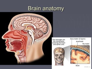
18. Subdural HematomaSubdural Hematoma
►Deceleration and acceleration or rotationalDeceleration and acceleration or rotational
forces that tear bridging veins can cause an acuteforces that tear bridging veins can cause an acute
subdural hematoma so it occurs in cases of widesubdural hematoma so it occurs in cases of wide
subdural space(old age & children)subdural space(old age & children)
Causes of subdural are:in minimal trauma in old age,Causes of subdural are:in minimal trauma in old age,
child abuse and ventricular decompression, maychild abuse and ventricular decompression, may
occur in patients receiving anticoagulants or patientsoccur in patients receiving anticoagulants or patients
with a coagulopathy condition.with a coagulopathy condition.
► The blood collects in the space between theThe blood collects in the space between the
arachnoid matter and the dura matter, Because thearachnoid matter and the dura matter, Because the
subdural space is not limited by the cranial sutures,subdural space is not limited by the cranial sutures,
blood can spread along the entire hemisphere andblood can spread along the entire hemisphere and
into the hemispheric fissure, limited only by the duralinto the hemispheric fissure, limited only by the dural
reflections .reflections .
► We have 3 major types :We have 3 major types :
Acute, subacute & chronicAcute, subacute & chronic
19. Acute Subdural HematomaAcute Subdural Hematoma
Crescent shaped;Crescent shaped;
Hyperdense, may contain hypodense fociHyperdense, may contain hypodense foci
due to serum, CSF or active bleedingdue to serum, CSF or active bleeding
20. ► In children, subduralIn children, subdural
hematomas occurring alonghematomas occurring along
the posterior interhemisphericthe posterior interhemispheric
fissure and the tentorium havefissure and the tentorium have
been described as commonbeen described as common
findings following violentfindings following violent
nonaccidental shaking (ie,nonaccidental shaking (ie,
shaken baby syndrome) .shaken baby syndrome) .
21. Subacute Subdural HematomaSubacute Subdural Hematoma
► Subacute SDH may be difficult to visualize by CTSubacute SDH may be difficult to visualize by CT
because as the hemorrhage is reabsorbed itbecause as the hemorrhage is reabsorbed it
becomesbecomes isodense to normal gray matterisodense to normal gray matter. A. A
subacute SDH should be suspected when yousubacute SDH should be suspected when you
identify shift of midline structures without an obviousidentify shift of midline structures without an obvious
mass. Giving contrast may help in difficult casesmass. Giving contrast may help in difficult cases
because the interface between the hematoma andbecause the interface between the hematoma and
the adjacent brain usually becomes more obviousthe adjacent brain usually becomes more obvious
due to enhancement of the dura and adjacentdue to enhancement of the dura and adjacent
vascular structures. Some of the notablevascular structures. Some of the notable
characteristics of subacute SDH are:characteristics of subacute SDH are:
- Compressed lateral ventricle- Compressed lateral ventricle
& or midline shift& or midline shift
- Effaced sulci .- Effaced sulci .
22.
23. Chronic Subdural HematomaChronic Subdural Hematoma
► Chronic SDH becomesChronic SDH becomes
low density as thelow density as the
hemorrhage is furtherhemorrhage is further
reabsorbed. It isreabsorbed. It is
usually uniformly lowusually uniformly low
density but may bedensity but may be
loculated. Rebleedingloculated. Rebleeding
often occurs andoften occurs and
causes mixed densitycauses mixed density
and fluid levels.and fluid levels.
24. Subarachnoid HemorrhageSubarachnoid Hemorrhage
►A subarachnoid hemorrhage occurs withA subarachnoid hemorrhage occurs with
injury of small arteries or veins on theinjury of small arteries or veins on the
surface of the brain. The ruptured vesselsurface of the brain. The ruptured vessel
bleeds into the space between the pia andbleeds into the space between the pia and
arachnoid matter. The most common causearachnoid matter. The most common cause
of subarachnoid hemorrhage is trauma .of subarachnoid hemorrhage is trauma .
►In the absence of significant trauma, theIn the absence of significant trauma, the
most common cause of subarachnoidmost common cause of subarachnoid
hemorrhage is the rupture of a cerebralhemorrhage is the rupture of a cerebral
aneurysm.aneurysm.
25. ► WhenWhen traumatictraumatic,,
subarachnoid hemorrhagesubarachnoid hemorrhage
occurs most commonlyoccurs most commonly
over the cerebralover the cerebral
convexities or adjacent toconvexities or adjacent to
otherwise injured brain (i.e.otherwise injured brain (i.e.
adjacent to a cerebraladjacent to a cerebral
contusion)contusion)
► If there is a large amountIf there is a large amount
of SAH particularly in theof SAH particularly in the
basilarcisterns,sulci&fissurbasilarcisterns,sulci&fissur
es the physician shouldes the physician should
consider whether aconsider whether a
ruptured aneurysmruptured aneurysm led toled to
the subsequent trauma.the subsequent trauma.
26. Quiz caseQuiz case
Young adult presented to the emergency department
after head trauma with GSC 3.
34. Cerebral ContusionCerebral Contusion
► Brain contusions commonly are identified in patients withBrain contusions commonly are identified in patients with
traumatic brain injury (traumatic brain injury (TBITBI) .) .
► The second mechanism is related toThe second mechanism is related to countercoupcountercoup acceleration oracceleration or
deceleration ,which causes the brain to strike the skull. In andeceleration ,which causes the brain to strike the skull. In an
event in which the head is in motion, cortical injury occursevent in which the head is in motion, cortical injury occurs
adjacent to the floor of the anterior or posterior cranial fossa, theadjacent to the floor of the anterior or posterior cranial fossa, the
sphenoid wing, the petrous ridge, the convexity of the skull, andsphenoid wing, the petrous ridge, the convexity of the skull, and
the falx or tentorium. The inferiorthe falx or tentorium. The inferior frontal and temporalfrontal and temporal lobes arelobes are
particularly vulnerableparticularly vulnerable
► Cerebral contusions are the most common primary intra-axialCerebral contusions are the most common primary intra-axial
injury. They often occur when the brain impacts an osseous ridgeinjury. They often occur when the brain impacts an osseous ridge
or a dural fold. The foci of punctate hemorrhage or edema areor a dural fold. The foci of punctate hemorrhage or edema are
located along gyral crests. The following are common locations:located along gyral crests. The following are common locations:
- Temporal lobe - anterior tip, inferior surface, sylvian region- Temporal lobe - anterior tip, inferior surface, sylvian region
- Frontal lobe - anterior pole, inferior surface- Frontal lobe - anterior pole, inferior surface
- Dorsolateral midbrain- Dorsolateral midbrain
- Inferior cerebellum- Inferior cerebellum
35. ► On CT, cerebral contusion appears as an ill-definedOn CT, cerebral contusion appears as an ill-defined
hypodense area mixed with foci of hemorrhage.hypodense area mixed with foci of hemorrhage.
Adjacent subarachnoid hemorrhage is common.Adjacent subarachnoid hemorrhage is common.
After 24-48 hoursAfter 24-48 hours, hemorrhagic transformation or, hemorrhagic transformation or
coalescence of petechial hemorrhages into acoalescence of petechial hemorrhages into a
rounded hematoma is commonrounded hematoma is common
► CT scans often demonstrate progression over timeCT scans often demonstrate progression over time
in the size and number of contusions and thein the size and number of contusions and the
amount of hemorrhage within the contusionsamount of hemorrhage within the contusions
► MRIMRI findings typically demonstrate the lesions fromfindings typically demonstrate the lesions from
the onset of injury, but many facilities cannot performthe onset of injury, but many facilities cannot perform
MRI on an emergent basisMRI on an emergent basis
► On MRI, contusions are isointense to hyperintenseOn MRI, contusions are isointense to hyperintense
on T1-weighted and hyperintense on T2-weightedon T1-weighted and hyperintense on T2-weighted
image& The signal intensity is increased in theimage& The signal intensity is increased in the
affected region on DWIs .affected region on DWIs .
38. Diffuse Axonal InjuryDiffuse Axonal Injury
► Diffuse axonal injury is often referred toDiffuse axonal injury is often referred to
as "shear injury". It is the most commonas "shear injury". It is the most common
cause of significant morbidity in CNScause of significant morbidity in CNS
trauma. Fifty percent of all primary intra-trauma. Fifty percent of all primary intra-
axial injuries are diffuse axonal injuries .axial injuries are diffuse axonal injuries .
► When shearing forces occurWhen shearing forces occur in areas ofin areas of
greater density differential,greater density differential, the axonsthe axons
suffer trauma; thissuffer trauma; this results inresults in edema andedema and
in axoplasmic leakage (which isin axoplasmic leakage (which is mostmost
severe during the first 2 weekssevere during the first 2 weeks followingfollowing
injury). The exact location of the shear-injury). The exact location of the shear-
strain injury depends on the plane ofstrain injury depends on the plane of
rotationrotation
► Immediate loss of consciousness isImmediate loss of consciousness is
typical of these injuries .typical of these injuries .
39. The true extent of axonal injury typically is worse than thatThe true extent of axonal injury typically is worse than that
visualized using current imaging techniques The CT of avisualized using current imaging techniques The CT of a
patient with diffuse axonal injury may be normal despite thepatient with diffuse axonal injury may be normal despite the
patient's presentation with a profound neurological deficit .patient's presentation with a profound neurological deficit .
► With CT, diffuse axonal injury may appear as ill-definedWith CT, diffuse axonal injury may appear as ill-defined
areas of high density or hemorrhage in characteristicareas of high density or hemorrhage in characteristic
locations.locations.
► One or more small intraparenchymalOne or more small intraparenchymal (petechial)(petechial)
hemorrhageshemorrhages less than 2 cm in diameter, locatedless than 2 cm in diameter, located in thein the
cerebral hemispheres at the grey white interfacecerebral hemispheres at the grey white interface as wellas well
as corpus callosum &brainstem.as corpus callosum &brainstem.
► One may also observe smallOne may also observe small focal areas of low densityfocal areas of low density
on CT scans; these correspond to areas of edemaon CT scans; these correspond to areas of edema
40.
41. Intraventricular HemorrhageIntraventricular Hemorrhage
► Traumatic intraventricularTraumatic intraventricular
hemorrhage is associatedhemorrhage is associated
with diffuse axonal injury,with diffuse axonal injury,
deep gray matter injury,deep gray matter injury,
and brainstem contusion.and brainstem contusion.
An isolated intraventricularAn isolated intraventricular
hemorrhage may be due tohemorrhage may be due to
rupture of subependymalrupture of subependymal
veins .veins .
42. Cerebral EdemaCerebral Edema
Severe brain edemaSevere brain edema
or a large intracranialor a large intracranial
hemorrhage mayhemorrhage may
cause downwardcause downward
brain displacementbrain displacement
and coning, which isand coning, which is
usually fatalusually fatal
48. StrokeStroke
Stroke is a clinical term for sudden, focalStroke is a clinical term for sudden, focal
neurological deficitneurological deficit
Hemorrhagic strokesHemorrhagic strokes
► due to rupture of adue to rupture of a
cerebral blood vesselcerebral blood vessel
that causes bleedingthat causes bleeding
into or around theinto or around the
brain .brain .
► account for 16% of allaccount for 16% of all
strokes .strokes .
ischemic strokeischemic stroke
► caused by blockage ofcaused by blockage of
blood flow in a majorblood flow in a major
cerebral blood vessel,cerebral blood vessel,
usually due to a bloodusually due to a blood
clot .clot .
► account for about 84%account for about 84%
of all strokes.of all strokes.
49. Hemorrhagic StrokeHemorrhagic Stroke
Hemorrhagic strokes account for 16% of all strokesHemorrhagic strokes account for 16% of all strokes
► Intracerebral hge is theIntracerebral hge is the
most common,most common,
accounting for 10% ofaccounting for 10% of
all strokes .all strokes .
► Subarachnoid hge,Subarachnoid hge,
due to rupture of adue to rupture of a
cerebral aneurysm,cerebral aneurysm,
accounts for 6% ofaccounts for 6% of
strokes overall.strokes overall.
50. Now Dudes tell me what are the reasons of cerebralNow Dudes tell me what are the reasons of cerebral
hemorrhage!???hemorrhage!???
1.1. Hypertensive hemorrhageHypertensive hemorrhage ..
2.2. Amyloid angiopathy.Amyloid angiopathy.
3.3. Ruptured vascular malformation.Ruptured vascular malformation.
4.4. Coagulopathy(A fluid level within the hematoma) .Coagulopathy(A fluid level within the hematoma) .
5.5. Hemorrhage into a tumor .Hemorrhage into a tumor .
6.6. Venous infarction.Venous infarction.
51.
52.
53. Subarachnoid HemorrhageSubarachnoid Hemorrhage
► Common aneurysm locations include theCommon aneurysm locations include the
anterior and posterior communicating arteries,anterior and posterior communicating arteries,
the middle cerebral artery bifurcation and the tipthe middle cerebral artery bifurcation and the tip
of the basilar artery.of the basilar artery.
► Subarachnoid hemorrhage typically presents asSubarachnoid hemorrhage typically presents as
thethe "worst headache of life""worst headache of life" for the patient .for the patient .
54. Ischemic strokeIschemic stroke
Ischemic strokes are caused by thrombosis, embolism ofIschemic strokes are caused by thrombosis, embolism of
thrombosis, hypoperfusion and lacunar infarctions(1%thrombosis, hypoperfusion and lacunar infarctions(1%((
► A thrombotic strokeA thrombotic stroke
(53%)occurs when a(53%)occurs when a
blood clot forms in situblood clot forms in situ
within a cerebral arterywithin a cerebral artery
and blocks or reducesand blocks or reduces
the flow of bloodthe flow of blood
through the arterythrough the artery
► An embolic strokeAn embolic stroke
(30%) occurs when a(30%) occurs when a
detached clot flowsdetached clot flows
into and blocks ainto and blocks a
cerebral arterycerebral artery
55. ► A CT is 58% sensitive forA CT is 58% sensitive for
infarction within the firstinfarction within the first
24 hours (Bryan et al,24 hours (Bryan et al,
1991). MRI is 82%1991). MRI is 82%
sensitive. If the patient issensitive. If the patient is
imaged greater than 24imaged greater than 24
hours after the event,hours after the event,
both CT and MR areboth CT and MR are
greater than 90%greater than 90%
sensitive.sensitive.
► After a stroke, edemaAfter a stroke, edema
progresses, and brainprogresses, and brain
density decreasesdensity decreases
proportionately.proportionately.
56. Diffuse Hypodensity and SulcalDiffuse Hypodensity and Sulcal
EffacementEffacement
► Hypodensity in greaterHypodensity in greater
than one-third of thethan one-third of the
middle cerebral arterymiddle cerebral artery
territory is generallyterritory is generally
considered to be aconsidered to be a
contra-indication tocontra-indication to
thrombolytic therapy.thrombolytic therapy.
57. Hyperdense Vessel SignHyperdense Vessel Sign
► A hyperdense vessel isA hyperdense vessel is
defined as a vesseldefined as a vessel
denser than itsdenser than its
counterpart andcounterpart and
denser than any non-denser than any non-
calcified vessel ofcalcified vessel of
similar size.similar size.
► This sign indicatesThis sign indicates
poor outcome andpoor outcome and
poor response to IV-poor response to IV-
TPA therapy.TPA therapy.
58. Basilar ThrombosisBasilar Thrombosis
► Thrombosis of theThrombosis of the
basilar artery is abasilar artery is a
common finding incommon finding in
stroke patients. CTstroke patients. CT
findings include afindings include a
dense basilar arterydense basilar artery
without contrastwithout contrast
injection.injection.
61. Subacute InfarctionSubacute Infarction
-Increasing mass effect-Increasing mass effect
- Wedge shaped low- Wedge shaped low
densitydensity
- Hgic transformation- Hgic transformation
After 4 - 7 days the CTAfter 4 - 7 days the CT
- Gyral enhancement- Gyral enhancement
- Persistent mass effect- Persistent mass effect
In 1-8 weeks:In 1-8 weeks:
- Mass effect resolves- Mass effect resolves
- Enhancement may- Enhancement may
persistpersist
83. Left temporopareital subdural hematoma
Right temporopareital subarachnoid hemorrhage
Subfalcine brain herniation
Left uncal herniation compressing the left posterior
cerebral artery
AnswerAnswer
86. Post gunshot injury showed:
Intracranial right parietal bullet
Right frontopareital parenchymal hematoma
with adjacent subarachnoid hemorrhage
Comminuted right parietal bone fracture with
intracranial small bone fragments
Pneumocephalus
Right pareital subgaleal hematoma
AnswerAnswer
90. Right frontopareital large subdural hematoma
showing blood fluid level suggestive of acute
bleeding on top of chronic subdural hematoma
Subfalcine herniation
AnswerAnswer
92. DWI & Perfusion MRI studies of the brain
reveal:
Acute cerebral infraction showing diffusion
restriction, Diffusion/perfusion mismatch,
Large area of pneumbra on perfusion studies
AnswerAnswer