This document discusses intraparenchymal hemorrhage (IPH), which is bleeding within the brain tissue. IPH accounts for 8-13% of strokes and has a higher risk of death or disability than other types of strokes. The bleeding can disrupt or compress brain tissue. Common causes include hypertension, drug abuse, aneurysms, and cerebral amyloid angiopathy. Symptoms depend on the location and size of the bleeding in the brain and may include headaches, neurological deficits, and altered consciousness. Treatment involves controlling bleeding, reducing swelling and pressure in the brain, and managing symptoms. The case presented is an 81-year-old man with IPH likely due to hypertension.
intracranial hemorrhage- by KEMISA HASSEN ZAINABU IIHS_jinjaUGANDA (2).pptxHASSENZAINABUKEMISA
By the end of this we shall be able to know the following
definition.of intracranial hemorriage
Classification of intracranial hemorriage
Types of intracranial hemorriage.
Causes of intracranial hemorriage.
Signs and symptoms of intracranial hemorriage
Investigations specific management.
Complication.
Ongoing nursing care.
A stroke occurs when the blood supply to part of your brain is interrupted or severely reduced, depriving brain tissue of oxygen and food.
Within minutes, brain cells begin to die.
Stroke can be either ischemic or hemorrhagic.
Couples presenting to the infertility clinic- Do they really have infertility...Sujoy Dasgupta
Dr Sujoy Dasgupta presented the study on "Couples presenting to the infertility clinic- Do they really have infertility? – The unexplored stories of non-consummation" in the 13th Congress of the Asia Pacific Initiative on Reproduction (ASPIRE 2024) at Manila on 24 May, 2024.
Title: Sense of Taste
Presenter: Dr. Faiza, Assistant Professor of Physiology
Qualifications:
MBBS (Best Graduate, AIMC Lahore)
FCPS Physiology
ICMT, CHPE, DHPE (STMU)
MPH (GC University, Faisalabad)
MBA (Virtual University of Pakistan)
Learning Objectives:
Describe the structure and function of taste buds.
Describe the relationship between the taste threshold and taste index of common substances.
Explain the chemical basis and signal transduction of taste perception for each type of primary taste sensation.
Recognize different abnormalities of taste perception and their causes.
Key Topics:
Significance of Taste Sensation:
Differentiation between pleasant and harmful food
Influence on behavior
Selection of food based on metabolic needs
Receptors of Taste:
Taste buds on the tongue
Influence of sense of smell, texture of food, and pain stimulation (e.g., by pepper)
Primary and Secondary Taste Sensations:
Primary taste sensations: Sweet, Sour, Salty, Bitter, Umami
Chemical basis and signal transduction mechanisms for each taste
Taste Threshold and Index:
Taste threshold values for Sweet (sucrose), Salty (NaCl), Sour (HCl), and Bitter (Quinine)
Taste index relationship: Inversely proportional to taste threshold
Taste Blindness:
Inability to taste certain substances, particularly thiourea compounds
Example: Phenylthiocarbamide
Structure and Function of Taste Buds:
Composition: Epithelial cells, Sustentacular/Supporting cells, Taste cells, Basal cells
Features: Taste pores, Taste hairs/microvilli, and Taste nerve fibers
Location of Taste Buds:
Found in papillae of the tongue (Fungiform, Circumvallate, Foliate)
Also present on the palate, tonsillar pillars, epiglottis, and proximal esophagus
Mechanism of Taste Stimulation:
Interaction of taste substances with receptors on microvilli
Signal transduction pathways for Umami, Sweet, Bitter, Sour, and Salty tastes
Taste Sensitivity and Adaptation:
Decrease in sensitivity with age
Rapid adaptation of taste sensation
Role of Saliva in Taste:
Dissolution of tastants to reach receptors
Washing away the stimulus
Taste Preferences and Aversions:
Mechanisms behind taste preference and aversion
Influence of receptors and neural pathways
Impact of Sensory Nerve Damage:
Degeneration of taste buds if the sensory nerve fiber is cut
Abnormalities of Taste Detection:
Conditions: Ageusia, Hypogeusia, Dysgeusia (parageusia)
Causes: Nerve damage, neurological disorders, infections, poor oral hygiene, adverse drug effects, deficiencies, aging, tobacco use, altered neurotransmitter levels
Neurotransmitters and Taste Threshold:
Effects of serotonin (5-HT) and norepinephrine (NE) on taste sensitivity
Supertasters:
25% of the population with heightened sensitivity to taste, especially bitterness
Increased number of fungiform papillae
Knee anatomy and clinical tests 2024.pdfvimalpl1234
This includes all relevant anatomy and clinical tests compiled from standard textbooks, Campbell,netter etc..It is comprehensive and best suited for orthopaedicians and orthopaedic residents.
New Drug Discovery and Development .....NEHA GUPTA
The "New Drug Discovery and Development" process involves the identification, design, testing, and manufacturing of novel pharmaceutical compounds with the aim of introducing new and improved treatments for various medical conditions. This comprehensive endeavor encompasses various stages, including target identification, preclinical studies, clinical trials, regulatory approval, and post-market surveillance. It involves multidisciplinary collaboration among scientists, researchers, clinicians, regulatory experts, and pharmaceutical companies to bring innovative therapies to market and address unmet medical needs.
Title: Sense of Smell
Presenter: Dr. Faiza, Assistant Professor of Physiology
Qualifications:
MBBS (Best Graduate, AIMC Lahore)
FCPS Physiology
ICMT, CHPE, DHPE (STMU)
MPH (GC University, Faisalabad)
MBA (Virtual University of Pakistan)
Learning Objectives:
Describe the primary categories of smells and the concept of odor blindness.
Explain the structure and location of the olfactory membrane and mucosa, including the types and roles of cells involved in olfaction.
Describe the pathway and mechanisms of olfactory signal transmission from the olfactory receptors to the brain.
Illustrate the biochemical cascade triggered by odorant binding to olfactory receptors, including the role of G-proteins and second messengers in generating an action potential.
Identify different types of olfactory disorders such as anosmia, hyposmia, hyperosmia, and dysosmia, including their potential causes.
Key Topics:
Olfactory Genes:
3% of the human genome accounts for olfactory genes.
400 genes for odorant receptors.
Olfactory Membrane:
Located in the superior part of the nasal cavity.
Medially: Folds downward along the superior septum.
Laterally: Folds over the superior turbinate and upper surface of the middle turbinate.
Total surface area: 5-10 square centimeters.
Olfactory Mucosa:
Olfactory Cells: Bipolar nerve cells derived from the CNS (100 million), with 4-25 olfactory cilia per cell.
Sustentacular Cells: Produce mucus and maintain ionic and molecular environment.
Basal Cells: Replace worn-out olfactory cells with an average lifespan of 1-2 months.
Bowman’s Gland: Secretes mucus.
Stimulation of Olfactory Cells:
Odorant dissolves in mucus and attaches to receptors on olfactory cilia.
Involves a cascade effect through G-proteins and second messengers, leading to depolarization and action potential generation in the olfactory nerve.
Quality of a Good Odorant:
Small (3-20 Carbon atoms), volatile, water-soluble, and lipid-soluble.
Facilitated by odorant-binding proteins in mucus.
Membrane Potential and Action Potential:
Resting membrane potential: -55mV.
Action potential frequency in the olfactory nerve increases with odorant strength.
Adaptation Towards the Sense of Smell:
Rapid adaptation within the first second, with further slow adaptation.
Psychological adaptation greater than receptor adaptation, involving feedback inhibition from the central nervous system.
Primary Sensations of Smell:
Camphoraceous, Musky, Floral, Pepperminty, Ethereal, Pungent, Putrid.
Odor Detection Threshold:
Examples: Hydrogen sulfide (0.0005 ppm), Methyl-mercaptan (0.002 ppm).
Some toxic substances are odorless at lethal concentrations.
Characteristics of Smell:
Odor blindness for single substances due to lack of appropriate receptor protein.
Behavioral and emotional influences of smell.
Transmission of Olfactory Signals:
From olfactory cells to glomeruli in the olfactory bulb, involving lateral inhibition.
Primitive, less old, and new olfactory systems with different path
Pulmonary Thromboembolism - etilogy, types, medical- Surgical and nursing man...VarunMahajani
Disruption of blood supply to lung alveoli due to blockage of one or more pulmonary blood vessels is called as Pulmonary thromboembolism. In this presentation we will discuss its causes, types and its management in depth.
Tom Selleck Health: A Comprehensive Look at the Iconic Actor’s Wellness Journeygreendigital
Tom Selleck, an enduring figure in Hollywood. has captivated audiences for decades with his rugged charm, iconic moustache. and memorable roles in television and film. From his breakout role as Thomas Magnum in Magnum P.I. to his current portrayal of Frank Reagan in Blue Bloods. Selleck's career has spanned over 50 years. But beyond his professional achievements. fans have often been curious about Tom Selleck Health. especially as he has aged in the public eye.
Follow us on: Pinterest
Introduction
Many have been interested in Tom Selleck health. not only because of his enduring presence on screen but also because of the challenges. and lifestyle choices he has faced and made over the years. This article delves into the various aspects of Tom Selleck health. exploring his fitness regimen, diet, mental health. and the challenges he has encountered as he ages. We'll look at how he maintains his well-being. the health issues he has faced, and his approach to ageing .
Early Life and Career
Childhood and Athletic Beginnings
Tom Selleck was born on January 29, 1945, in Detroit, Michigan, and grew up in Sherman Oaks, California. From an early age, he was involved in sports, particularly basketball. which played a significant role in his physical development. His athletic pursuits continued into college. where he attended the University of Southern California (USC) on a basketball scholarship. This early involvement in sports laid a strong foundation for his physical health and disciplined lifestyle.
Transition to Acting
Selleck's transition from an athlete to an actor came with its physical demands. His first significant role in "Magnum P.I." required him to perform various stunts and maintain a fit appearance. This role, which he played from 1980 to 1988. necessitated a rigorous fitness routine to meet the show's demands. setting the stage for his long-term commitment to health and wellness.
Fitness Regimen
Workout Routine
Tom Selleck health and fitness regimen has evolved. adapting to his changing roles and age. During his "Magnum, P.I." days. Selleck's workouts were intense and focused on building and maintaining muscle mass. His routine included weightlifting, cardiovascular exercises. and specific training for the stunts he performed on the show.
Selleck adjusted his fitness routine as he aged to suit his body's needs. Today, his workouts focus on maintaining flexibility, strength, and cardiovascular health. He incorporates low-impact exercises such as swimming, walking, and light weightlifting. This balanced approach helps him stay fit without putting undue strain on his joints and muscles.
Importance of Flexibility and Mobility
In recent years, Selleck has emphasized the importance of flexibility and mobility in his fitness regimen. Understanding the natural decline in muscle mass and joint flexibility with age. he includes stretching and yoga in his routine. These practices help prevent injuries, improve posture, and maintain mobilit
Flu Vaccine Alert in Bangalore Karnatakaaddon Scans
As flu season approaches, health officials in Bangalore, Karnataka, are urging residents to get their flu vaccinations. The seasonal flu, while common, can lead to severe health complications, particularly for vulnerable populations such as young children, the elderly, and those with underlying health conditions.
Dr. Vidisha Kumari, a leading epidemiologist in Bangalore, emphasizes the importance of getting vaccinated. "The flu vaccine is our best defense against the influenza virus. It not only protects individuals but also helps prevent the spread of the virus in our communities," he says.
This year, the flu season is expected to coincide with a potential increase in other respiratory illnesses. The Karnataka Health Department has launched an awareness campaign highlighting the significance of flu vaccinations. They have set up multiple vaccination centers across Bangalore, making it convenient for residents to receive their shots.
To encourage widespread vaccination, the government is also collaborating with local schools, workplaces, and community centers to facilitate vaccination drives. Special attention is being given to ensuring that the vaccine is accessible to all, including marginalized communities who may have limited access to healthcare.
Residents are reminded that the flu vaccine is safe and effective. Common side effects are mild and may include soreness at the injection site, mild fever, or muscle aches. These side effects are generally short-lived and far less severe than the flu itself.
Healthcare providers are also stressing the importance of continuing COVID-19 precautions. Wearing masks, practicing good hand hygiene, and maintaining social distancing are still crucial, especially in crowded places.
Protect yourself and your loved ones by getting vaccinated. Together, we can help keep Bangalore healthy and safe this flu season. For more information on vaccination centers and schedules, residents can visit the Karnataka Health Department’s official website or follow their social media pages.
Stay informed, stay safe, and get your flu shot today!
Report Back from SGO 2024: What’s the Latest in Cervical Cancer?bkling
Are you curious about what’s new in cervical cancer research or unsure what the findings mean? Join Dr. Emily Ko, a gynecologic oncologist at Penn Medicine, to learn about the latest updates from the Society of Gynecologic Oncology (SGO) 2024 Annual Meeting on Women’s Cancer. Dr. Ko will discuss what the research presented at the conference means for you and answer your questions about the new developments.
Recomendações da OMS sobre cuidados maternos e neonatais para uma experiência pós-natal positiva.
Em consonância com os ODS – Objetivos do Desenvolvimento Sustentável e a Estratégia Global para a Saúde das Mulheres, Crianças e Adolescentes, e aplicando uma abordagem baseada nos direitos humanos, os esforços de cuidados pós-natais devem expandir-se para além da cobertura e da simples sobrevivência, de modo a incluir cuidados de qualidade.
Estas diretrizes visam melhorar a qualidade dos cuidados pós-natais essenciais e de rotina prestados às mulheres e aos recém-nascidos, com o objetivo final de melhorar a saúde e o bem-estar materno e neonatal.
Uma “experiência pós-natal positiva” é um resultado importante para todas as mulheres que dão à luz e para os seus recém-nascidos, estabelecendo as bases para a melhoria da saúde e do bem-estar a curto e longo prazo. Uma experiência pós-natal positiva é definida como aquela em que as mulheres, pessoas que gestam, os recém-nascidos, os casais, os pais, os cuidadores e as famílias recebem informação consistente, garantia e apoio de profissionais de saúde motivados; e onde um sistema de saúde flexível e com recursos reconheça as necessidades das mulheres e dos bebês e respeite o seu contexto cultural.
Estas diretrizes consolidadas apresentam algumas recomendações novas e já bem fundamentadas sobre cuidados pós-natais de rotina para mulheres e neonatos que recebem cuidados no pós-parto em unidades de saúde ou na comunidade, independentemente dos recursos disponíveis.
É fornecido um conjunto abrangente de recomendações para cuidados durante o período puerperal, com ênfase nos cuidados essenciais que todas as mulheres e recém-nascidos devem receber, e com a devida atenção à qualidade dos cuidados; isto é, a entrega e a experiência do cuidado recebido. Estas diretrizes atualizam e ampliam as recomendações da OMS de 2014 sobre cuidados pós-natais da mãe e do recém-nascido e complementam as atuais diretrizes da OMS sobre a gestão de complicações pós-natais.
O estabelecimento da amamentação e o manejo das principais intercorrências é contemplada.
Recomendamos muito.
Vamos discutir essas recomendações no nosso curso de pós-graduação em Aleitamento no Instituto Ciclos.
Esta publicação só está disponível em inglês até o momento.
Prof. Marcus Renato de Carvalho
www.agostodourado.com
For Better Surat #ℂall #Girl Service ❤85270-49040❤ Surat #ℂall #Girls
INTRAPARENCYMAL HAMORRAGE by mwebaza victor.doc
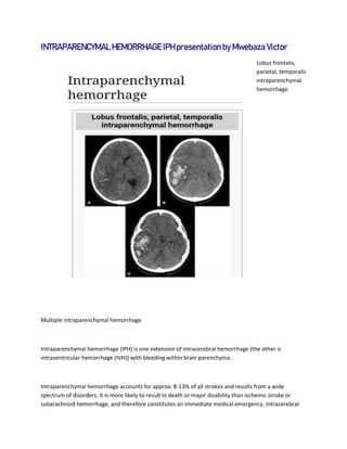
1. INTRAPARENCYMAL HEMORRHAGE IPH presentation by Mwebaza Victor
Lobus frontalis,
parietal, temporalis
intraparenchymal
hemorrhage
Multiple intraparenchymal hemorrhage
Intraparenchymal hemorrhage (IPH) is one extension of intracerebral hemorrhage (the other is
intraventricular hemorrhage (IVH)) with bleeding within brain parenchyma.
Intraparenchymal hemorrhage accounts for approx. 8-13% of all strokes and results from a wide
spectrum of disorders. It is more likely to result in death or major disability than ischemic stroke or
subarachnoid hemorrhage, and therefore constitutes an immediate medical emergency. Intracerebral
2. hemorrhages and accompanying edema may disrupt or compress adjacent brain tissue, leading to
neurological dysfunction. Substantial displacement of brain parenchyma may cause elevation of
intracranial pressure (ICP) and potentially fatal herniation syndromes.
Pathophysiology
Nontraumatic intraparenchymal hemorrhage most commonly results from hypertensive damage to
blood vessel walls e.g.: - hypertension - eclampsia - drug abuse, but it also may be due to autoregulatory
dysfunction with excessive cerebral blood flow e.g.: - reperfusion injury - hemorrhagic transformation -
cold exposure - rupture of an aneurysm or arteriovenous malformation (AVM) - arteriopathy (e.g.
cerebral amyloid angiopathy, moyamoya) - altered hemostasis (e.g. thrombolysis, anticoagulation,
bleeding diathesis) - hemorrhagic necrosis (e.g. tumor, infection) - venous outflow obstruction (e.g.
cerebral venous sinus thrombosis). Nonpenetrating and penetrating cranial trauma can also be common
causes of intracerebral hemorrhage.
Physical Symptoms
Clinical manifestations of intraparenchymal hemorrhage are determined by the size and location of
hemorrhage, but may include the following:
Hypertension, fever, or cardiac arrhythmias
Nuchal rigidity
Subhyaloid retinal hemorrhages
Altered level of consciousness
Anisocoria, Nystagmus
Focal neurological deficits
Putamen - Contralateral hemiparesis, contralateral sensory loss, contralateral conjugate gaze paresis,
homonymous hemianopsia, aphasia, neglect, or apraxia
Thalamus - Contralateral sensory loss, contralateral hemiparesis, gaze paresis, homonymous hemianopia,
miosis, aphasia, or confusion
Lobar - Contralateral hemiparesis or sensory loss, contralateral conjugate gaze paresis, homonymous
hemianopia, abulia, aphasia, neglect, or apraxia
Caudate nucleus - Contralateral hemiparesis, contralateral conjugate gaze paresis, or confusion
3. Brain stem - Tetraparesis, facial weakness, decreased level of consciousness, gaze paresis, ocular
bobbing, miosis, or autonomic instability
Cerebellum - Ataxia, usually beginning in the trunk, ipsilateral facial weakness, ipsilateral sensory loss,
gaze paresis, skew deviation, miosis, or decreased level of consciousness
Causes
Hypertension
Arteriovenous malformation
Aneurysm rupture
Cerebral amyloid angiopathy
Intracranial neoplasm
Coagulopathy
Hemorrhagic transformation of an ischemic infarct
Cerebral venous thrombosis
Sympathomimetic drug abuse
Moyamoya
Sickle cell disease
Eclampsia or postpartum vasculopathy
Infection
Vasculitis
Neonatal intraventricular hemorrhage
Trauma
Diagnosis
Computed tomography (CT scan): A CT scan may be normal if it is done soon after the onset of
symptoms. A CT scan is the best test to look for bleeding in or around your brain. In some hospitals, a
perfusion CT scan may be done to see where the blood is flowing and not flowing in your brain.
4. Magnetic resonance imaging (MRI scan): A special MRI technique (diffusion MRI) may show evidence of
an ischemic stroke within minutes of symptom onset. In some hospitals, a perfusion MRI scan may be
done to see where the blood is flowing and not flowing in your brain.
Angiogram: a test that looks at the blood vessels that feed the brain. An angiogram will show whether
the blood vessel is blocked by a clot, the blood vessel is narrowed, or if there is an abnormality of a
blood vessel known as an aneurysm.
Carotid duplex: A carotid duplex is an ultrasound study that assesses whether or not you have
atherosclerosis (narrowing) of the carotid arteries. These arteries are the large blood vessels in your
neck that feed your brain.
Transcranial Doppler (TCD): Transcranial Doppler is an ultrasound study that assesses whether or not
you have atherosclerosis (narrowing) of the blood vessels inside of your brain. It can also be used to see
if you have emboli (blood clots) in your blood vessels.
Treatment
Intracerebral hemorrhages is a severe condition requiring prompt medical attention. Treatment goals
include lifesaving interventions, supportive measures, and control of symptoms. Treatment depends on
the location, extent, and cause of the bleeding. Often, treatment can reverse the damage that has been
done.
A craniotomy is sometimes done to remove blood, abnormal blood vessels, or a tumor. Medications
may be used to reduce swelling, prevent seizures, lower blood pressure, and control pain.
Acute intraparenchymal hemorrhage
Clinical History: An 81-year-old diabetic male presents with a change in mental status.
Findings: Axial CT images through the brain demonstrate two areas of hyperdensity, one measuring
approximately 2 x 1.5 cm and a second measuring approximately 3 x 4 cm. There is some surrounding
5. edema with mass effect upon the left ventricle. In addition, there is extension into the ventricular
system.
Diagnosis: Acute intraparenchymal hemorrhage.
Discussion: There are many causes for intraparenchymal hemorrhage. A common cause is secondary to
hypertension. These most commonly occur within the basal ganglia, subcortical white matter,
cerebellum, thalamus and pons. Hemorrhage into the posterior fossa with mass effect or extension into
the ventricular system carries poor prognosis.
In younger patients, vascular malformations, specifically AVMs and cavernous angiomas are more
common causes for hemorrhage. In addition, venous malformations are associated with hemorrhage.
In the elderly population, amyloid angiopathy is associated with cerebral infarcts as well as hemorrhage
in superficial locations, rather than deep white matter or basal ganglia. These are usually described as
"lobar". These bleedings are not associated with systemic amyloidosis.
Hemorrhagic neoplasms are more complex, heterogeneous bleeds often with associated edema. These
hemorrhages are related to tumor necrosis, vascular invasion and neovascularity. Glioblastomas are the
most common primary malignancies to hemorrhage while thyroid, renal cell carcinoma, melanoma, and
lung cancer are the most common causes of hemorrhage from metastatic disease.
Other causes of intraparenchymal hemorrhage include hemorrhagic transformation of infarction which
is usually in a classic vascular distribution and is seen in approximately
24 to 48 hours following the ischemic event. This hemorrhage
rarely extends into the ventricular system.
Gallery
Caption1