Learn PD-L1 in 3 minutes | Programmed cell death 1 ligand 1, CD274, B7-H1

Does your next experiment involve Programmed cell death 1 ligand 1 (PD-L1)? This is a presentation about PD-L1/CD274 intended for scientists who are designing controls and performing immunoassays detecting PD-L1/CD274. It contains useful info such as Western blot band size, protein expression, and interesting facts. Anti-PD-L1 (CD274) Rabbit Monoclonal Antibody (M00109): https://www.bosterbio.com/anti-pd-l1-cd274-rabbit-monoclonal-antibody-m00109-boster.html References: Uniprot.org, ProteinAtlas.org, PMID: 10581077, PMC2193311, PMID: 14515254 Learn more about PD-L1/CD274 (infographic and discussion): https://www.bosterbio.com/bosterbio-gene-info-cards/CD274 Boster Biological Technology Website: www.bosterbio.com Email: support@bosterbio.com

Recommended
Recommended
More Related Content
Similar to Learn PD-L1 in 3 minutes | Programmed cell death 1 ligand 1, CD274, B7-H1
Similar to Learn PD-L1 in 3 minutes | Programmed cell death 1 ligand 1, CD274, B7-H1 (20)
Recently uploaded
Recently uploaded (20)
Learn PD-L1 in 3 minutes | Programmed cell death 1 ligand 1, CD274, B7-H1
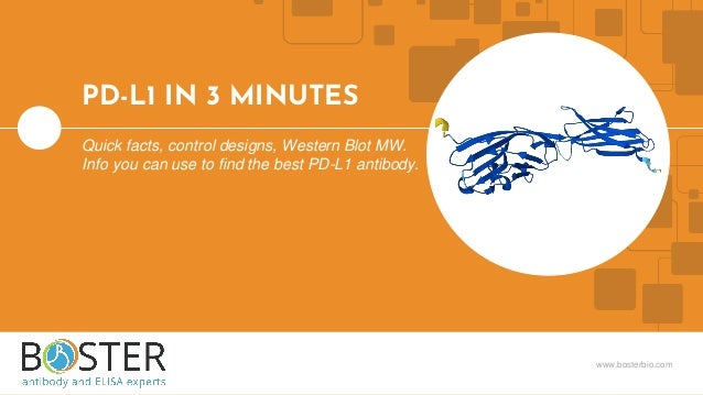
- 1. www.bosterbio.com PD-L1 IN 3 MINUTES Quick facts, control designs, Western Blot MW. Info you can use to find the best PD-L1 antibody.
- 2. FACTS 01. Function, structure, etc. TISSUES 02. Expressing tissues/cells WESTERN BLOT 03. Expected band sizes HISTORY 04. …and medical significance TABLE OF CONTENTS
- 3. 3 ● Programmed cell death 1 ligand 1 (PD-L1) or B7-H1 ● Encoded by the CD274 gene ● Cell surface receptor that regulates T-cell activation and immune responses ● Binds PD-1 receptor to inhibit T-cell activation ● 290 AAs long ● Western blot: ~36-50 kDa 01. FACTS AlphaFold predicted structure Image source: https://alphafold.ebi.ac.uk/entry/Q9NZQ7
- 4. 4 02. TISSUES Image source: https://www.proteinatlas.org/ENSG00000120217-CD274/tissue
- 5. 5 03. PD-L1 WESTERN BLOT 11102 images available on PMC: https://www.ncbi.nlm.nih.gov/pmc/?term=pd-l1+western+blot&report=imagesdocsum After reviewing several WB images, we observe PD-L1 band at ~36-50 kDa. PMC6500975 Figure 3B & D Boster Bio PD-L1 Antibody (Rabbit Monoclonal) Western blot image PMC5954021 Figure 1A & C PMC7596815 Figure 5B Cell Signaling Technologies PD-L1 (D4H1Z) Rabbit mAb #60475
- 6. 6 PD-L1 was identified as a co- stimulatory receptor involved in negatively regulating immune responses. 04. HISTORY Multiple PD-L1 and PD-1 antibodies are approved by the US FDA for the treatment of several cancer types. 1999 CLINICAL POTENTIAL
- 7. 3942 B Valley Ave Pleasanton, CA 94566 Tel: (888) 466-3604 Email: support@bosterbio.com www.bosterbio.com
Editor's Notes
- Does your next experiment involve PDL1? This video is for scientists who want to run Western blot, IHC, flow or other immunoassays against PDL1.
- We will cover some basic facts, protein expression, and PDL1’s expected behavior in Western blot. All information in this video is based on public information and no proprietary experimental evidence was used.
- Topic 1: Quick facts about PDL1. Programmed cell death 1 ligand 1, or PDL1, is part of the B7 family of cell surface receptors which regulate immune responses through either costimulatory or coinhibitory signaling. PDL1 is 290 amino acids long and appears at between 35 and 50 kilodaltons on Western blots.
- Topic 2: Where is PDL1 expressed? High levels of PDL1 have been found in the bone marrow and lymphoid tissues, connective and soft tissues, and the respiratory system, with the highest levels observed in female reproductive tissues. PDL1 is expressed at lower levels in the seminal vesicles, skeletal muscle, and colon. RNA expression has also been reported for PDL1 in the brain, eye, endocrine system, skin, urinary bladder, and pancreas. You can find more information at protein atlas dot org including which cell lines express PDL1 and which do not. This information will come in handy when designing positive and negative controls for western blot and immunohistochemistry.
- Topic 3: PDL1 in Western blotting. Antibody specificity can make or break an experiment. After looking through Western blot images from publications and antibody companies, we can see that typically one band can be expected in a PDL1 Western blot at around 50 kilodaltons. Check out the PMC link for more images, which is also provided in the description box.
- Topic 4: Some interesting facts about PDL1. Overall, PDL1 is an important molecule for proper immune function. Since the discovery of PDL1 in 1999, significant progress has been made in understanding the role of PDL1 in the immune evasion of cancer cells. The development of PDL1 and PD1 targeting therapies has been a major breakthrough in the treatment of numerous cancer types leading to the 2018 Nobel Prize in Medicine being awarded to Tasuka Honjo and James P. Allison “for their discovery of cancer therapy by inhibition of negative immune regulation.”