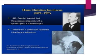
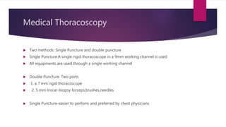
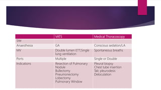
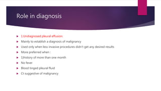
Thoracoscopy was developed in the early 1900s to break adhesions in tuberculosis patients and diagnose pleural lesions. There are two main types - medical thoracoscopy using one or two ports, and VATS which uses multiple ports and often single lung ventilation. Thoracoscopy is used to diagnose undiagnosed pleural effusions, establish malignancy, or for pleurodesis in malignant effusions. It is also used to treat parapneumonic effusions, pneumothorax by treating bullae or creating pleurodesis, and can remove retained blood clots in haemothorax. Complications are generally low but include prolonged air leak, bleeding, pneumonia and empyema.
2. History:
Developed by Jacobeus in early 1900s to break adhesions in tuberculosis patients
Also was used to localize and diagnose benign and malignant lesions of pleura
Later it was used extensively to assist in diagnosis of pleural effusion
5. Medical Thoracoscopy
Two methods: Single Puncture and double puncture
Single Puncture:A single rigid thoracoscope in a 9mm working channel is used
All equipments are used through a single working channel
Double Puncture: Two ports
1. a 7 mm rigid thoracoscope
2. 5 mm trocar-biopsy forceps,brushes,needles
Single Puncture-easier to perform and preferred by chest physicians
6. VATS Medical Thoracoscopy
Site
Anaesthesia GA Conscious sedation/LA
MV Double lumen ETT,Single
lung ventilation
Spontaneous breaths
Ports Multiple Single or Double
Indications Resection of Pulmonary
Nodule
Bullectomy
Pneumonectomy
Lobectomy
Pulmonary Window
Pleural biopsy
Chest tube insertion
Talc pleurodesis
Deloculation
7.
8. Role in diagnosis
1.Undiagnosed pleural effusion:
Mainly to establish a diagnosis of malignancy
Used only when less invasive procedures didn’t get any desired results
More preferred when :
1)history of more than one month
No fever
Blood tinged pleural fluid
Ct suggestive of malignancy
9. 2.Malignant pleural effusions:
Mainly for pleurodesis
Especially in loculated effusions
Ovarian cancer and pleural effusion-because the amount of material in pleural
effusion,dictate surgical therapy
10. 3.Parapneumonic pleural effusions
If not drained by thoracocentesis or ICD
CT Scan should be done prior to know the exact location of the empyema and
need for intervention
If fibropurulent material cannot be drained-convert to a open thoracotomy
11. If there is undrained empyema,there are usually four basic alternatives:
A)insert one more ICD tube
B)Instill fibrinolytics and dna ase intrapleurally
C)thoracoscopy
D)Thoracotomy
12. 4.Pneumothorax
Mainly done for two objectives:
A)to treat the bullous disease responsible for pneumothorax
B)to create a pleurodesis
Bullae- treated by endoscopic stapling device
Earlier-Electrocoagulation and roeder loops was used
Howover using a loop can have high chance of recurrence.
13. Mostly VATS is used, But medical thoracoscopy with talc insufflation is also being
used
ACCP Guidelines :
thoracoscopy preferred for primary spontaneous pneumothorax ,usually after an
ipsilateral recurrence
Bullectomy and parietal pleural ablation also has been recommended
Secondary spontaneous pneumothorax-always indicated
14. BTS-chemical pleurodesis to be performed only if the patient is unwilling for
surgical intervention
Indications :
A)Secondary ipsilateral pneumothorax
B)First contralateral pneumothorax
C)Bilateral spontaneous pneumothorax
D)Persisitent Air Leaks (5-7 days of tube drainage)
E)Professions at risk (pilots,divers)
15. 5.Haemothorax
Usually thoracotomy is more preferred to thoracoscopy
Can be used for removing retained blood clots
If retained blood clots are more than 30 percentage usually thoracotomy is
preferred
16. Complications
Prolonged Air Leak – 3.2 percentage
Significant bleeding –only in 1 percent
Pneumonia-1.1 %
Empyema – 0.6 %
Complications of medical thoracoscopy is less than VATS