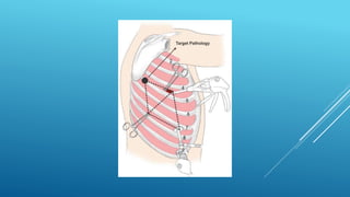
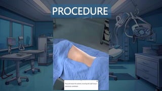
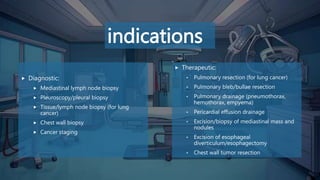
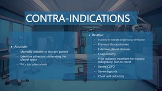
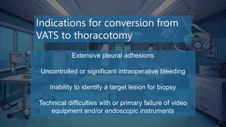
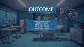
VIDEO-ASSISTED
THORACIC SURGERY
(VATS)
Prepared by:
 Racheen S. Haji
University of zakho
Faculty of medical sciences
School of medicine
Surgical department
DEFINITION
DEFINITION
(VATS): is minimally invasive thoracic surgery that does not use a formal thoracotomy
incision, it is principally employed in the management of (pulmonary, mediastinal, and
pleural pathology.
Its benefits:
• Can be performed in a short time period.
• Less postoperative morbidity.
• Earlier return to normal activity.
The standard VATS pleurodesis procedure requires three ports placed
• at the 7th intercostal space mid-axillary line.
• 4th intercostal space anterior axillary line
• the last port just anterior to the tip of the scapula.
(Triangulated fashion)
PROCEDURE
PROCEDURE
EQUIPMENT
• Double sided endotracheal tube
• Thoracoscope, with lens and video camera
• A light source and cable
• Long-blade diathermy pen
• Endoscopic biopsy forceps
• Endoscopic staple-transection devices
• Rigid or flexible trocar cannula
• Thoracotomy tray
• Chest tube drainage device with water seal
• Suction source and tubing
INDICATIONS
indications
 Diagnostic:
 Mediastinal lymph node biopsy
 Pleuroscopy/pleural biopsy
 Tissue/lymph node biopsy (for lung
cancer)
 Chest wall biopsy
 Cancer staging
 Therapeutic:
• Pulmonary resection (for lung cancer)
• Pulmonary bleb/bullae resection
• Pulmonary drainage (pneumothorax,
hemothorax, empyema)
• Pericardial effusion drainage
• Excision/biopsy of mediastinal mass and
nodules
• Excision of esophageal
diverticulum/esophagectomy
• Chest wall tumor resection
CONTRA-
INDICATIONS
CONTRA-INDICATIONS
 Absolute:
• Markedly unstable or shocked patient
• Extensive adhesions obliterating the
pleural space
• Prior talc pleurodesis
 Relative:
• Inability to tolerate single-lung ventilation
• Previous thoracotomies
• Extensive pleural diseases
• Coagulopathy
• Prior radiation treatment for thoracic
malignancy; plan to resect
• Severe COPD
• Severe hypoxia
• Chest wall deformity
COMPLICATIONS
COMPLICATIONS
Postoperative air leak
Post-operative pain
Hypoxemia
Atelectasis
Bleeding
Wound infection
Intercostal nerve damage
Tumor implantation following VATS
Indications for conversion from
VATS to thoracotomy
Extensive pleural adhesions
Uncontrolled or significant intraoperative bleeding
Inability to identify a target lesion for biopsy
Technical difficulties with or primary failure of video
equipment and/or endoscopic instruments
OUTCOME
OUTCOME
overall survival rates of:
100% after 1 year
85.9% after 2 years
65.3% after 3 years
55.9% after 4 years
THANKS FOR YOUR ATTENTIONS.
Any question…

Video-assisted thoracic surgery (VATS)

  • 1.
    VIDEO-ASSISTED THORACIC SURGERY (VATS) Prepared by: Racheen S. Haji University of zakho Faculty of medical sciences School of medicine Surgical department
  • 3.
  • 4.
    DEFINITION (VATS): is minimallyinvasive thoracic surgery that does not use a formal thoracotomy incision, it is principally employed in the management of (pulmonary, mediastinal, and pleural pathology. Its benefits: • Can be performed in a short time period. • Less postoperative morbidity. • Earlier return to normal activity.
  • 5.
    The standard VATSpleurodesis procedure requires three ports placed • at the 7th intercostal space mid-axillary line. • 4th intercostal space anterior axillary line • the last port just anterior to the tip of the scapula. (Triangulated fashion)
  • 7.
  • 8.
  • 9.
    EQUIPMENT • Double sidedendotracheal tube • Thoracoscope, with lens and video camera • A light source and cable • Long-blade diathermy pen • Endoscopic biopsy forceps • Endoscopic staple-transection devices • Rigid or flexible trocar cannula • Thoracotomy tray • Chest tube drainage device with water seal • Suction source and tubing
  • 18.
  • 19.
    indications  Diagnostic:  Mediastinallymph node biopsy  Pleuroscopy/pleural biopsy  Tissue/lymph node biopsy (for lung cancer)  Chest wall biopsy  Cancer staging  Therapeutic: • Pulmonary resection (for lung cancer) • Pulmonary bleb/bullae resection • Pulmonary drainage (pneumothorax, hemothorax, empyema) • Pericardial effusion drainage • Excision/biopsy of mediastinal mass and nodules • Excision of esophageal diverticulum/esophagectomy • Chest wall tumor resection
  • 20.
  • 21.
    CONTRA-INDICATIONS  Absolute: • Markedlyunstable or shocked patient • Extensive adhesions obliterating the pleural space • Prior talc pleurodesis  Relative: • Inability to tolerate single-lung ventilation • Previous thoracotomies • Extensive pleural diseases • Coagulopathy • Prior radiation treatment for thoracic malignancy; plan to resect • Severe COPD • Severe hypoxia • Chest wall deformity
  • 22.
  • 23.
    COMPLICATIONS Postoperative air leak Post-operativepain Hypoxemia Atelectasis Bleeding Wound infection Intercostal nerve damage Tumor implantation following VATS
  • 24.
    Indications for conversionfrom VATS to thoracotomy Extensive pleural adhesions Uncontrolled or significant intraoperative bleeding Inability to identify a target lesion for biopsy Technical difficulties with or primary failure of video equipment and/or endoscopic instruments
  • 25.
  • 26.
    OUTCOME overall survival ratesof: 100% after 1 year 85.9% after 2 years 65.3% after 3 years 55.9% after 4 years
  • 27.
    THANKS FOR YOURATTENTIONS. Any question…