This document provides an overview of motor imagery (MI), also known as mental imagery. It discusses the history, theories, types, neurophysiology, frameworks for practice, and supporting evidence of MI. MI involves mentally simulating a motor action without physical movement. It is used in rehabilitation to improve motor skills after injuries. Studies show MI activates similar brain regions as physical practice and can improve motor performance. The document reviews evidence that MI training can benefit recovery from stroke, spinal cord injury, Parkinson's disease, and other conditions. Outcomes used to measure MI ability include movement and imagery questionnaires.
3. Introduction
• MI also known as mental imagery
• Imagining of an action without its physical execution; using all of the senses.
• It is an active process during which the representation of an action is internally
reproduced within working memory without any overt (observable) output.
Used
• To treat people who have health problems, including injury to the spinal cord,
stroke, Parkinson's disease, or fibromyalgia.
• Walking, Relearning daily activities, Awareness of affected limb, etc.
4. • Studies have shown that this "mental rehearsal/motor imagery practice” i.e,
repetition or rehearsing of imagined motor acts with the intention of improving
their physical execution actually stimulates the brain areas responsible for
making the weaker arm or leg move.
• In a study Jeannerod, stated that conscious MI and unconscious motor
preparation share common mechanics and are functionally equivalent.
• So the mental practice using MI training results in improvement in motor
performance.
5. History
• Johann Friedrich Herbart in 1825, proposed that the imagery of perceptual
effects can elicit the related movements.
• William James in 1890, wrote “that every representation of a movement
awakens in some degree the actual movement”.
• In rehabilitation, Fansler CL in 1980s stated that mental practice can improve
the motor performance, in his study “Effects of mental practice on balance in
elderly women.”
6. • In sports, Studies have shown that specific training can increase the amount
and the efficiency of kinaesthetic imagery.
• And also explained that by practicing imagery in the same way as we practice
the skill itself, the desired neural pathways are strengthened.
• Motor imagery is a cognitive tool strategically used by athletes for learning
and optimising their specific movement tasks.
• Boschker, 2001 stated, the main aim of motor imagery is to enhance specific
motor actions.
• Dancers, for example, use motor imagery to exercise the memorisation of
long sequences and to improve movement quality in terms of spatiotemporal
adaptation and artistic expression.
7. Theories of mental imagery
• Abstract/Symbolic theories:
• Mental “images” aren’t images at all
• Mental imagery consists of the manipulation of abstract symbols in the brain
(like computer code)
• Analog/Pictoral theories:
• Mental imagery is similar to perception.
• These are current dominating theories.
8. Other theories of mental imagery
• Psychoneuromuscular theories
• Imagery programs muscles for action
• Imagery facilitates the learning of motor skills because imagined events
innervate the muscles as physical practice, they strengthen neural pathways.
• Symbolic theory :
• Breaking task into symbols, represent things by symbols.
9. • Bioinformational theory :
• Basketball free hit actions actually helps in proper execution of action.
• Triple code model:
• Primary importance is placed in the psychophysiology of imagery
• Image : sensory experience
• Somatic : psychological response
• Meaning of the image (personal)
11. Types of motor imagery
• Kinaesthetic motor imagery (internal )(i.e. imagining the feeling
associated with performing a movement)
• Visual motor imagery (external)(i.e. imagining the movement itself)
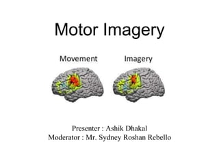
12. Neurophysiology of motor imagery
• Premotor cortex and rostral part of the posterior SMA were activated
bilaterally.
• Mental imagery activation is 30% of the level seen in actual performance
motor cortex.
13. • Recent research works suggest that motor imagery of different body parts has
significant relation with the specific areas (in homunculus) as shown in the fig
below.
14. • When we perform particular movement or imagine the movement, motor imagery
could produce ERD (event related desynchronisation) in the corresponding
hemisphere and ERS (event related synchronisation) in the opposite hemisphere.
17. Neuroscience and MI
• Technology used to research the brain while an imagery task is
performed.
• PET scan
• SPECT
• fMRI = measures hemodynamic response to neural activity.
• ;- because of their spatial and temporal resolution
• Because these technique have been applied to the study of
mapping the neural basis of motor imagery.
18. Developing a framework
• The subjects may imagine the movement in:
• 3rd person perspective (or external imagery)
• 1st person perspective (or internal imagery)
• 3 pillars in developing imagery framework
• The patient - pt’s choose activities
• Evidence - mental practice may alter neural function
• Therapist - Utilise 5-step outline to keep intervention dynamic.
19. • 5 steps outline to intervention
1. Suitable candidate
2. Nature of treatment
3. Teach
4. implant, incorporate, monitor
5. PT reduces support gradually
20. Four W’s of imagery in rehabilitation
• Where
• When
• Why
• What
21. Evidence supporting effect of motor imagining
• Motor imagery is practiced to improve motor performance and to learn
motor tasks.
• Abundant evidence on the positive effects of MI practice on motor
performance and learning has been published.
• For individuals who are healthy or those who have health-related
problems, the rehearsal or practice of imagery tasks has been proven to be
either beneficial by itself or in addition to physical practice.
22. For healthy individual and athletes
• Studies of healthy individuals have shown enhancement of the performance of
various aspects of motor control because of MI practice.
• Including gains in strength of selected muscle groups, improved speed in arm
pointing capacity, increased range of motion of the hip joint when MI was added to
proprioceptive neuromuscular facilitation, and improved postural control in elderly
people.
23. • Recently, the use of MI practice to facilitate the mastering of professional
skills such as nursing and surgery has been demonstrated.
• For imagery applied in the sports, positive effects have been reported in
speed, performance accuracy, muscle strength, and motor skill
performance.
24. MI for rehabilitation
• In conditions like stroke, spinal cord injury (SCI), Parkinson disease (PD), and
people with intractable pain.
25. 1. Stroke
• Sharma et al in her systemic review in 2006, characterized motor imagery as a
“backdoor” to accessing the motor system and rehabilitation at all stages of stroke
recovery because “it is not dependent on residual functions yet still incorporates
voluntary drive”
• The literature suggests the encouraging effect of motor imagery training on motor
recovery after stroke.
• Based on the available literature in healthy volunteers, robust (vigorous) activation of
the nonprimary motor structures, but only weak and inconsistent activation of M1,
occurs during motor imagery.
• Provided appropriate methodology, motor imagery may provide a valuable tool to
access the motor network and improve outcome after stroke.
26. MI in UE recovery of function with stroke patients
2. Seong-Sik kim, PT, PhD and , Byoung-Hee Lee, PT, PhD conducted a
RCT ,2015
• Population : 24 patients, control group 12 and motor imagery group 12.
• Intervention : The MI group participated in 30 minutes of MI training on
each of the 18 tasks (9 hours total) related to their daily living activities.
• After the 4-week intervention period, the FMA - Upper Extremity
outcomes and Wolf Motor Function Test outcomes were compared.
27. • Results : The post-test score of the motor imagery group on the outcomes was
significantly higher than that of the control group.
• Conclusion : Motor imagery training has a positive influence on upper
extremity performance by improving functional mobility during stroke
rehabilitation. These results suggest that motor imagery training is feasible and
beneficial for improving upper extremity function in stroke patients.
28. MI for gait training
1. Ruth Dickstein, Ayelet Dunsky, Emanuel Marcovitz conducted a case report
• A 69 year old man with left hemiparesis received MI gait practice for 6 weeks.
Intervention focused on task-oriented gait and on impairments of the affected lower
limb.
• Outcomes: temporal-distance stride parameters and sagittal kinematics of the knee
joint were taken.
• Preintervention, midterm, postintervention, and follow-up measurements were
taken.
29. • Intervention : 1st 4 weeks focused on the specific gait impairments and on
improving speed and symmetry, last 2 weeks to the performance of
functional task-oriented gait activities
• Results : At 6 weeks postintervention, the patient had a 23% increase in
gait speed and a 13% reduction in double-support time. An increase in
range of motion of the knees, No changes in gait symmetry were noted.
• Conclusion : MI may be useful for the enhancement of walking ability in
patients following stroke.
30.
31.
32. 2. Gyuchang Lee, PT, PhD and collegues conducted a study to investigate the effects of motor imagery
training on improvement of gait ability of patients with chronic stroke
• The motor imagery training was performed using imagination of normal gait
movement.
• Participants were randomly allocated to two groups: a motor imagery training
group 13 and a control group 11.
• Both groups received treadmill training for 30 minutes 3 sessions per week
for 6 weeks. The motor imagery training group practiced additional motor
imagery training for 15 minutes. Measures were evaluated by gait ability.
33. • Results : The outcomes significantly improved by motor imagery training
were— gait speed, step length paretic side, step length of non-paretic
side, stride length of paretic side, stride length of non-paretic side, single
limb support period of paretic side, and double limb support period of
both sides.
• Conclusion: The motor imagery training improved gait ability. These
results suggest that motor imagery training is feasible and suitable for
individuals with stroke.
34. 3. Gait rehabilitation of chronic post-stroke hemiparesis
• 17 post stroke patients, MI training only
• Intervention : 15-20 min sessions, 3/week for 6 weeks
• Results : increased walking speed, stride length, and single-leg stance time
(affected LE)
• Conclusion : Improved mobility and dynamic balance.
35. 2. Spinal cord injury
1. Study done by Steven C. Cramer and collegues “Effects of motor imagery
training after chronic, complete spinal cord injury”
• Subjects : 10 subjects with SCI and complete paraplegia and 10 healthy
controls underwent assessment before and after 7 days of motor imagery
training to tongue and to foot.
• Results : Motor imagery training significantly improved the behavioral
outcome measure, speed of movement, in non-paralyzed muscles.
36. • Training was also associated with increased fMRI activation in left putamen, an
area associated with motor learning, during attempted right foot movement in both
groups, despite foot movements being present in controls and absent in subjects
with SCI.
• The current study found that motor imagery training improves motor performance
and alters brain function in subjects with complete SCI despite lack of voluntary
motor control and peripheral feedback.
• Motor imagery might be of value as one component of a restorative intervention.
37. 2. Study conducted by Alkadhi H at el., “What disconnection tells about motor
imagery: evidence from paraplegic patients”
• They investigated patients with complete SCI to find out how the complete
disruption of motor efferents and sensory afferents influences brain activation
during motor imagery of the disconnected feet.
• Subjects : 8 SCI patients underwent behavioural assessment and fMRI.
• Results : When compared to a healthy population, stronger activity was detected
in primary and all non-primary motor cortical areas and subcortical regions. In
paraplegic patients the primary motor cortex was consistently activated, even to
the same degree as during movement execution in the controls.
38. • In paraplegics the extent of activation in the primary motor cortex and in non-
primary motor areas was significantly correlated with the vividness of
movement imagery, as assessed by an interview.
• The present findings provide new insights on the neuroanatomy of motor
imagery and the possible role of kinaesthetic feedback in the suppression of
cortical motor output required during covert movements.
39. 3. Parkinson disease
• For individuals with PD, the ability to apply MI is controversial
• The authors interpreted these findings as disordered imagery ability resulting
from deficits in dopamine inputs to the basal ganglia in patients with PD
• In patients with PD treated with dopaminergic stimulation, imagery-related
enhancement in several sites of the brain was noted during the “on” phase but not
the “off” phase.
40. 1. Study done by Ruth Tamir, Ruth Dickstein, Moshe Huberman in 2007,
“Integration of Motor Imagery and Physical Practice in Group Treatment Applied
to Subjects With Parkinson’s Disease”
• Compared group treatment using a combination of physical and motor imagery
practice with group treatment using only physical practice in subjects with PD.
• Subjects : 23 patients with idiopathic PD, 12 received combined therapy,
whereas 11 received physical therapy alone. 1-h sessions held twice a week for
12 weeks.
• Comparable motor tasks provided to both groups included functional tasks, and
relaxation exercises.
41. • Outcome measures: the time required to complete sequences of movements, the
performance of balance tasks, impairment and functional scores on the Unified
Parkinson’s Disease Rating Scale (UPDRS), and specific cognitive abilities
(Stroop and clock drawing tests).
• Results: the experimental group (EG) exhibited significantly faster performance
of movement sequences than the control group. Subjects of EG demonstrated
higher gains in the mental and motor subsets of the UPDRS and in the cognitive
tests. Both groups improved on the ADL scale.
• Conclusions. The combination of motor imagery and real practice may be
effective in the treatment of PD, especially for reducing bradykinesia.
42. Damage to Fronto-Parietal Networks Impairs Motor Imagery Ability after
Stroke
• Aim: To identify brain regions that are mandatory for preserved motor imagery
ability after stroke.
• Subjects : 37 patients with hemiplegia after a first time stroke participated.
• Outcomes : Motor Imagery questionnaire and temporal congruence test. A voxelwise
lesion symptom mapping approach was used to identify neural correlates of motor
imagery in this cohort within the first year post-stroke.
43. • Results: Poor motor imagery vividness — lesions in the left putamen, left ventral
premotor cortex and long association fibers linking parieto-occipital regions with
the dorsolateral premotor and prefrontal areas. Poor temporal congruence —
rostrally located white matter of the superior corona radiata.
• Conclusion: This study confirms the association between white matter tract lesions
and impaired motor imagery ability, thus emphasising the importance of an intact
fronto-parietal network for motor imagery also the crucial role of the basal ganglia
and premotor cortex when performing motor imagery tasks.
57. References
• Dickstein R, Deutsch JE. Motor imagery in physical therapist practice. Physical
therapy. 2007 Jul 1;87(7):942-53.
• Malouin F, Richards CL. Mental practice for relearning locomotor skills. Physical
therapy. 2010 Feb 1;90(2):240-51.
• Iseki K, Hanakawa T, Shinozaki J, Nankaku M, Fukuyama H. Neural
mechanisms involved in mental imagery and observation of gait. Neuroimage.
2008 Jul 1;41(3):1021-31.
• Oostra KM, Van Bladel A, Vanhoonacker AC, Vingerhoets G. Damage to fronto-
parietal networks impairs motor imagery ability after stroke: a voxel-based lesion
symptom mapping study. Frontiers in behavioral neuroscience. 2016 Feb 1;10:5.
58. • Isaac A, Marks DF, Russell DG. An instrument for assessing imagery of
movement: the vividness of movement imagery questionnaire (VMIQ). Journal
of Mental Imagery . 1986;10:23–30
• Braun S, Kleynen M, Schols J, Schack T, Beurskens A, Wade D. Using mental
practice in stroke rehabilitation: a framework. Clinical rehabilitation. 2008
Jul;22(7):579-91.
• Cramer SC, Orr EL, Cohen MJ, Lacourse MG. Effects of motor imagery
training after chronic, complete spinal cord injury. Exp Brain Res .
2006;177:233–242.
• Sharma N, Pomeroy VM, Baron JC. Motor imagery: a backdoor to the motor
system after stroke? Stroke . 2006;37:1941–1952.
59. • Stevens J, Stoykov ME. Using motor imagery in the rehabilitation of hemiparesis.
Arch Phys Med Rehabil . 2003;84:1090–1092.
• Malouin F, Richards CL, Doyon J, et al. . Training mobility tasks after stroke with
combined mental and physical practice: a feasibility study. Neurorehabil Neural
Repair . 2004;18:66–75
• lewis, D.E., O’Reilly, M. J., Khuu, S. K., & Pearson, J. (2013). conditioning the
mind’s eye associative learning with volunatry mental imagery. clinical psychological
science, 2167702613484716
• parkinson Tamir R, Dickstein R, Huberman M. Integration of motor imagery and
physical practice in group treatment applied to subjects with Parkinson's disease.
Neurorehabil Neural Repair . 2007;21:68–75.
• Hall CR, Pongrac J. Movement Imagery Questionnaire . London, Ontario, Canada:
Department of Physical Education, University of Western Ontario; 1983.