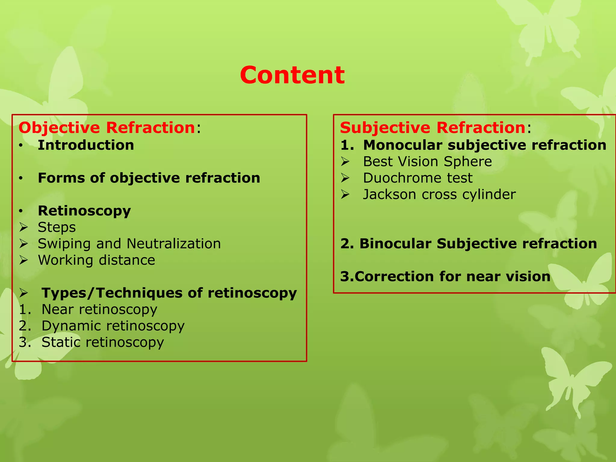
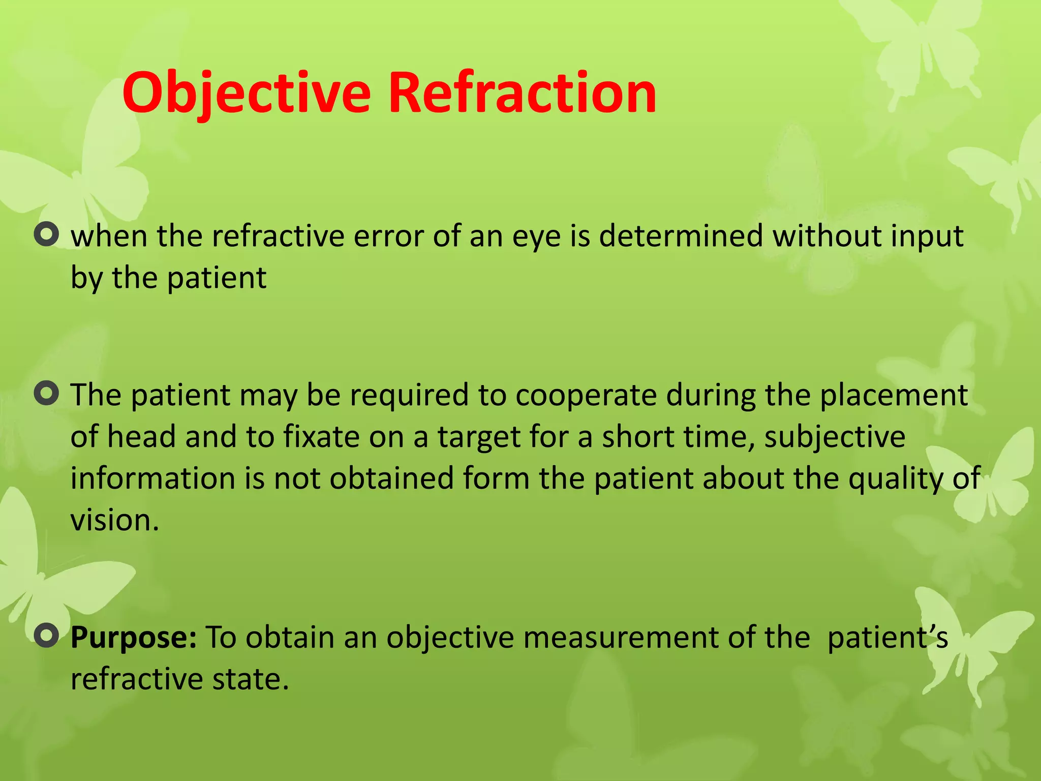
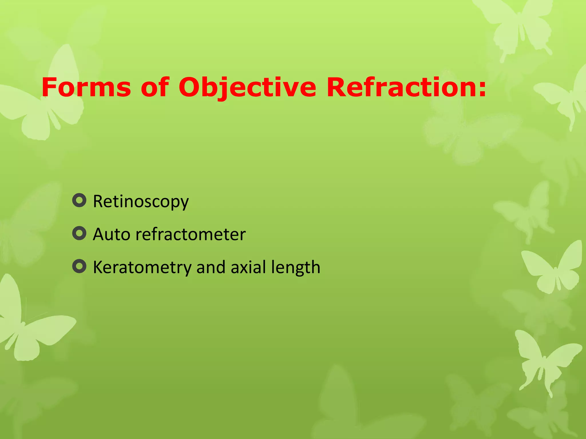
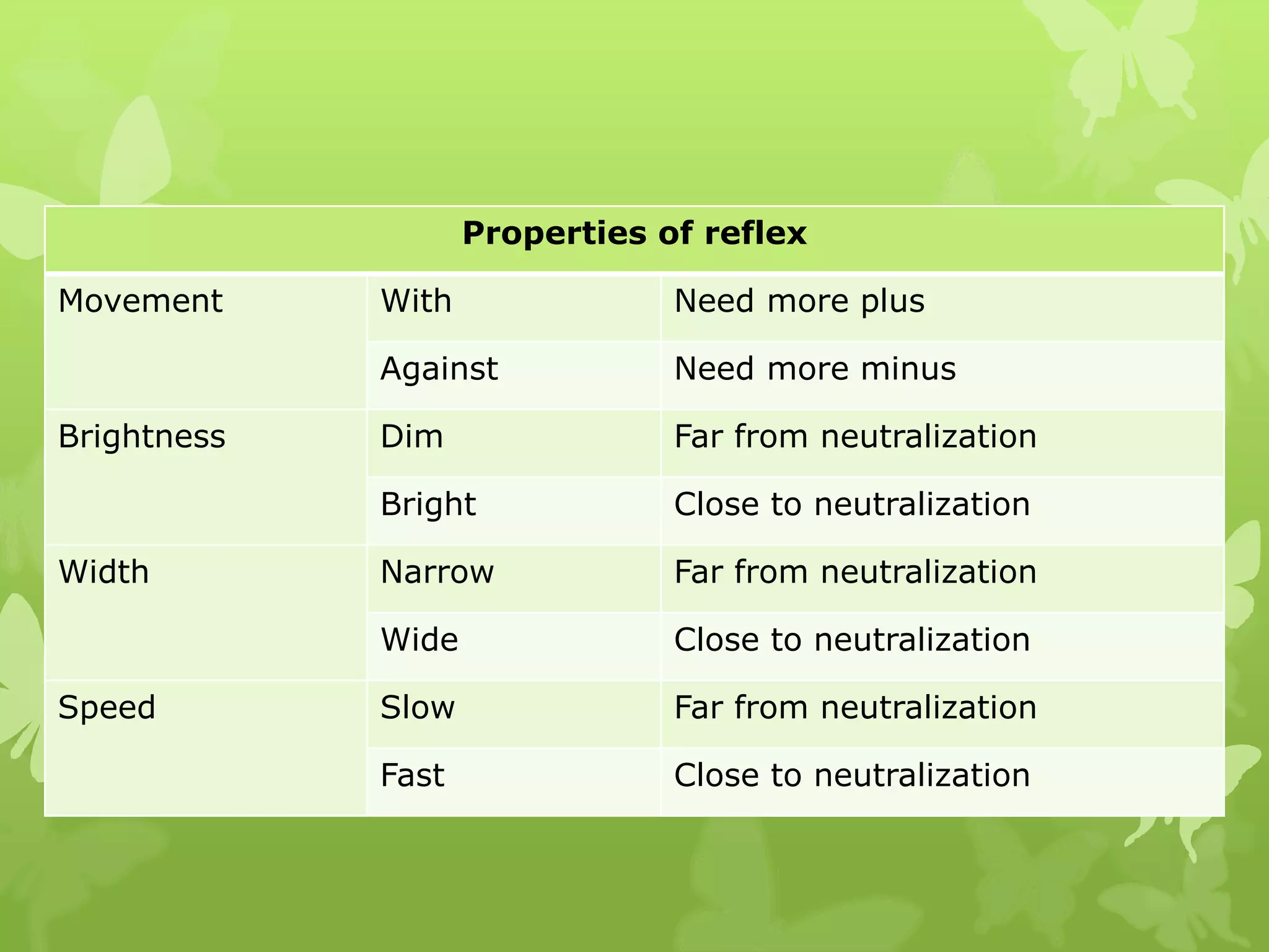
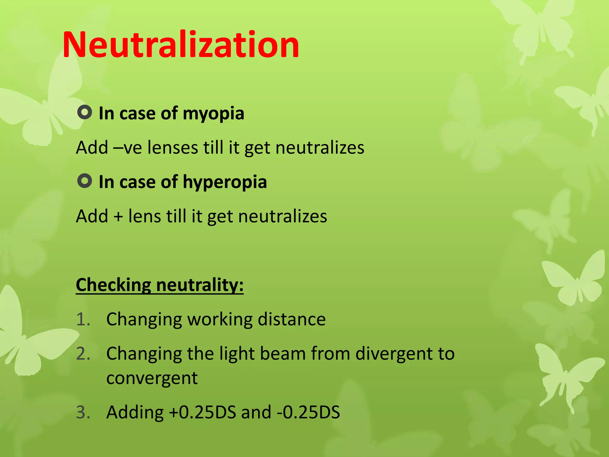
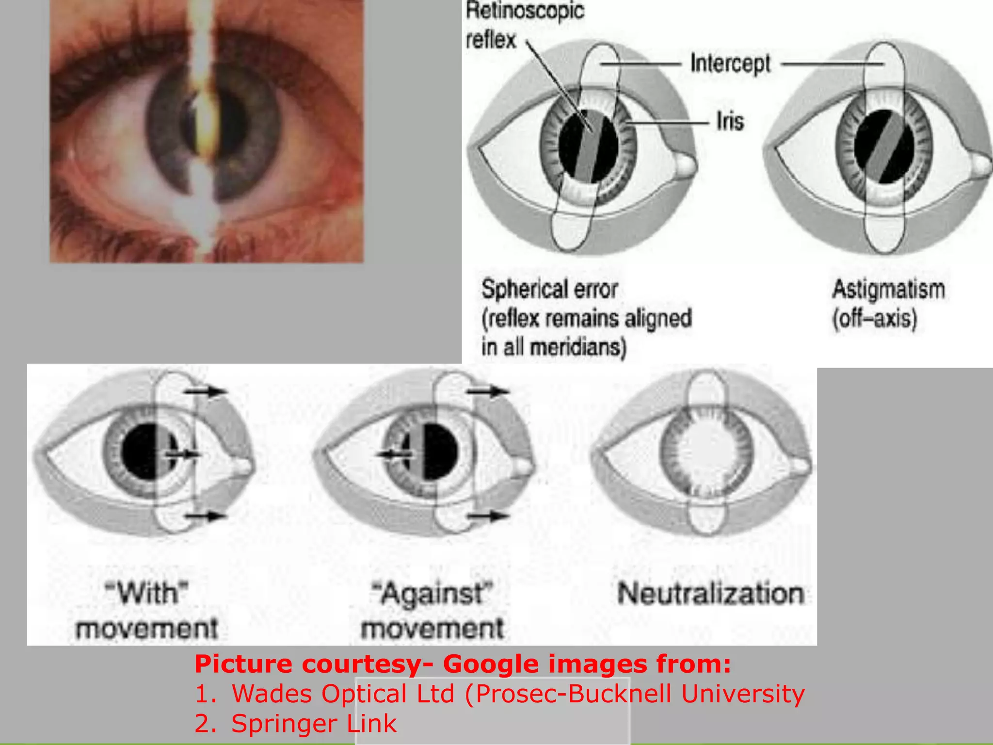
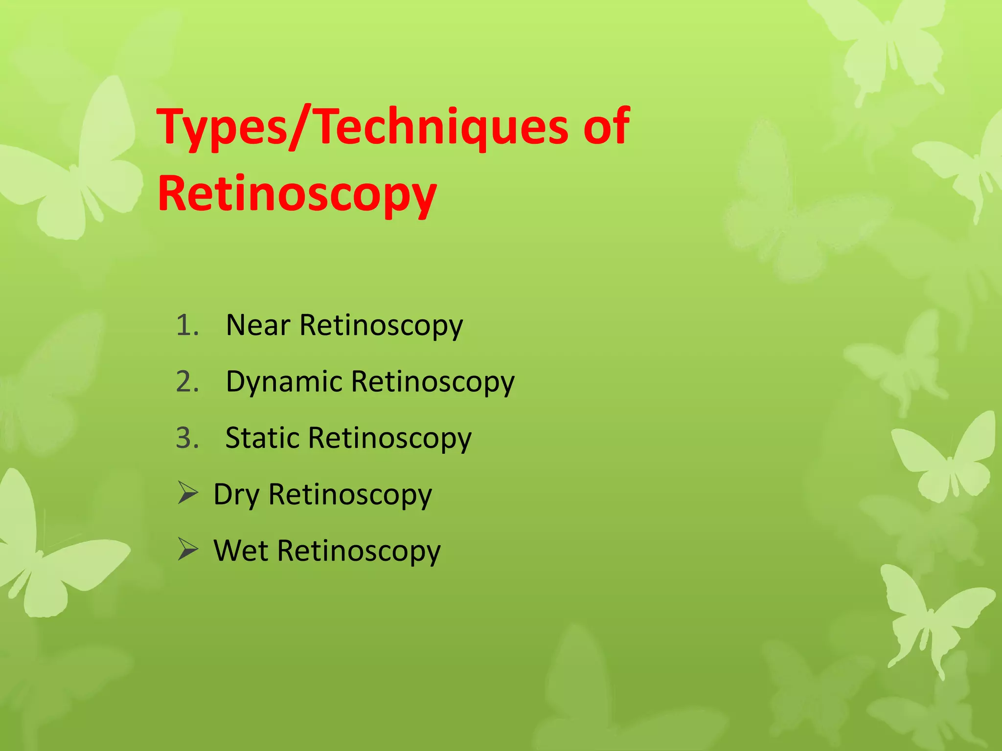
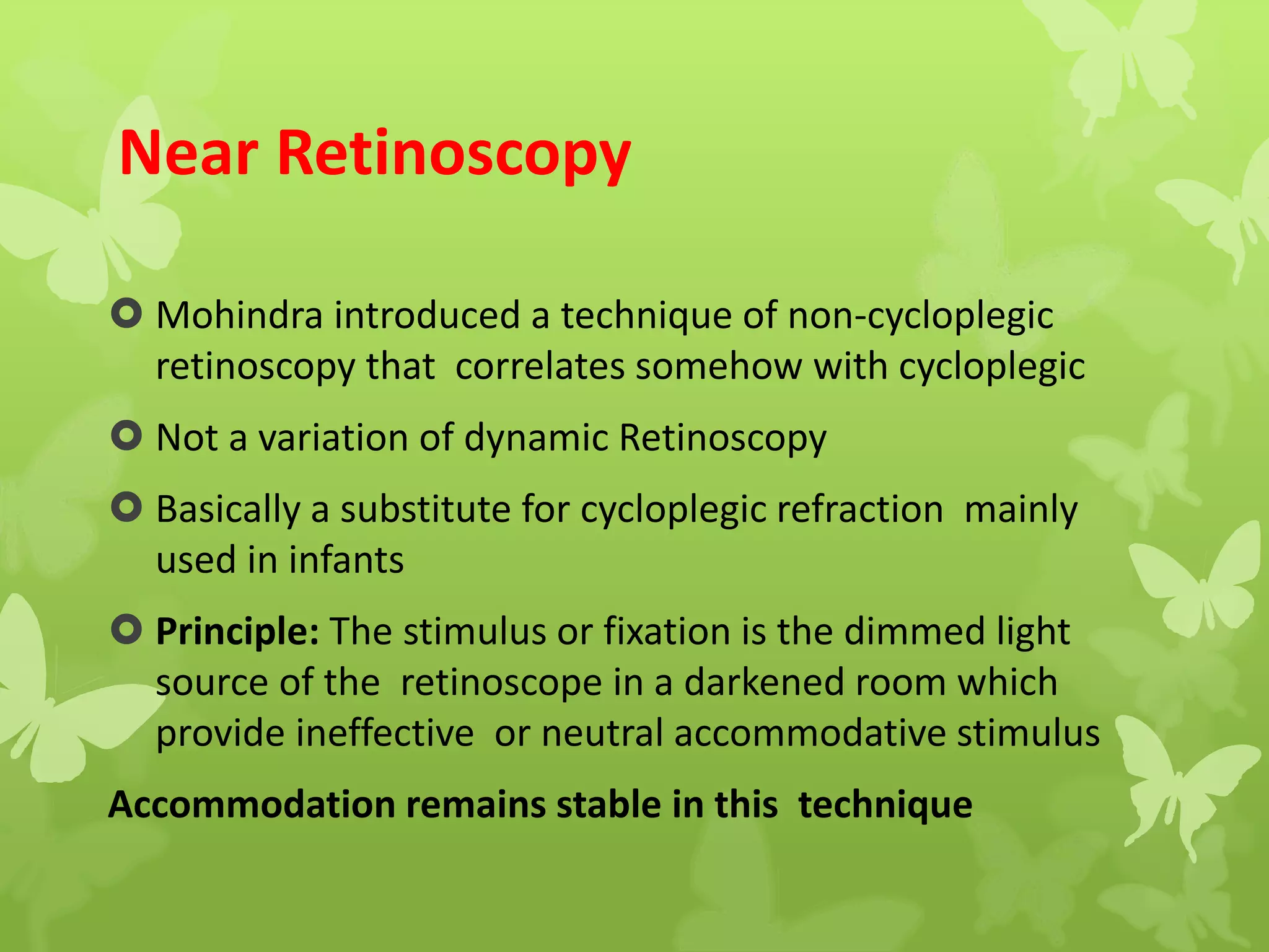
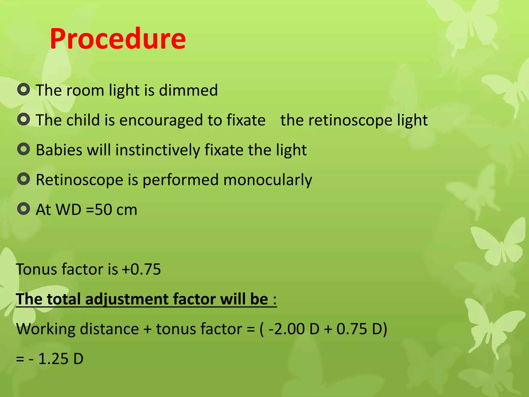
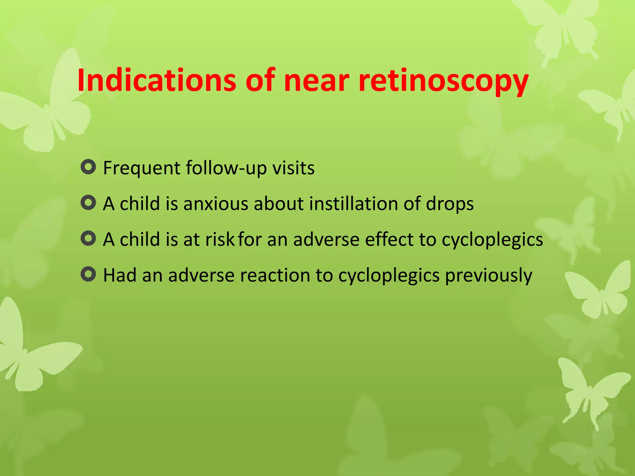
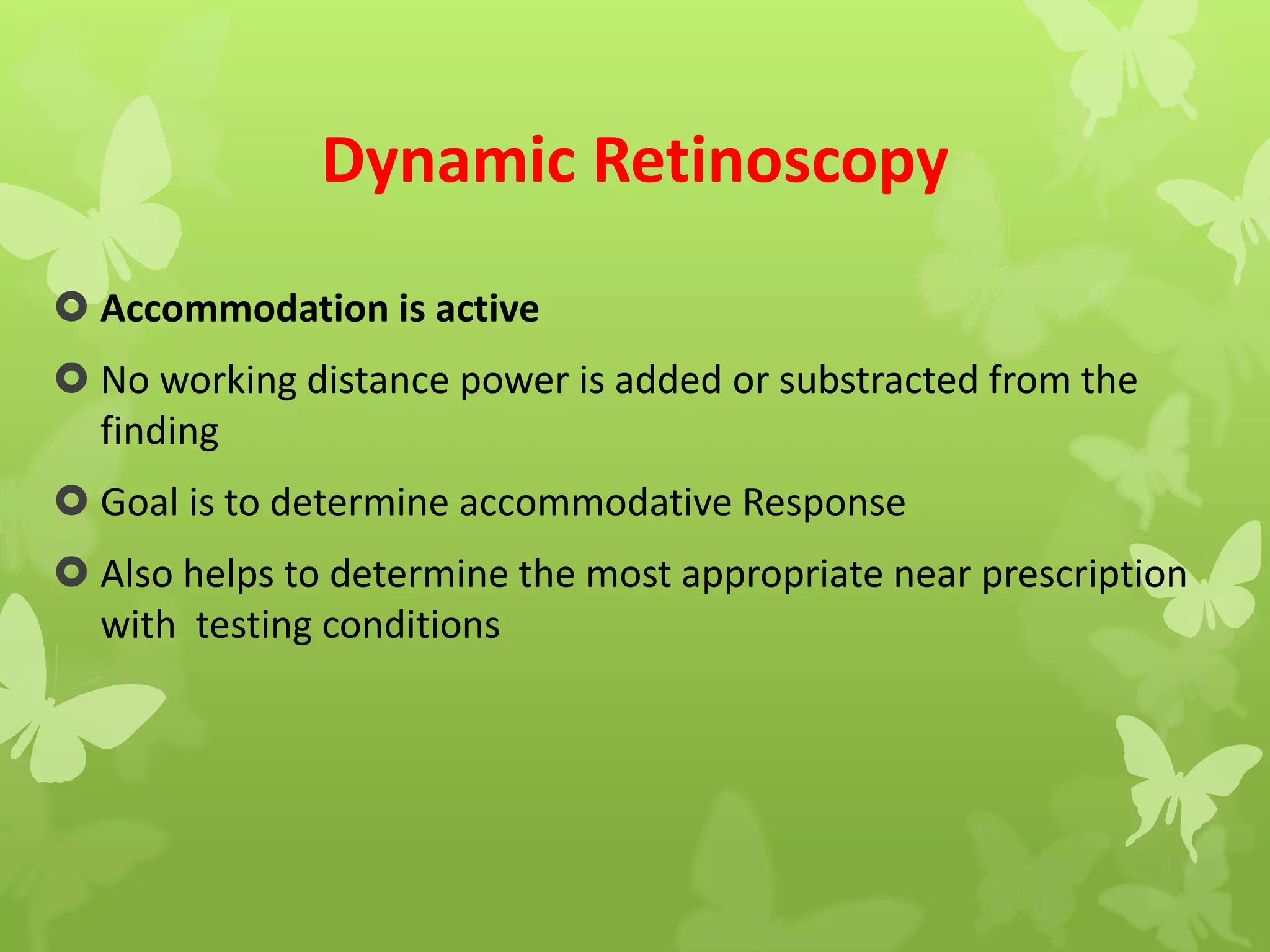
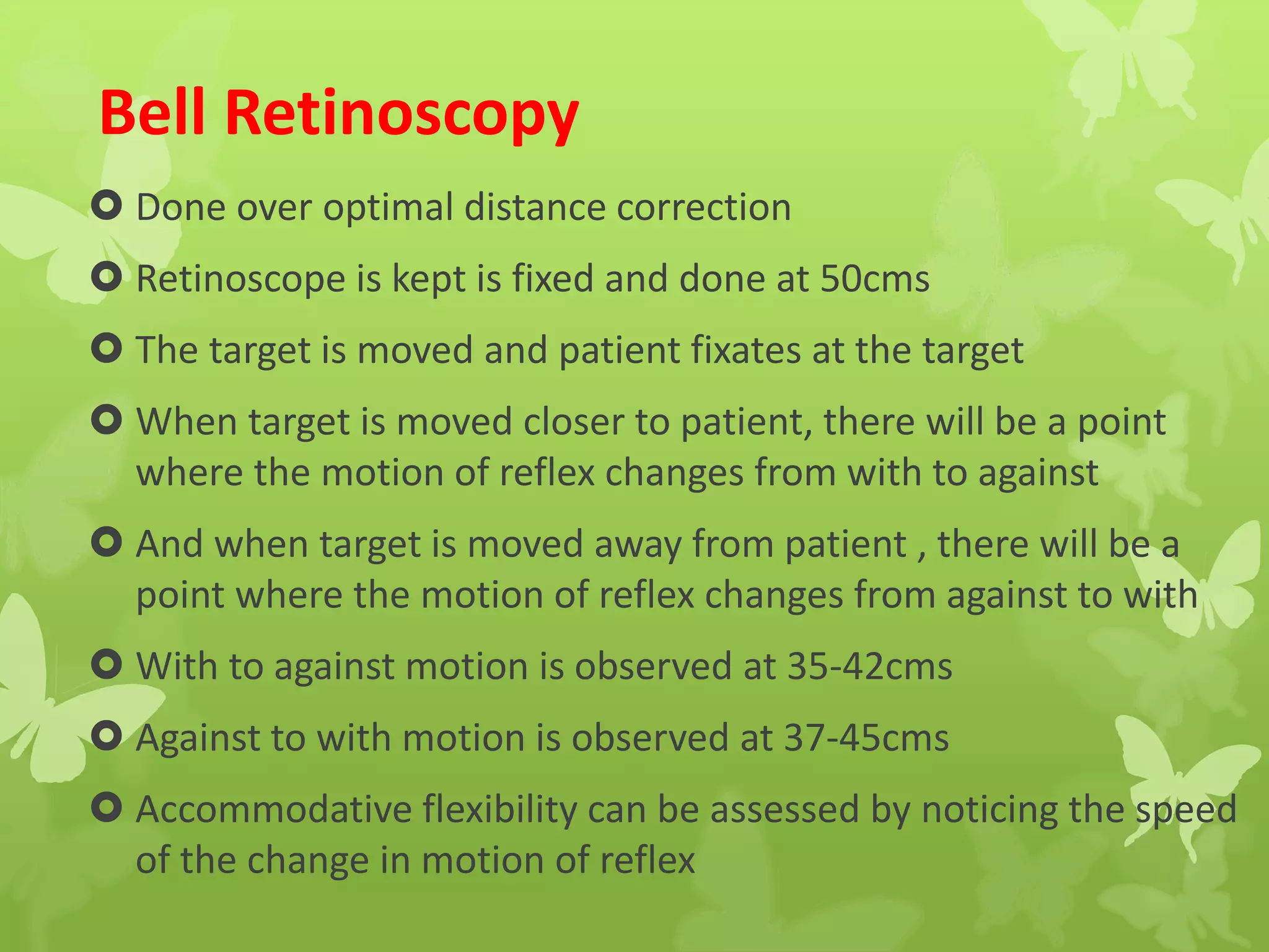
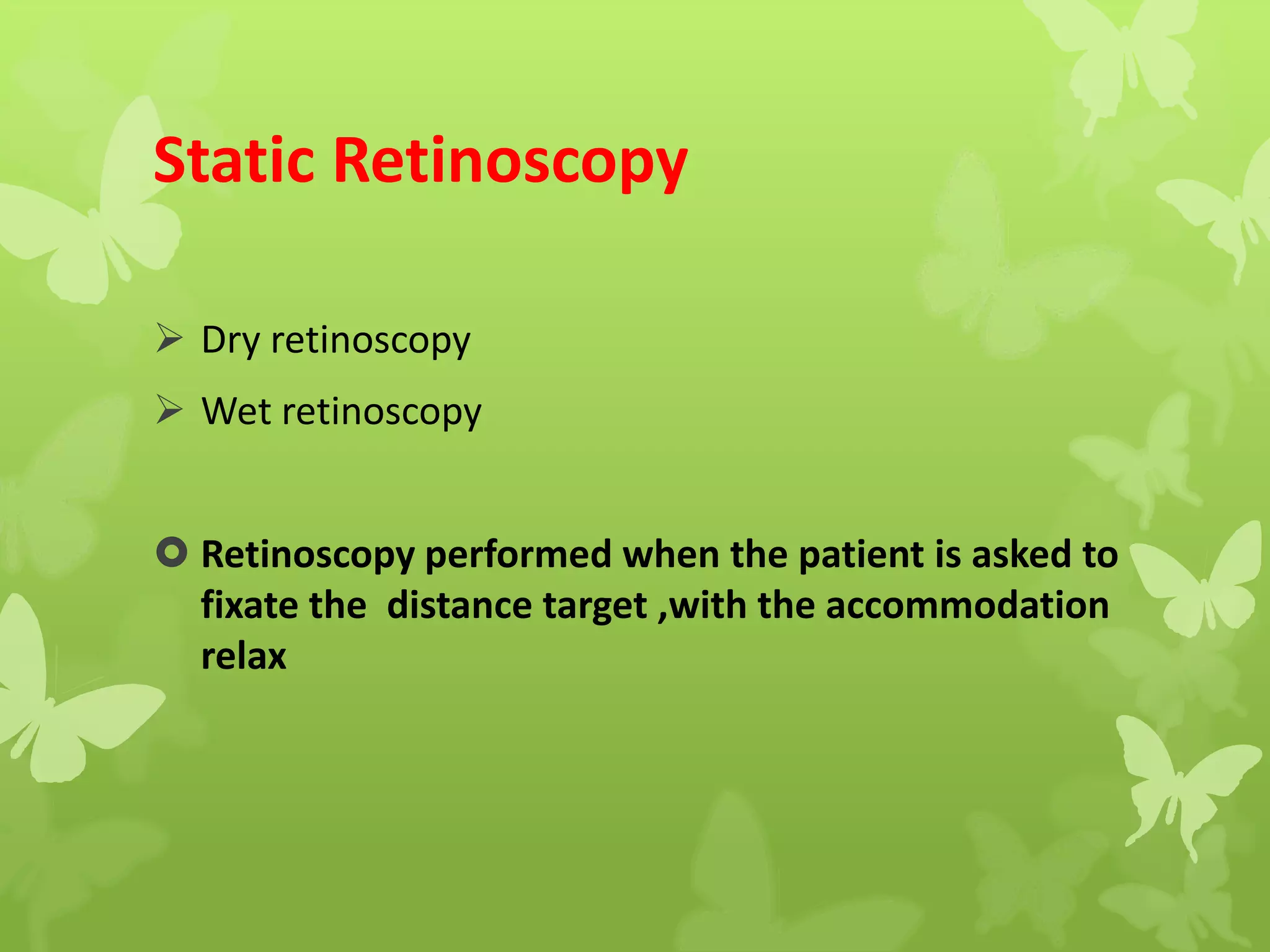
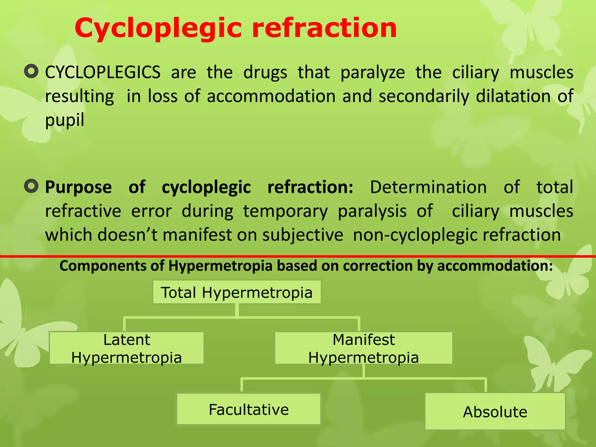
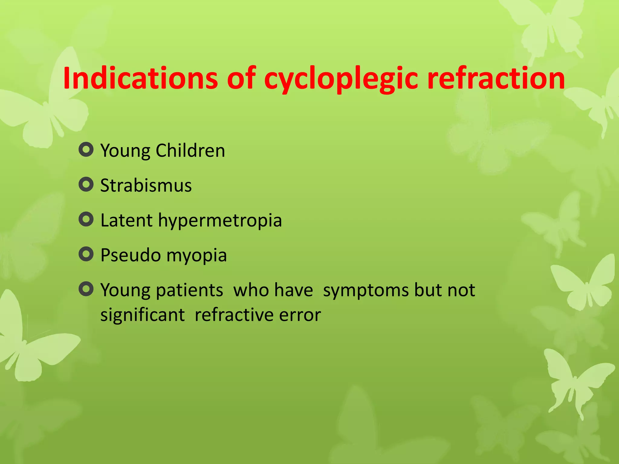
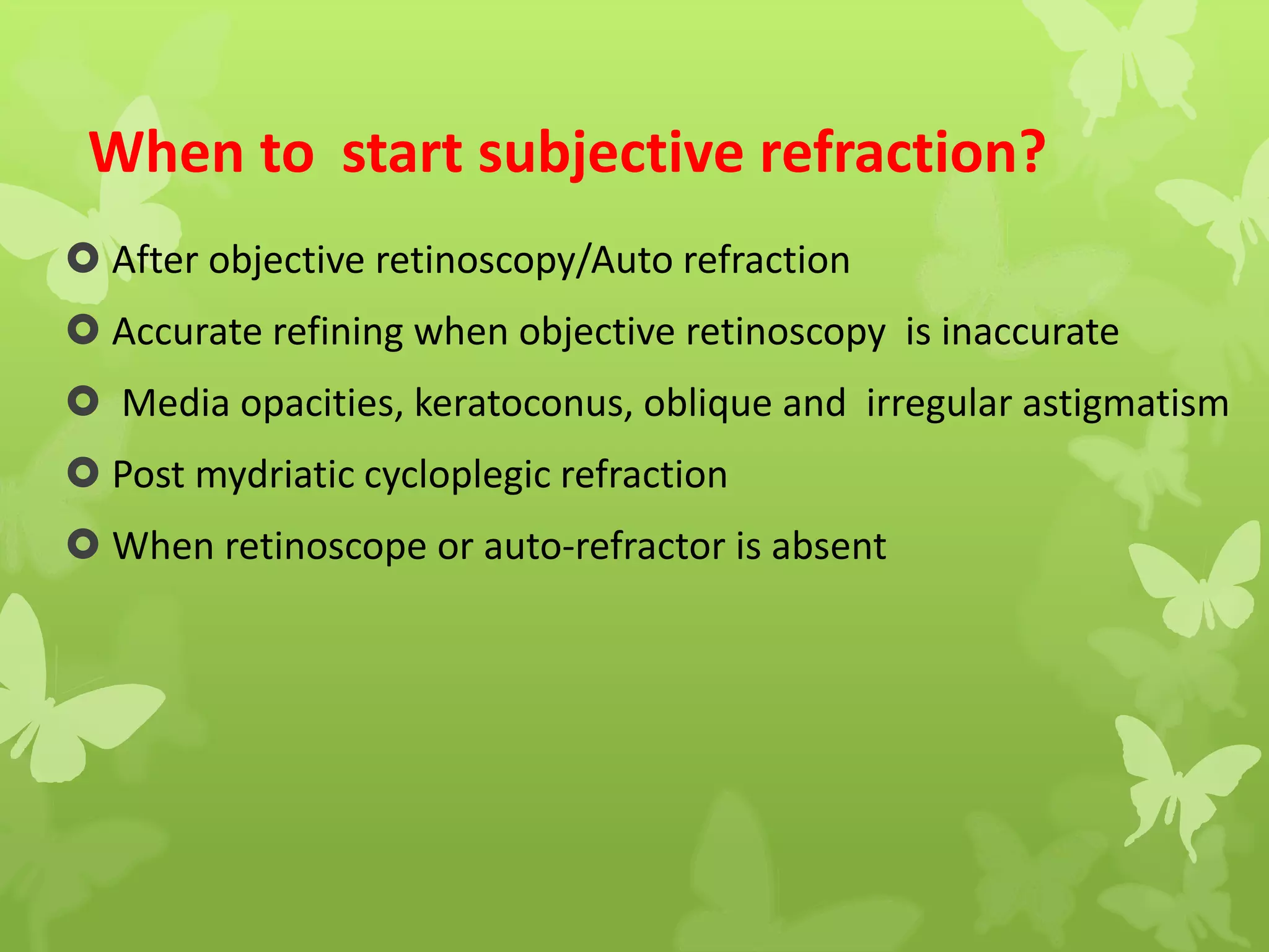
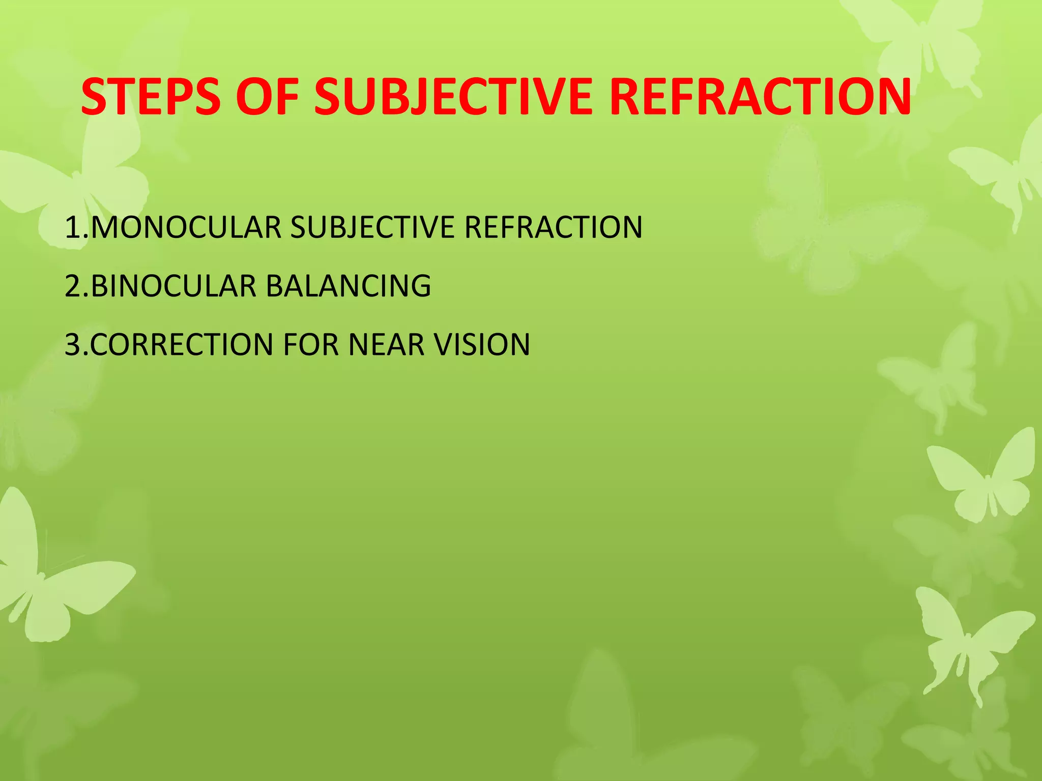
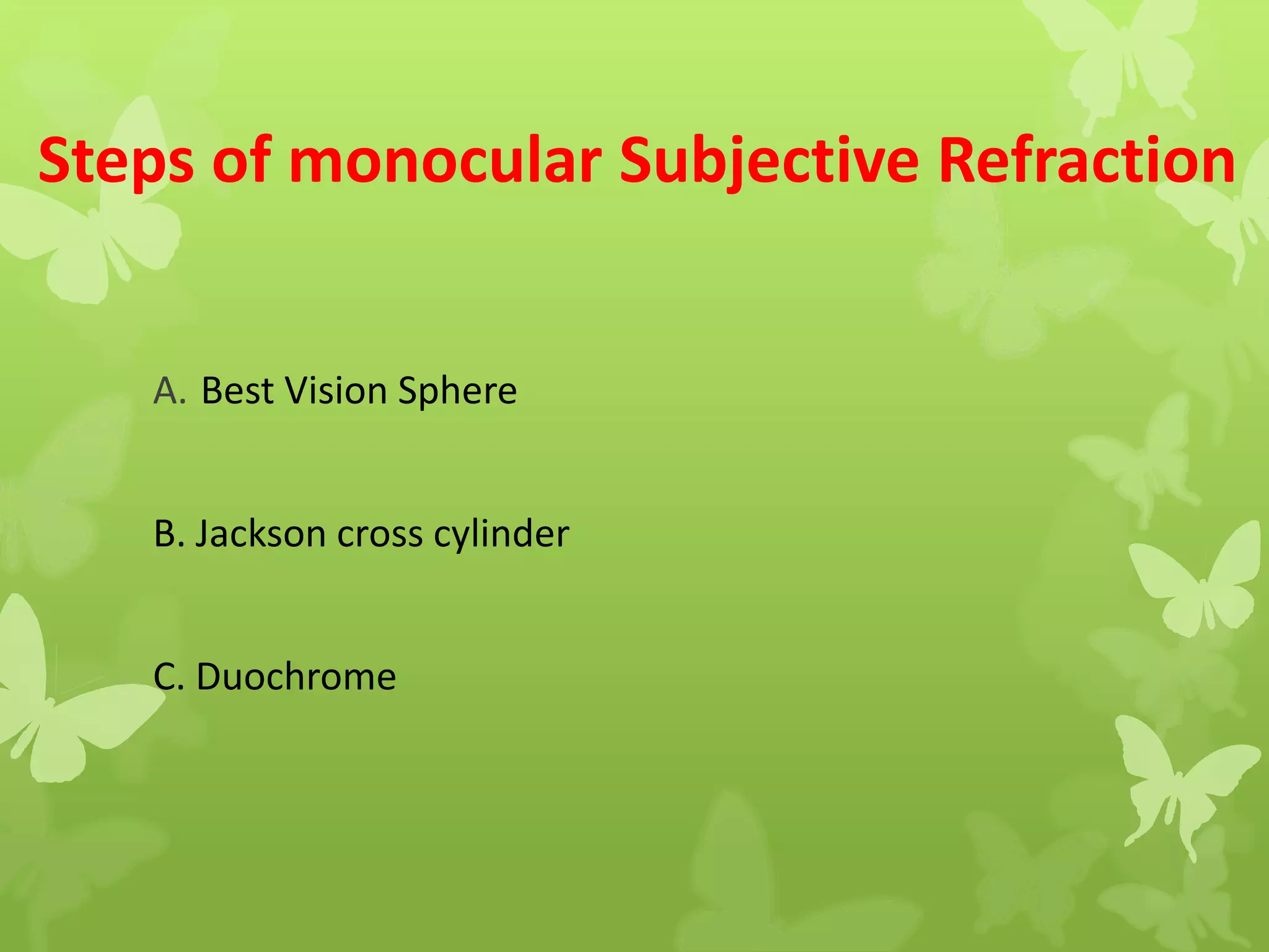
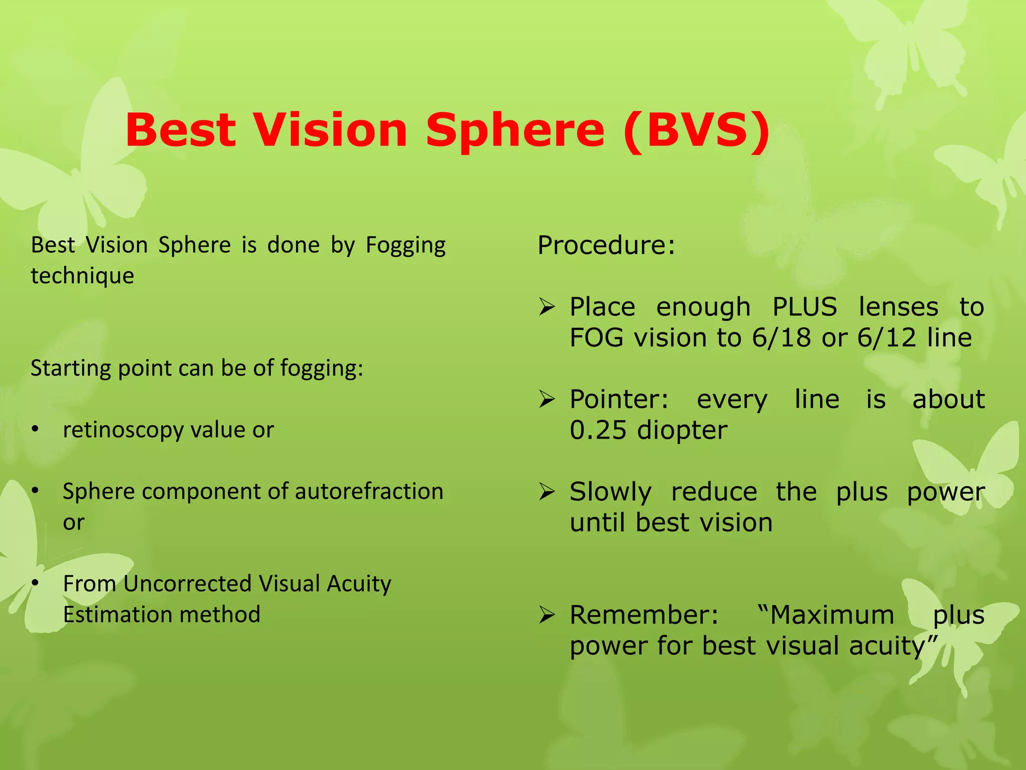
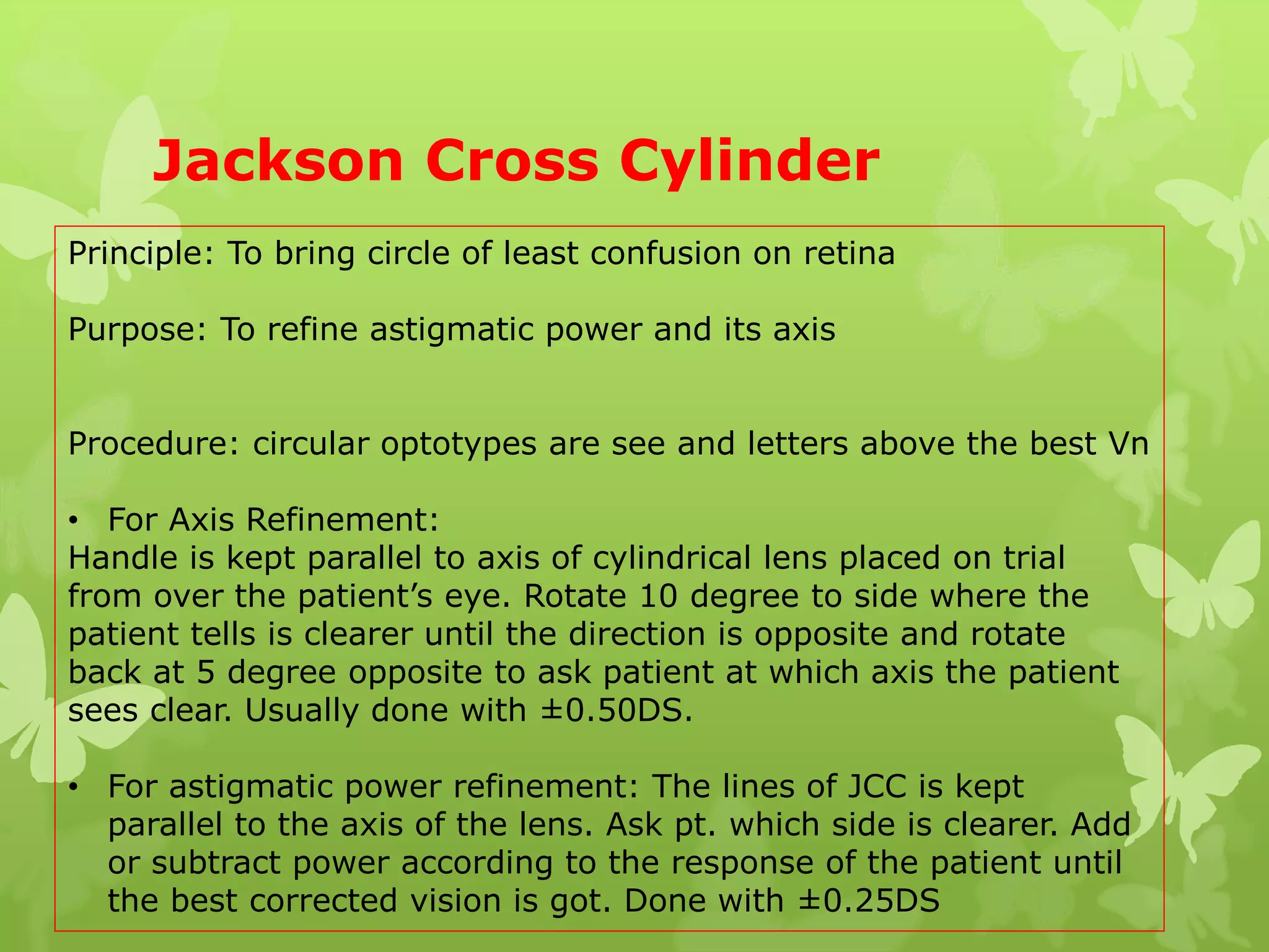
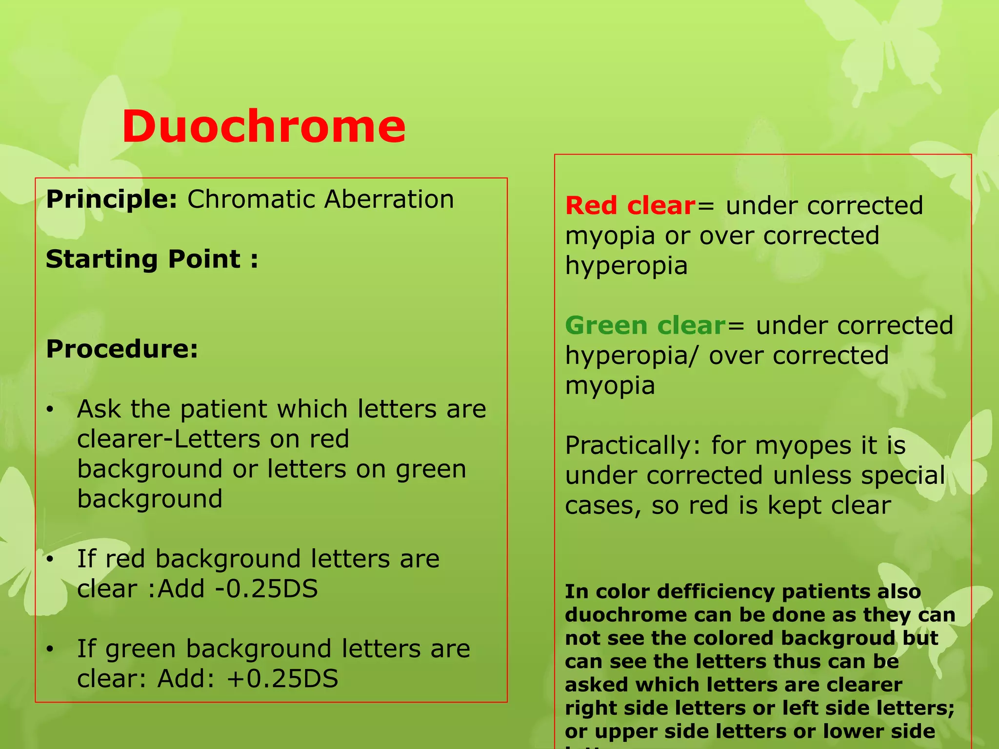
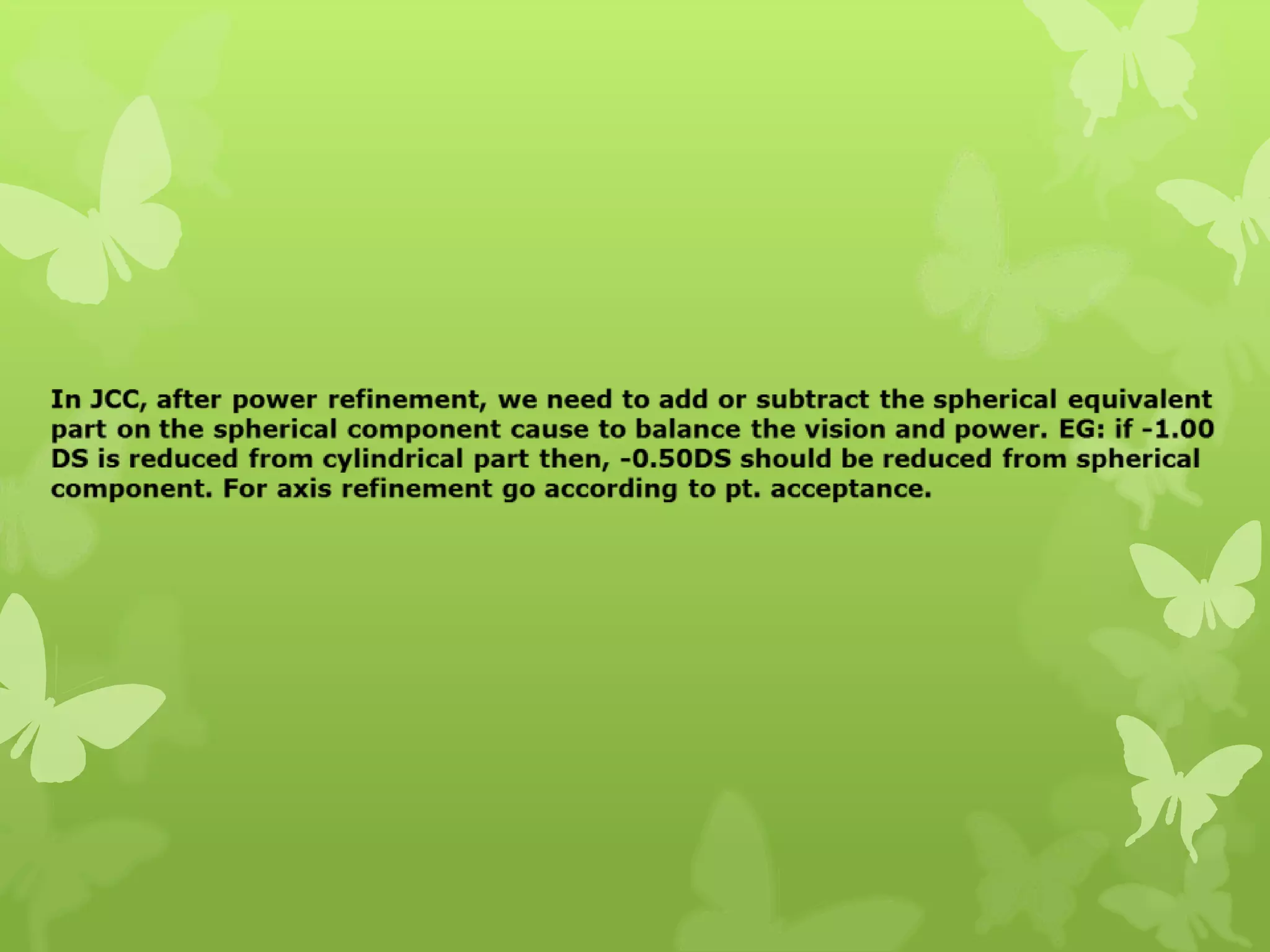
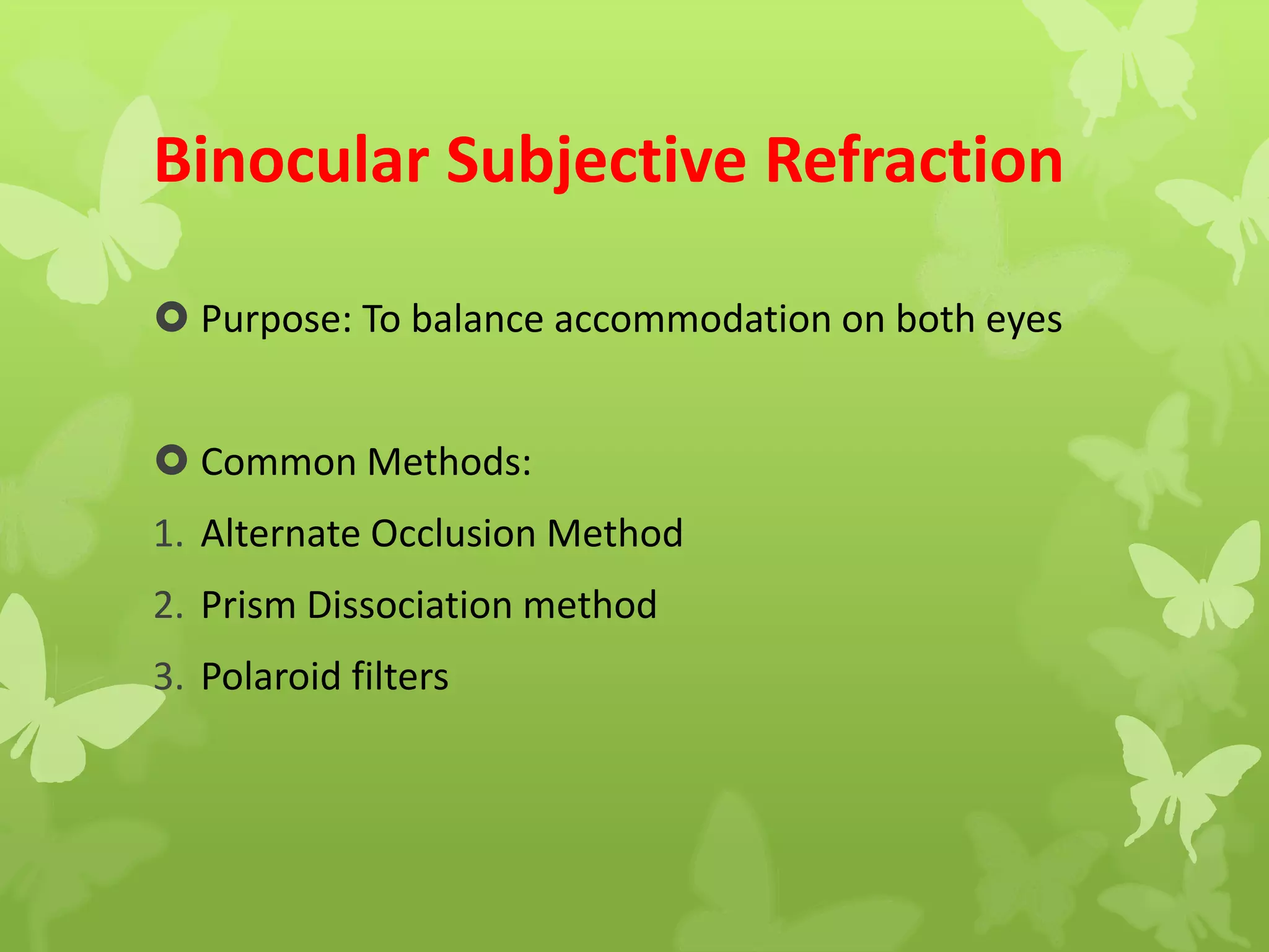
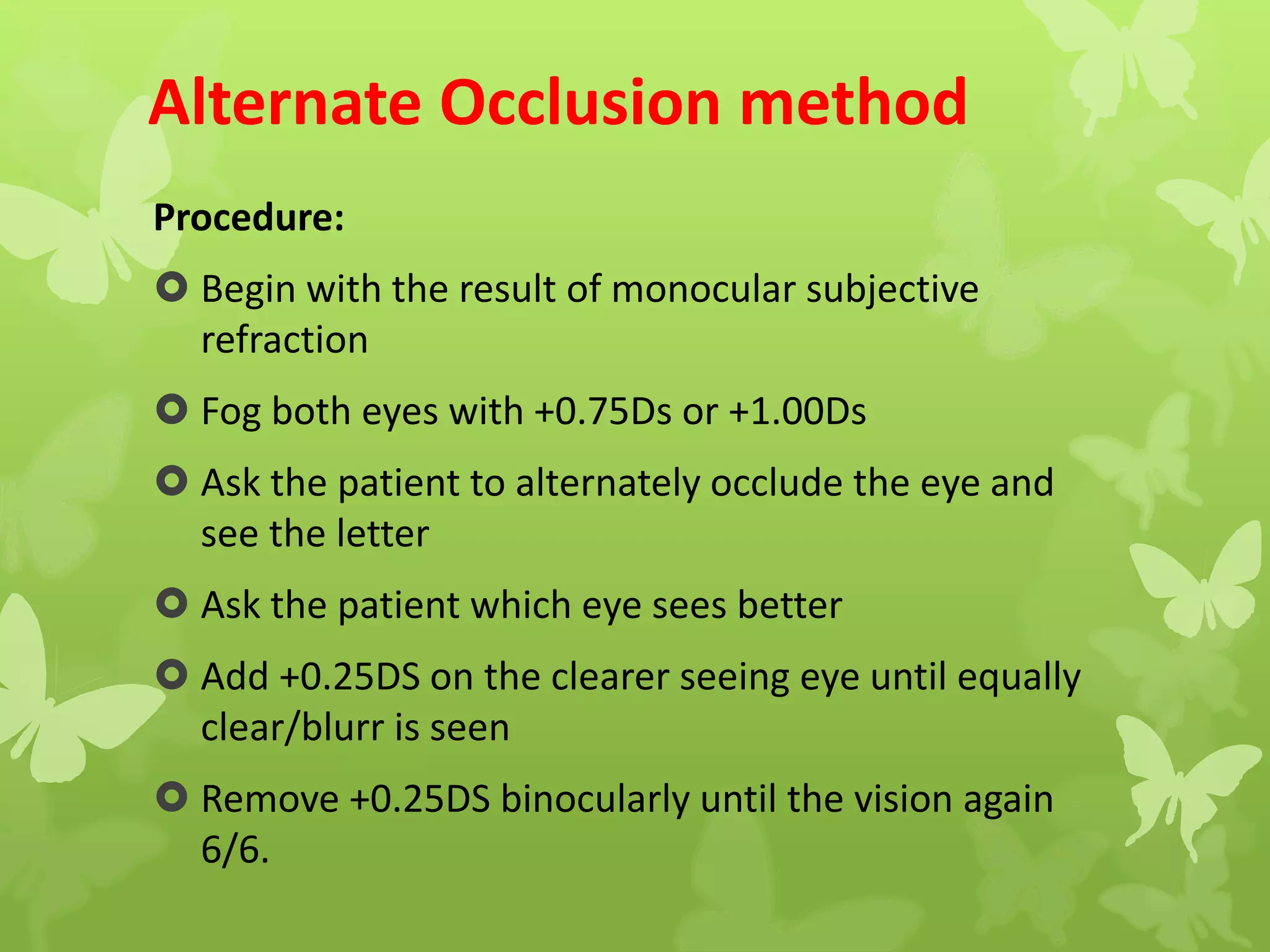
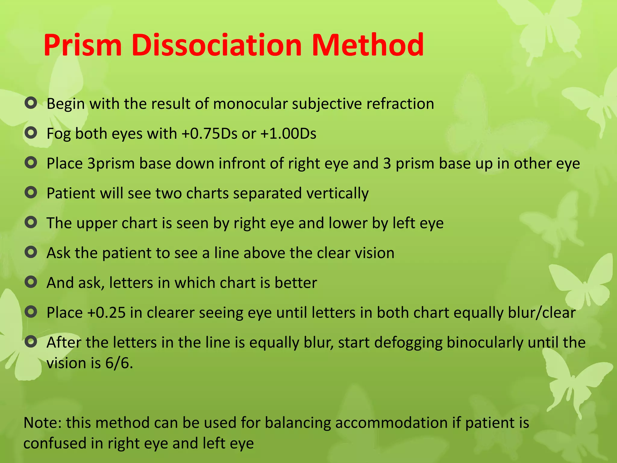
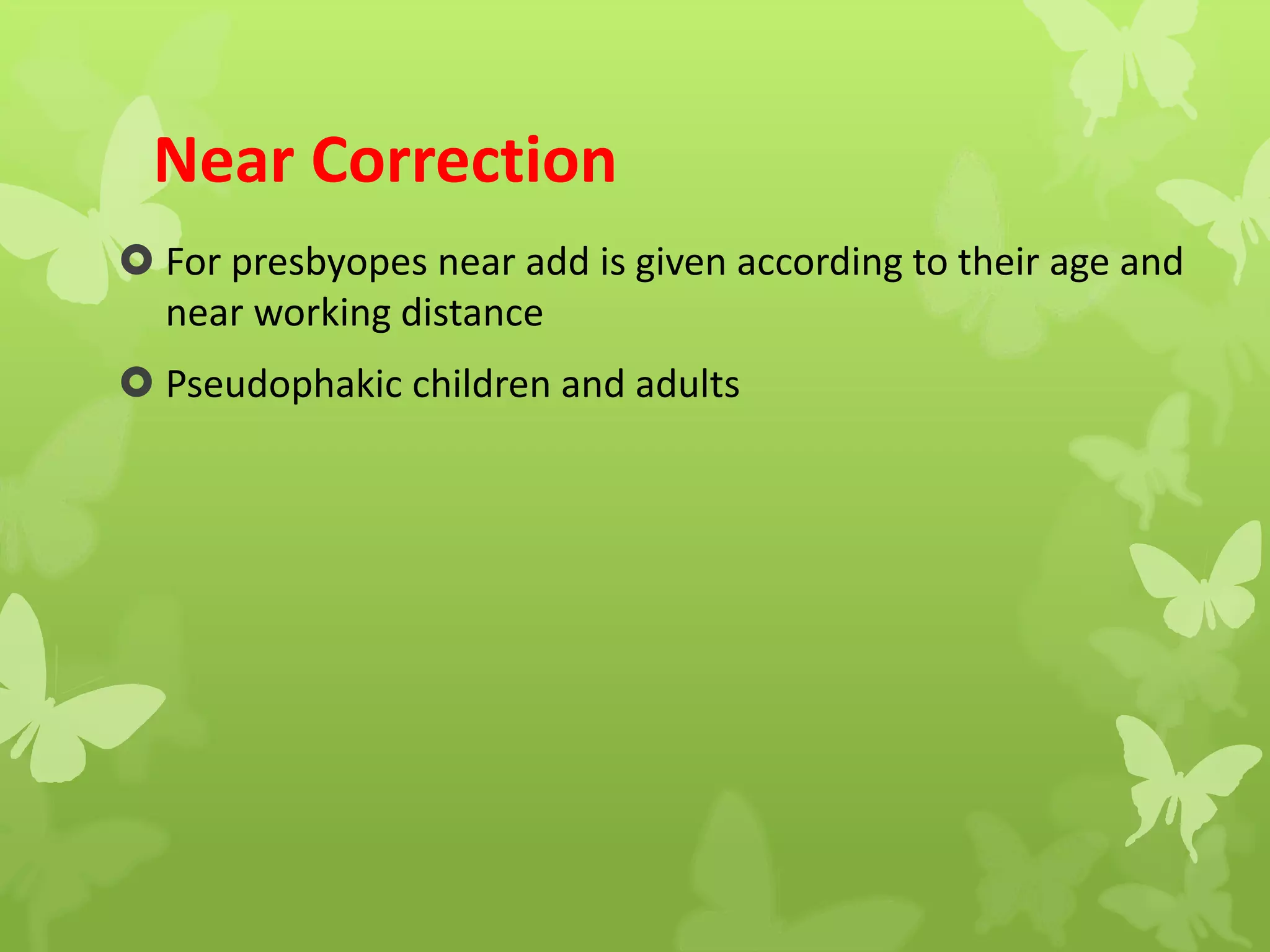
Objective Refraction and
Subjective Refraction
By:
Anu Musyakhwo
M.Optom
Content
Objective Refraction:
• Introduction
• Forms of objective refraction
• Retinoscopy
 Steps
 Swiping and Neutralization
 Working distance
 Types/Techniques of retinoscopy
1. Near retinoscopy
2. Dynamic retinoscopy
3. Static retinoscopy
Subjective Refraction:
1. Monocular subjective refraction
 Best Vision Sphere
 Duochrome test
 Jackson cross cylinder
2. Binocular Subjective refraction
3.Correction for near vision
Objective Refraction
 when the refractive error of an eye is determined without input
by the patient
 The patient may be required to cooperate during the placement
of head and to fixate on a target for a short time, subjective
information is not obtained form the patient about the quality of
vision.
 Purpose: To obtain an objective measurement of the patient’s
refractive state.
Forms of Objective Refraction:
 Retinoscopy
 Auto refractometer
 Keratometry and axial length
Retinoscopy
 Common used synonyms of retinoscpy are
“Skiascopy” and “skiametry” and other synonyms
occasionally seen in literature are “umbrascopy”
“pupilloscpy” “retinoskiascopy
 Principle of retinoscopy: Foucault principle –
To convert the observed eye into a degree of
myopia that the image formed by the fundus
infront of observed eye coincides with the
effective pupil of the examiner (peephole of
retinoscope)
 To bring the far point of patient’s eye into the
peephole of retinoscope
Requirements
 Semi-dim room
 Retinoscope
 Trial lenses
 Trial frame
 Visual acuity chart
Working Distance
 Is the distance between the Retinoscope and the patient’s eye
 It should be compensated on the gross retinoscopy to get net
retinoscopy value and start subjective refraction
 It can be calculated by this formula : D= 1/F Where F is = the power
of the lens .
E.g. F= 1.50D; so the working distance is 0.65mm
1.50 /65cm (arm’s length ) so = 0.66 X 100=66 cm
AND also,
 The power factor that is to compensated to get the net retinoscopy
can be calculated by using formula:
 Power to be compensated =1/working distance in metre
Eg: At 50cm(0.5meter) the power compensated is 1/0.5= 2Diopter
Fixation target for the patient
1. For static retinoscopy: 20/200 or 6/60 in Snellen chart.
2. For dynamic retinoscopy: retinoscope head
3. For near retinoscopy: depends on the type of retinoscopy done
Procedure
 Tell the patient about the examination
 Ask the patient to fixate on target
 The pt.’s right eye should be examined with the examiner’s
right eye with the retinoscope in right hand & vice versa.
 WD should be maintained an arm length distance (66 /50cm)
 The examiner should keep both eyes open.
 The examiner should stay as close to visual axis as possible.
 The examiner should not obstruct the view of target.
 Start sweeping the meridia (at 90, 180,45 and 135 degree)
 To sweep horizontal meridian, make the reflex vertical
 To sweep vertical meridian, make the reflex horizontal
For spherical refractive error:
 A single lens neutralizes all the meridia
For regular astigmatic error:
 Swipe all the meridia, find meridia which have
power and after neutralizing a meridian go to the
meridian 90 degree to the neutralized meridian.
Neutralize other meridian also. Can be done using:
1. Two sphere lenses
2. One spherical lens and other cylindrical lens
Skewing and straddling
Skew phenomenon: if we swipe the streak off axis, the
reflex will tend to travel along the correct axis. This
guide us to back on the correct axis.
Straddling the axis: if there is regular astigmatism,
when one meridian has been neutralized, the meridian
exactly 90deg away will have the strongest & most
defined reflex. The streak turned to 45deg off axis both
side & if the axis correct with of reflex will same in this 2
position.
Properties of reflex
Movement With Need more plus
Against Need more minus
Brightness Dim Far from neutralization
Bright Close to neutralization
Width Narrow Far from neutralization
Wide Close to neutralization
Speed Slow Far from neutralization
Fast Close to neutralization
Neutralization
 In case of myopia
Add –ve lenses till it get neutralizes
 In case of hyperopia
Add + lens till it get neutralizes
Checking neutrality:
1. Changing working distance
2. Changing the light beam from divergent to
convergent
3. Adding +0.25DS and -0.25DS
Picture courtesy- Google images from:
1. Wades Optical Ltd (Prosec-Bucknell University
2. Springer Link
Types/Techniques of
Retinoscopy
1. Near Retinoscopy
2. Dynamic Retinoscopy
3. Static Retinoscopy
 Dry Retinoscopy
 Wet Retinoscopy
Near Retinoscopy
 Mohindra introduced a technique of non-cycloplegic
retinoscopy that correlates somehow with cycloplegic
 Not a variation of dynamic Retinoscopy
 Basically a substitute for cycloplegic refraction mainly
used in infants
 Principle: The stimulus or fixation is the dimmed light
source of the retinoscope in a darkened room which
provide ineffective or neutral accommodative stimulus
Accommodation remains stable in this technique
Procedure
 The room light is dimmed
 The child is encouraged to fixate the retinoscope light
 Babies will instinctively fixate the light
 Retinoscope is performed monocularly
 At WD =50 cm
Tonus factor is +0.75
The total adjustment factor will be :
Working distance + tonus factor = ( -2.00 D + 0.75 D)
= - 1.25 D
Indications of near retinoscopy
 Frequent follow-up visits
 A child is anxious about instillation of drops
 A child is at riskfor an adverse effect to cycloplegics
 Had an adverse reaction to cycloplegics previously
Dynamic Retinoscopy
 Accommodation is active
 No working distance power is added or substracted from the
finding
 Goal is to determine accommodative Response
 Also helps to determine the most appropriate near prescription
with testing conditions
Types of dynamic retinoscopy
Frequently used in clinical practice are:
 Monocular Estimation Method (MEM)
 Nott retinoscopy
 Bell retinoscopy
Monocular Estimation Method(MEM)
 MEM is done over patient’s optimal distance refractive
correction
 The viewing target is the letters in MEM card or the retinoscope
head at patient’s working distance (at 40cm)
 Dim the room light
 Ask the patient to maintain fixation. In children ask them to
read the letters in the card to maintain fixation and
accommodation
 Perform retinoscopy in usual manner but lenses should be
placed infront of the eye for least time
 Record dioptric power of the lens that provides neutrality
 Repeat the procedure on left eye
 The normal dioptric value during MEM is +0.50 to +0.75DS, the
plus value above this indicates lag of accommodation and
negative lenses indicate lead of accommodation
Nott Retinoscopy
 is done over patient’s optimal distance refractive correction
 Dim or turn off the lights but use additional illumination so that
patient can see the target
 Fixation target (near chart) is placed at patient’s near working
distance at 40cms and retinoscopy is done from 50cms
 ask the patient to focus on letters on card
 Perform retinoscopy as quickly as possible
Contd.
 If neutrality is not obtained at 50cms, change the working
distance of retinoscopy. (further away if with movement at
50cms or closer if against movement is seen, until neutrality is
obtained)
 Repeat the procedure in left eye
 Interpretation: record the dioptric difference between near
chart and positon of retinoscope when neutrality is obtained. If
the neutrality point is behind the chart then there is lag of
accommodation but if the neutrality point is infront the chart
then there is lead of accommodation.
 For instance: if near chart is at 40cms and the neutrality point is
at 57cms then the accommodative lag is: +2.50D-1.75DS
=+0.75DS
Bell Retinoscopy
 Done over optimal distance correction
 Retinoscope is kept is fixed and done at 50cms
 The target is moved and patient fixates at the target
 When target is moved closer to patient, there will be a point
where the motion of reflex changes from with to against
 And when target is moved away from patient , there will be a
point where the motion of reflex changes from against to with
 With to against motion is observed at 35-42cms
 Against to with motion is observed at 37-45cms
 Accommodative flexibility can be assessed by noticing the speed
of the change in motion of reflex
Static Retinoscopy
 Dry retinoscopy
 Wet retinoscopy
 Retinoscopy performed when the patient is asked to
fixate the distance target ,with the accommodation
relax
Cycloplegic refraction
 CYCLOPLEGICS are the drugs that paralyze the ciliary muscles
resulting in loss of accommodation and secondarily dilatation of
pupil
 Purpose of cycloplegic refraction: Determination of total
refractive error during temporary paralysis of ciliary muscles
which doesn’t manifest on subjective non-cycloplegic refraction
Total Hypermetropia
Latent
Hypermetropia
Manifest
Hypermetropia
Facultative Absolute
Components of Hypermetropia based on correction by accommodation:
Indications of cycloplegic refraction
 Young Children
 Strabismus
 Latent hypermetropia
 Pseudo myopia
 Young patients who have symptoms but not
significant refractive error
Additional Indications
 Every nonverbal and non communicative children
 Patient with high heterophoria
 Accommodative esotropia ( atropine is best choice)
 Accommodative asthenopia
 Poor reliability b/w dry retinoscopy objective
finding with subjective finding.
Selection of Cycloplegics Drugs
Table courtesy: Roya Attar. All About Red Caps: Mydriatics and
Cycloplegics. Optometry Times. American Academy of
Ophthalmology
Dry Retinoscopy
 Without use of cycloplegics
 Patient is asked to look at distance for relaxing
accommodation
 Mostly in adults above 14-16 years
Subjective Refraction
 Examiner communicates with the patient and using patient’s
responses to the vision provided with various lenses
determines the optical correction that best suits the patient
 When a subjective refraction is not possible, limited or
unreliable it is preferable to have more than one assessment of
objective refraction correction (eg. Retinoscopy,
autorefraction)
Purpose
 To find the strongest plus lens or the weakest minus lens
which allows the patient to obtain the best possible visual
acuity
 Modification is needed: to find the combination of positive
and negative lenses to get the actual acceptance for
correction of pt. refractive error
When to start subjective refraction?
 After objective retinoscopy/Auto refraction
 Accurate refining when objective retinoscopy is inaccurate
 Media opacities, keratoconus, oblique and irregular astigmatism
 Post mydriatic cycloplegic refraction
 When retinoscope or auto-refractor is absent
STEPS OF SUBJECTIVE REFRACTION
1.MONOCULAR SUBJECTIVE REFRACTION
2.BINOCULAR BALANCING
3.CORRECTION FOR NEAR VISION
Steps of monocular Subjective Refraction
A. Best Vision Sphere
B. Jackson cross cylinder
C. Duochrome
Best Vision Sphere (BVS)
Best Vision Sphere is done by Fogging
technique
Starting point can be of fogging:
• retinoscopy value or
• Sphere component of autorefraction
or
• From Uncorrected Visual Acuity
Estimation method
Procedure:
 Place enough PLUS lenses to
FOG vision to 6/18 or 6/12 line
 Pointer: every line is about
0.25 diopter
 Slowly reduce the plus power
until best vision
 Remember: “Maximum plus
power for best visual acuity”
Jackson Cross Cylinder
Principle: To bring circle of least confusion on retina
Purpose: To refine astigmatic power and its axis
Procedure: circular optotypes are see and letters above the best Vn
• For Axis Refinement:
Handle is kept parallel to axis of cylindrical lens placed on trial
from over the patient’s eye. Rotate 10 degree to side where the
patient tells is clearer until the direction is opposite and rotate
back at 5 degree opposite to ask patient at which axis the patient
sees clear. Usually done with ±0.50DS.
• For astigmatic power refinement: The lines of JCC is kept
parallel to the axis of the lens. Ask pt. which side is clearer. Add
or subtract power according to the response of the patient until
the best corrected vision is got. Done with ±0.25DS
Duochrome
Principle: Chromatic Aberration
Starting Point :
Procedure:
• Ask the patient which letters are
clearer-Letters on red
background or letters on green
background
• If red background letters are
clear :Add -0.25DS
• If green background letters are
clear: Add: +0.25DS
Red clear= under corrected
myopia or over corrected
hyperopia
Green clear= under corrected
hyperopia/ over corrected
myopia
Practically: for myopes it is
under corrected unless special
cases, so red is kept clear
In color defficiency patients also
duochrome can be done as they can
not see the colored backgroud but
can see the letters thus can be
asked which letters are clearer
right side letters or left side letters;
or upper side letters or lower side
Binocular Subjective Refraction
 Purpose: To balance accommodation on both eyes
 Common Methods:
1. Alternate Occlusion Method
2. Prism Dissociation method
3. Polaroid filters
Alternate Occlusion method
Procedure:
 Begin with the result of monocular subjective
refraction
 Fog both eyes with +0.75Ds or +1.00Ds
 Ask the patient to alternately occlude the eye and
see the letter
 Ask the patient which eye sees better
 Add +0.25DS on the clearer seeing eye until equally
clear/blurr is seen
 Remove +0.25DS binocularly until the vision again
6/6.
Prism Dissociation Method
 Begin with the result of monocular subjective refraction
 Fog both eyes with +0.75Ds or +1.00Ds
 Place 3prism base down infront of right eye and 3 prism base up in other eye
 Patient will see two charts separated vertically
 The upper chart is seen by right eye and lower by left eye
 Ask the patient to see a line above the clear vision
 And ask, letters in which chart is better
 Place +0.25 in clearer seeing eye until letters in both chart equally blur/clear
 After the letters in the line is equally blur, start defogging binocularly until the
vision is 6/6.
Note: this method can be used for balancing accommodation if patient is
confused in right eye and left eye
Near Correction
 For presbyopes near add is given according to their age and
near working distance
 Pseudophakic children and adults
References
 Primary care of optometry , page 183
 Clinical procedure in primary eye care, Butterworth
 Borish’s Clinical Refraction
 Clinical refraction ,Chapter 18
 Pediatric Refraction and spectacle prescription, Srijana Lamichhane Moderator Dr.
Bipin Koirala, Slideshare.
 Objective, Subjective and Cycloplegic Refraction. Gauri S Shrestha. Slideshare.
Objective Refraction and Subjective Refraction

Objective Refraction and Subjective Refraction

  • 1.
    Objective Refraction and SubjectiveRefraction By: Anu Musyakhwo M.Optom
  • 2.
    Content Objective Refraction: • Introduction •Forms of objective refraction • Retinoscopy  Steps  Swiping and Neutralization  Working distance  Types/Techniques of retinoscopy 1. Near retinoscopy 2. Dynamic retinoscopy 3. Static retinoscopy Subjective Refraction: 1. Monocular subjective refraction  Best Vision Sphere  Duochrome test  Jackson cross cylinder 2. Binocular Subjective refraction 3.Correction for near vision
  • 3.
    Objective Refraction  whenthe refractive error of an eye is determined without input by the patient  The patient may be required to cooperate during the placement of head and to fixate on a target for a short time, subjective information is not obtained form the patient about the quality of vision.  Purpose: To obtain an objective measurement of the patient’s refractive state.
  • 4.
    Forms of ObjectiveRefraction:  Retinoscopy  Auto refractometer  Keratometry and axial length
  • 5.
    Retinoscopy  Common usedsynonyms of retinoscpy are “Skiascopy” and “skiametry” and other synonyms occasionally seen in literature are “umbrascopy” “pupilloscpy” “retinoskiascopy
  • 6.
     Principle ofretinoscopy: Foucault principle – To convert the observed eye into a degree of myopia that the image formed by the fundus infront of observed eye coincides with the effective pupil of the examiner (peephole of retinoscope)  To bring the far point of patient’s eye into the peephole of retinoscope
  • 7.
    Requirements  Semi-dim room Retinoscope  Trial lenses  Trial frame  Visual acuity chart
  • 8.
    Working Distance  Isthe distance between the Retinoscope and the patient’s eye  It should be compensated on the gross retinoscopy to get net retinoscopy value and start subjective refraction  It can be calculated by this formula : D= 1/F Where F is = the power of the lens . E.g. F= 1.50D; so the working distance is 0.65mm 1.50 /65cm (arm’s length ) so = 0.66 X 100=66 cm AND also,  The power factor that is to compensated to get the net retinoscopy can be calculated by using formula:  Power to be compensated =1/working distance in metre Eg: At 50cm(0.5meter) the power compensated is 1/0.5= 2Diopter
  • 9.
    Fixation target forthe patient 1. For static retinoscopy: 20/200 or 6/60 in Snellen chart. 2. For dynamic retinoscopy: retinoscope head 3. For near retinoscopy: depends on the type of retinoscopy done
  • 10.
    Procedure  Tell thepatient about the examination  Ask the patient to fixate on target  The pt.’s right eye should be examined with the examiner’s right eye with the retinoscope in right hand & vice versa.  WD should be maintained an arm length distance (66 /50cm)  The examiner should keep both eyes open.  The examiner should stay as close to visual axis as possible.  The examiner should not obstruct the view of target.  Start sweeping the meridia (at 90, 180,45 and 135 degree)  To sweep horizontal meridian, make the reflex vertical  To sweep vertical meridian, make the reflex horizontal
  • 11.
    For spherical refractiveerror:  A single lens neutralizes all the meridia For regular astigmatic error:  Swipe all the meridia, find meridia which have power and after neutralizing a meridian go to the meridian 90 degree to the neutralized meridian. Neutralize other meridian also. Can be done using: 1. Two sphere lenses 2. One spherical lens and other cylindrical lens
  • 12.
    Skewing and straddling Skewphenomenon: if we swipe the streak off axis, the reflex will tend to travel along the correct axis. This guide us to back on the correct axis. Straddling the axis: if there is regular astigmatism, when one meridian has been neutralized, the meridian exactly 90deg away will have the strongest & most defined reflex. The streak turned to 45deg off axis both side & if the axis correct with of reflex will same in this 2 position.
  • 13.
    Properties of reflex MovementWith Need more plus Against Need more minus Brightness Dim Far from neutralization Bright Close to neutralization Width Narrow Far from neutralization Wide Close to neutralization Speed Slow Far from neutralization Fast Close to neutralization
  • 14.
    Neutralization  In caseof myopia Add –ve lenses till it get neutralizes  In case of hyperopia Add + lens till it get neutralizes Checking neutrality: 1. Changing working distance 2. Changing the light beam from divergent to convergent 3. Adding +0.25DS and -0.25DS
  • 15.
    Picture courtesy- Googleimages from: 1. Wades Optical Ltd (Prosec-Bucknell University 2. Springer Link
  • 16.
    Types/Techniques of Retinoscopy 1. NearRetinoscopy 2. Dynamic Retinoscopy 3. Static Retinoscopy  Dry Retinoscopy  Wet Retinoscopy
  • 17.
    Near Retinoscopy  Mohindraintroduced a technique of non-cycloplegic retinoscopy that correlates somehow with cycloplegic  Not a variation of dynamic Retinoscopy  Basically a substitute for cycloplegic refraction mainly used in infants  Principle: The stimulus or fixation is the dimmed light source of the retinoscope in a darkened room which provide ineffective or neutral accommodative stimulus Accommodation remains stable in this technique
  • 18.
    Procedure  The roomlight is dimmed  The child is encouraged to fixate the retinoscope light  Babies will instinctively fixate the light  Retinoscope is performed monocularly  At WD =50 cm Tonus factor is +0.75 The total adjustment factor will be : Working distance + tonus factor = ( -2.00 D + 0.75 D) = - 1.25 D
  • 19.
    Indications of nearretinoscopy  Frequent follow-up visits  A child is anxious about instillation of drops  A child is at riskfor an adverse effect to cycloplegics  Had an adverse reaction to cycloplegics previously
  • 20.
    Dynamic Retinoscopy  Accommodationis active  No working distance power is added or substracted from the finding  Goal is to determine accommodative Response  Also helps to determine the most appropriate near prescription with testing conditions
  • 21.
    Types of dynamicretinoscopy Frequently used in clinical practice are:  Monocular Estimation Method (MEM)  Nott retinoscopy  Bell retinoscopy
  • 22.
    Monocular Estimation Method(MEM) MEM is done over patient’s optimal distance refractive correction  The viewing target is the letters in MEM card or the retinoscope head at patient’s working distance (at 40cm)  Dim the room light  Ask the patient to maintain fixation. In children ask them to read the letters in the card to maintain fixation and accommodation  Perform retinoscopy in usual manner but lenses should be placed infront of the eye for least time  Record dioptric power of the lens that provides neutrality  Repeat the procedure on left eye  The normal dioptric value during MEM is +0.50 to +0.75DS, the plus value above this indicates lag of accommodation and negative lenses indicate lead of accommodation
  • 23.
    Nott Retinoscopy  isdone over patient’s optimal distance refractive correction  Dim or turn off the lights but use additional illumination so that patient can see the target  Fixation target (near chart) is placed at patient’s near working distance at 40cms and retinoscopy is done from 50cms  ask the patient to focus on letters on card  Perform retinoscopy as quickly as possible
  • 24.
    Contd.  If neutralityis not obtained at 50cms, change the working distance of retinoscopy. (further away if with movement at 50cms or closer if against movement is seen, until neutrality is obtained)  Repeat the procedure in left eye  Interpretation: record the dioptric difference between near chart and positon of retinoscope when neutrality is obtained. If the neutrality point is behind the chart then there is lag of accommodation but if the neutrality point is infront the chart then there is lead of accommodation.  For instance: if near chart is at 40cms and the neutrality point is at 57cms then the accommodative lag is: +2.50D-1.75DS =+0.75DS
  • 25.
    Bell Retinoscopy  Doneover optimal distance correction  Retinoscope is kept is fixed and done at 50cms  The target is moved and patient fixates at the target  When target is moved closer to patient, there will be a point where the motion of reflex changes from with to against  And when target is moved away from patient , there will be a point where the motion of reflex changes from against to with  With to against motion is observed at 35-42cms  Against to with motion is observed at 37-45cms  Accommodative flexibility can be assessed by noticing the speed of the change in motion of reflex
  • 26.
    Static Retinoscopy  Dryretinoscopy  Wet retinoscopy  Retinoscopy performed when the patient is asked to fixate the distance target ,with the accommodation relax
  • 27.
    Cycloplegic refraction  CYCLOPLEGICSare the drugs that paralyze the ciliary muscles resulting in loss of accommodation and secondarily dilatation of pupil  Purpose of cycloplegic refraction: Determination of total refractive error during temporary paralysis of ciliary muscles which doesn’t manifest on subjective non-cycloplegic refraction Total Hypermetropia Latent Hypermetropia Manifest Hypermetropia Facultative Absolute Components of Hypermetropia based on correction by accommodation:
  • 28.
    Indications of cycloplegicrefraction  Young Children  Strabismus  Latent hypermetropia  Pseudo myopia  Young patients who have symptoms but not significant refractive error
  • 29.
    Additional Indications  Everynonverbal and non communicative children  Patient with high heterophoria  Accommodative esotropia ( atropine is best choice)  Accommodative asthenopia  Poor reliability b/w dry retinoscopy objective finding with subjective finding.
  • 30.
    Selection of CycloplegicsDrugs Table courtesy: Roya Attar. All About Red Caps: Mydriatics and Cycloplegics. Optometry Times. American Academy of Ophthalmology
  • 31.
    Dry Retinoscopy  Withoutuse of cycloplegics  Patient is asked to look at distance for relaxing accommodation  Mostly in adults above 14-16 years
  • 32.
    Subjective Refraction  Examinercommunicates with the patient and using patient’s responses to the vision provided with various lenses determines the optical correction that best suits the patient  When a subjective refraction is not possible, limited or unreliable it is preferable to have more than one assessment of objective refraction correction (eg. Retinoscopy, autorefraction)
  • 33.
    Purpose  To findthe strongest plus lens or the weakest minus lens which allows the patient to obtain the best possible visual acuity  Modification is needed: to find the combination of positive and negative lenses to get the actual acceptance for correction of pt. refractive error
  • 34.
    When to startsubjective refraction?  After objective retinoscopy/Auto refraction  Accurate refining when objective retinoscopy is inaccurate  Media opacities, keratoconus, oblique and irregular astigmatism  Post mydriatic cycloplegic refraction  When retinoscope or auto-refractor is absent
  • 35.
    STEPS OF SUBJECTIVEREFRACTION 1.MONOCULAR SUBJECTIVE REFRACTION 2.BINOCULAR BALANCING 3.CORRECTION FOR NEAR VISION
  • 36.
    Steps of monocularSubjective Refraction A. Best Vision Sphere B. Jackson cross cylinder C. Duochrome
  • 37.
    Best Vision Sphere(BVS) Best Vision Sphere is done by Fogging technique Starting point can be of fogging: • retinoscopy value or • Sphere component of autorefraction or • From Uncorrected Visual Acuity Estimation method Procedure:  Place enough PLUS lenses to FOG vision to 6/18 or 6/12 line  Pointer: every line is about 0.25 diopter  Slowly reduce the plus power until best vision  Remember: “Maximum plus power for best visual acuity”
  • 38.
    Jackson Cross Cylinder Principle:To bring circle of least confusion on retina Purpose: To refine astigmatic power and its axis Procedure: circular optotypes are see and letters above the best Vn • For Axis Refinement: Handle is kept parallel to axis of cylindrical lens placed on trial from over the patient’s eye. Rotate 10 degree to side where the patient tells is clearer until the direction is opposite and rotate back at 5 degree opposite to ask patient at which axis the patient sees clear. Usually done with ±0.50DS. • For astigmatic power refinement: The lines of JCC is kept parallel to the axis of the lens. Ask pt. which side is clearer. Add or subtract power according to the response of the patient until the best corrected vision is got. Done with ±0.25DS
  • 39.
    Duochrome Principle: Chromatic Aberration StartingPoint : Procedure: • Ask the patient which letters are clearer-Letters on red background or letters on green background • If red background letters are clear :Add -0.25DS • If green background letters are clear: Add: +0.25DS Red clear= under corrected myopia or over corrected hyperopia Green clear= under corrected hyperopia/ over corrected myopia Practically: for myopes it is under corrected unless special cases, so red is kept clear In color defficiency patients also duochrome can be done as they can not see the colored backgroud but can see the letters thus can be asked which letters are clearer right side letters or left side letters; or upper side letters or lower side
  • 41.
    Binocular Subjective Refraction Purpose: To balance accommodation on both eyes  Common Methods: 1. Alternate Occlusion Method 2. Prism Dissociation method 3. Polaroid filters
  • 42.
    Alternate Occlusion method Procedure: Begin with the result of monocular subjective refraction  Fog both eyes with +0.75Ds or +1.00Ds  Ask the patient to alternately occlude the eye and see the letter  Ask the patient which eye sees better  Add +0.25DS on the clearer seeing eye until equally clear/blurr is seen  Remove +0.25DS binocularly until the vision again 6/6.
  • 43.
    Prism Dissociation Method Begin with the result of monocular subjective refraction  Fog both eyes with +0.75Ds or +1.00Ds  Place 3prism base down infront of right eye and 3 prism base up in other eye  Patient will see two charts separated vertically  The upper chart is seen by right eye and lower by left eye  Ask the patient to see a line above the clear vision  And ask, letters in which chart is better  Place +0.25 in clearer seeing eye until letters in both chart equally blur/clear  After the letters in the line is equally blur, start defogging binocularly until the vision is 6/6. Note: this method can be used for balancing accommodation if patient is confused in right eye and left eye
  • 44.
    Near Correction  Forpresbyopes near add is given according to their age and near working distance  Pseudophakic children and adults
  • 45.
    References  Primary careof optometry , page 183  Clinical procedure in primary eye care, Butterworth  Borish’s Clinical Refraction  Clinical refraction ,Chapter 18  Pediatric Refraction and spectacle prescription, Srijana Lamichhane Moderator Dr. Bipin Koirala, Slideshare.  Objective, Subjective and Cycloplegic Refraction. Gauri S Shrestha. Slideshare.