Acinar cell types in the pancreas of frugivorous bat rousettus leschenaulti
•
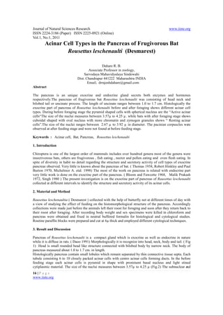
1 like•327 views


Recommended
More Related Content
What's hot
What's hot (20)
Similar to Acinar cell types in the pancreas of frugivorous bat rousettus leschenaulti
Similar to Acinar cell types in the pancreas of frugivorous bat rousettus leschenaulti (20)
More from Alexander Decker
More from Alexander Decker (20)
Recently uploaded
Recently uploaded (20)
Acinar cell types in the pancreas of frugivorous bat rousettus leschenaulti
- 1. Journal of Natural Sciences Research www.iiste.org ISSN 2224-3186 (Paper) ISSN 2225-0921 (Online) Vol.1, No.1, 2011 Acinar Cell Types in the Pancreas of Frugivorous Bat Rousettus leschenaulti (Desmarest) Dahare R. B. Associate Professor in zoology, Sarvodaya Mahavidyalaya Sindewahi Dist. Chandrapur 441222 Maharashtra INDIA Email; drrajeshdahare@gmail.com Abstract The pancreas is an unique exocrine and endocrine gland secrets both enzymes and hormones respectively.The pancreas of frugivorous bat Rousettus leschenaulti was consisting of head neck and bilobed tail or uncinate process. The length of uncinate ranges between 1.0 to 1.7 cm. Histologically the exocrine part of pancreas of Rousettus leschenaulti before and after foraging shows different acinar cell types. During before foraging stage the pyramid shaped cells with spherical nucleus are the “Active acinar cells“The size of the nuclui measures between 3.57µ to 4.25 µ. while bats with after foraging stage shows cuboidal shaped with oval nucleus with more chromatin and zymogen granules shows “ Resting acinar cells”.The size of the nuclei ranges between 2.67 µ to 3.92 µ in diameter. The pacinian corpuscles were observed at after feeding stage and were not found at before feeding stage. Keywords : Acinar cell, Bat, Pancreas, Rousettus leschenaulti 1. Introduction Chiroptera is one of the largest order of mammals includes over hundred genera most of the genera were insectivorous bats, others are frugivorous , fish eating , nector and pollen eating and even flesh eating. In spite of diversity in habit no detail regarding the structure and secretory activity of cell types of exocrine pancreas observed. Very little is known about the pancreas of bat. ( Thomas 1938, Robert Hinkley and Paul Burton 1970, Michelmor A. etal. 1998) The most of the work on pancreas is related with endocrine part very little work is done on the exocrine part of the pancreas. ( Bloom and Fawcette 1968, Malik Prakash 1972, Singh 1980 ) The present investigation is on the exocrine part of pancreas of Rausettus leschenaulti collected at different intervals to identify the structure and secretory activity of its acinar cells. 2. Material and Method Rausettus leschenaultia ( Desmarest ) collected with the help of butterfly net at different times of day with a view of studying the effect of feeding on the histomorphological structure of the pancreas. Accordingly collections were made just before the animals left their roost for foraging and soon after they return back to their roost after foraging. After recording body weight and sex specimens were killed in chloroform and pancreas were obtained and fixed in neutral buffered formalin for histological and cytological studies. Routine paraffin blocks were prepared and cut at 6µ thick and employed different cytological techniques. 3. Result and Discussion Pancreas of Rousettus leschenaulti is a compact gland which is exocrine as well as endocrine in nature while it is diffuse in rats. ( Daoo 1991) Morphologically it is recognize into head, neck, body and tail. ( Fig 1) Head is small rounded bead like structure connected with bilobed body by narrow neck. The body of pancreas measured about 1.0 to 1.7 cm. in length. Histologically pancreas contain small lobules which remain separated by thin connective tissue septa. Each tubule consisting 6 to 10 closely packed acinar cells with centro acinar cells forming ducts. In the before feeding stage each acinar cells is pyramid in shape with prominent basal nucleus and light stined cytiplasmic material. The size of the nuclui measures between 3.57µ to 4.25 µ (Fig.2) The subnuclear and 16 | P a g e www.iiste.org
- 2. Journal of Natural Sciences Research www.iiste.org ISSN 2224-3186 (Paper) ISSN 2225-0921 (Online) Vol.1, No.1, 2011 lateronuclear areas of cytoplasm are chromophilic in their staining property. The active acinar cells are observed during before foraging stage. It is observed by Bloom and Fawcett 1968 and Singh 1980 in domestic animals. The inter lobuler and intercalated ducts in most part of exocrine part is empty with minimum secretion during this stage. Similarly no pacinian corpuscles are observed during this stage The Rousettus leschenaultia collected when they come back to their roost in the morning after foraging. The pancreas of these bats not shows significance difference in shape size as compare to before foraging stage. Histologically exocrine pancreas shows large number of pacinian corpuscles. (Fig.3) As it is observed by Alfred Troutmann and Josef Fiebtger 2002. Acinar cells appear cuboidal in shape with basally situated oval nuclei ( Fig. 4) with a size of 2.67 µ to 3.92 µ in diameter . The chromatin material in nucleus appears dense. The lateronuclear region appears chromophilic due to presence of rough endoplasmic region. The apical region of acinar cells shows eosinophilic zymogen granules. At after foraging stage resting acinar cells are observed. Which are also observed by Singh 1980 in many domestic animals. 4. Acknowledgement The authors are highly thankful to Dr. I. H. Jeevaji Reader in Zoology for guidance and Dr. M. M. Gadegone Director Institute of Science Nagpur India for his encouragement and laboratory facilities. References Alfred Trautmann and Josef Fiebtger (2002) Fundamentals of the histology of domestic animals . Green world Publication, Lucknow, India : 216-219 Bloom and Fawcett D. W. ( 1968 ) A text book of histology . W. B. Saunders company Philadelphia Daoo J. V. (1991) Influence of whole body irradiation on haematology metabolism and ultrastructure of pancreatic islet in wister rat . Ph.D Thesis Institute of science Bombay. Malik M. R. and Prakash P. (1972) Comparative histology of the pancreas of buffalo and ox “ A Note”. Indian J. animal sci. Vol 42: 681-682 Michelmore A. J. Keegan D. J. Kramer B. (1998) Immunocytochemical identification of endocrine cell in the pancreas of the fruit bat Rousettus aegyptiacus. Gen. Comp. Endocrinol June 110 (3) : 319 – 325 Robert Hickling and Paul Barton (1970) Fine structure of Pancreatic islet cells of normal and alloxan treated bat ( Eptesicus fuscus) Anat. Rec. 166 : 67-86 Singh L. P. (1980) Note on the acinar cell type in the pancreas of domestic animals. Indian J. Animal Sci. 50 ( 90 ) : 169- 172. Thomas T. B. (1935) Cellular components of the mammalian islet of langerhans. Am. J. Anat. 62 : 31- 57 17 | P a g e www.iiste.org
- 3. Journal of Natural Sciences Research www.iiste.org ISSN 2224-3186 (Paper) ISSN 2225-0921 (Online) Vol.1, No.1, 2011 Fig. 1 Pancreas of Rousettus leschenaulti shows head neck and tail or uncinate Fig. 2 Tranaverse section of pancreas of Rousettus leschenaulti at before foraging HE X 567 Fig. 3 Transverse section of pancreas during after foraging shows pacinian corpuscles. HE X 256 Fig. 4 Transverse section of pancreas during after foraging shows oval nuclei HE X 567 18 | P a g e www.iiste.org