SlideShare a Scribd company logo
1 of 27
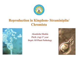
Akanksha Shukla
Ph.D. (Ag) 1st year
Deptt. Of Plant Pathology
• Eukaryotic kingdom created by Cavalier- Smith in 1981
• The members of this kingdom were earlier placed under former kingdom
Protista.
• The members are characterized by:-
1. Unicellular, filamentous or colonial nature
2. Presence of Tinsel type flagellum. D.N. Patterson used the term
Straminopiles in 1989 for the organisms bearing unique flagellum hair
structure i.e. mastigonemes
3. Photosynthetic or heterotrophic nutrition
4. Cell wall made of cellulose and glucan, chitin is absent
5. Mitochondria with tubular cristae
6. Presence of golgi bodies and peroxisomes
7. Storage product is mycolaminarin, and
8. Diploid nuclei
• These fungi are placed in 3 phyla (Dictionary of the Fungi, Kirk et.al., 2008):
– Oomycota
– Hypochytriomycota
– Labyrinthulomycota
• Flagella which are entirely smooth or bear a coat of fine fibrillar surface
material visible only by high-resolution electron microscopy (Andersen et
al., 1991) are commonly called whiplash flagella.
• A second type of flagellum is decorated with hair-like structures .This is the
tinsel or straminipilous flagellum (Dick, 1997). The hairs are called tripartite
tubular hairs (TTHs) because they are divided into three parts.
Fig: Ultrastructure of flagella in Straminipila. (a)Whiplash flagellum of Pythium
monospermum (Oomycota).The tip is narrower than the main body of the flagellum
because the two central microtubules are longer than the nine outer doublets. Arrows
indicate the coating of the flagellum with very fine hairs. (b) Tinsel flagellum of Achlya
colorata (Oomycota) with numerous TTHs. Each TTH ends in two fibres, one longer and
thicker than the other (arrows)
• The name Oomycota means “egg fungi” and refers to Oogamous type of
sexual reproduction.
• The characteristics that set apart Oomycota from true fungi are as follows:-
• Phylum Oomycota contains 1 Class and 8 Orders (Kirk et.al., 2008)
Distinguishing
Characters
Pseudofungi True Fungi
1. Lysine biosynthetic
pathway
Diaminopimelic acid
pathway
Alpha aminoadipic acid
pathway
2. Sterol Fucosterol Ergosterol
3. Reserve Product Mycolaminarin
(β -1-3 glucans)
Glycogen
4. Sugar alcohol Absent Present
 Two types of zoospore may be produced
and, if so, the auxiliary (primary) zoospore
is the first formed. It is grapeseed shaped,
with both flagella inserted apically and it
encysts soon after its formation.
Encystment is by withdrawal of the flagella,
so that a tuft of tripartite tubular hairs is left
behind on the surface of the developing cyst
.
 The cyst germinates to give rise to the
principal (secondary) zoospore, which is by
far the more common type and also the
more vigorous swimmer. This typical and
readily recognized oomycete zoospore is
uniform in appearance across the phylum
The Oomycota are characterized by motile asexual spores (zoospores) which
are produced in spherical or elongated zoosporangia. They are heterokont,
possessing one straminipilous and one whiplash-type flagellum.
Fig:
(a) Auxiliary (primary) zoospore
(b) Principal (secondary) zoospore.
 Sexual reproduction in Oomycota is
oogamous i.e. it involves larger non-
motile gamete called egg or oophere
contained in globular oogonia and
smaller motile male gamete formed in
club-shaped antheridia.
 In some primitive oomycetes, the
gametangia are not differentiated into
male and female. The holocarpic thalli
of different sizes act as gametangia and
fuse to form zygote.
 Heterothallic species of Oomycota
display relative sexuality, i.e. a strain
can produce antheridia in combination
with a second strain but oogonia when
paired against a third
Fig: Schematic drawing and
terminology of sexual reproductive
organs in the Oomycota
• The mature oospore contains three major pools of storage
compounds
1. The oospore wall often appears stratified, and this is due in part to a
polysaccharide reserve compartment, the endospore, which is
located between the plasma membrane and the outer spore wall
epispore. Upon germination, the endospore is thought to coat the
emerging germ tube with wall material, and some material may
also be taken up by endocytosis.
2. A large storage vacuole inside the oospore protoplast is called the
ooplast. It arises by fusion of dense-body vesicles and, like them,
contains mycolaminarin and phosphate.
3. The ooplast contributes membrane precursor material during the
process of oospore germination. The third storage compartment
consists of one or several lipid droplets which provide the
endogenous energy supply required for germination.
Life Cycle- Vegetative hyphae are diploid and coenocytic. Asexual reproduction is by means of
diplanetic (auxiliary and principal) zoospores. The principal zoospore state is polyplanetic. sexual
reproduction is initiated by the formation of antheridia and oogonia. Each oogonium contains several
oospheres. Karyogamy occurs soon after fertilization of an oosphere by an antheridial nucleus. The
oospore may germinate by means of a germsporangium or a hyphal tip. Open and closed circles
represent haploid nuclei of opposite mating type; diploid nuclei are larger and half-filled. Key events
in the life cycle are meiosis (M), plasmogamy (P) and karyogamy (K).
 The life cycle of the Oomycota is of the haplomitotic type, i.e. mitosis occurs
only between karyogamy and meiosis. All vegetative structures of Oomycota
are therefore diploid. This is in contrast to the Eumycota in which vegetative
nuclei are usually haploid, the first division after karyogamy being meiotic.
 Meiosis occurs in the male antheridia and in the female oogonia, and is
followed by plasmogamy (fusion between the protoplasts) and karyogamy
(fusion of haploid nuclei). Numerous meioses can occur synchronously, so
that true sexual reproduction can actually happen within the same protoplast
In some species, after a period of rest the
oospores, whilst still inside the oogonium,
germinate. A hypha, called a germ tube, grows
out from the oospore, punctures the oogonium
wall and grows out, forming a club-shaped tip,
which eventually becomes compartmentalised
by the formation of a cross-wall at its base. The
end compartment develops into
a germsporangium, which produces germ
zoospores (which are diploid) which are
released when ripe and disperse and eventually
encyst and germinate to produce new mycelia.
Families-
1. Saprolegniaceae
2. Leptolegniaceae
The Saprolegniales are the best-known group of aquatic fungi, often termed
the water moulds. Members of this group are abundant in wet soils, lake
margins and freshwater, mainly as saprotrophs on plant and animal debris.
The Saprolegniales are the only order within the Oomycota to produce both
auxiliary and principal zoospores, although both forms are not produced in
all genera.
The production of two distinct motile stages is termed diplanetism also called
as dimorphism
Depending on environmental conditions, the cysts of principal zoospores may
germinate either by means of a germ tube developing into a hypha or by the
emergence of a new principal zoospore. The repetition of the same type of
motile spore is called polyplanetism.
(a) Apex of vegetative hypha.
(b-d) Stages in the development of
zoosporangia.
(e) Release of zoospores.
(f) Proliferation of zoosporangium. A
second zoosporangium is developing
within the empty one.
(g) Auxiliary zoospore (first motile stage).
(h) Cyst formed at the end of the first
motile stage (auxiliary cyst).
(i,j) Germination of auxiliary cyst to
release a second motile stage (principal
zoospores).
These have the typical reniformshape.
(k-m) Principal zoospores.
(n) Principal zoospore at the moment of
encystment.
(o) Principal cyst.
(p) Principal cyst germinating by means of
a germ tube.
Fig: Zoosporangial proliferation -
• Members of the genus Saprolegnia
characterized to date are homothallic, i.e.
a culture derived from a single zoospore
will give rise to a mycelium forming both
oogonia and antheridia.
• Sexual reprduction occurs by
Gametangial Contact
• The oogonium is globular ,bigger and has
a comparatively thicker wall than he
antheridium, which is club shaped.
• At maturity oogonium has several
oospheres ( eggs), the mature antheridium
is multinucleate
• Plasmogamy involves gametangial
contact. Several antheridium may
surround the oogonium. Fertilization tube
originate from antheridium which
penetrate the oogonia and release he male
nuclei.
• Each fertilized oosphere secretes a thick
smooth wall and develops into an oospore,
inside the oogonium.
Species of Pythium grow in water and soil as saprotrophs, but under suitable
conditions, e.g. where seedlings are grown crowded together in poorly
drained soil, they can become parasitic, causing diseases such as pre-
emergence killing, damping off and foot rot.
Fig: Pythium mycelium in the rotting tissue of a cress seedling
hypocotyl. Note the spherical sporangium initial and the absence of
haustoria.
(a) Pythium debaryanum.
Spherical sporangium with
short tube and a vesicle
containing zoospores.
(b- k) Pythium
aphanidermatum.
(b) Lobed sporangium showing
a long tube and the vesicle
which is beginning to
expand.
(c-g) Further stages in the
enlargement of the vesicle,
and differentiation of
zoospores
(h) Enlarged vesicle showing
the zoospores. Flagella are
also visible.
(i) Zoospores. (j) Encystment of
zoospore showing a shed
flagellum. (k) Germination
of a zoospore cyst
 Zoospores of Pythium spp. are always of the principal type.
Pythium middletonii.
(a) Sporangium shortly before
discharge. The thickened tip of
the papilla which consists of a
cap of cell wall material.
(b) Inflation of the vesicle
begins.
(c,d) Protoplasm is retreating
from the sporangium. The
shrinkage in sporangium
diameter
(e) Zoospores have differentiated
within the vesicle, with flagella
visible between the vesicle wall
and the zoospores.
(f) Zoospores escape following
the rupture of the vesicle wall.
The whole process of discharge
takes about 20min.
Fig: Stages in zoospore discharge
• Most species of Pythium are homothallic, i.e. oogonia and antheridia are
readily formed in cultures derived from single zoospores. However, some
heterothallic species are known, e.g. P. sylvaticum, P. heterothallicum and
P. splendens.
• Oogonia arise as terminal or intercalary spherical swellings which become
cut off from the adjacent mycelium by cross-wall formation.
• The antheridia arise as club-shaped swollen hyphal tips, often as branches
of the oogonial stalk (monoclinous) or sometimes from separate hyphae
(diclinous).
• The young oogonium is multinucleate and the cytoplasm within it
differentiates into a multinucleate central mass, the ooplasm from which the
oosphere develops, and a peripheral mass, the periplasm, also containing
several nuclei. The periplasm does not contribute to the formation of the
oosphere.
• Meiotic divisions are synchronous in the antheridium and the oogonium
Fig: Oogonia and oospores of Pythium.
(a) Pythium debaryanum.
(b) Pythiumm amillatum.
Oogonium showing spiny outgrowths of oogonial wall.
(c) Pythium ultimum.
(d, e) Germination of oospores of P. ultimum (after Drechsler,1960).
• Phytophthora (Gr.: ‘plant destroyer’) species being highly destructive plant
pathogens.
• Most species form an aseptate mycelium
• Within the host, the mycelium is intercellular, but haustoria may be formed.
• The sporangia of Phytophthora spp. are usually pear-shaped or lemon-shaped
and arise on simple or branched sporangiophores.
• The first sporangium is terminal, but the hypha bearing it may push it to one
side and form further Sporangia by sympodial growth .
• On the host plant, the sporangiophores may emerge through the stomata.
• Mature sporangia of most species have a terminal papilla which appears as a
plug.
• sporangia may germinate either directly by means of a germ tube, or by
releasing zoospore.
• Thick-walled asexual spherical chlamydospores have also been described for
many Phytophthora spp.
Fig: (a) Sporangiophores penetrating a stoma of
a potato leaf.
(b)Zoospores and zoospore cysts, one formed
inside a zoosporangium.
(c) Intercellular mycelium from a potato tuber
showing the finger-like haustoria penetrating the
cell walls.
Fig: Reproductive structures-
(a) Sporangia
(b) Chlamydospore
• Sexual reproduction is infrequent in P. infestans, for the reason that it is
heterothallic and the + and – strains are not reported from the same place.
a) P. infestans, P. cinnamomi and P. erythroseptica, the oogonium, during its
development, penetrates and grows through the antheridium (Hemmes,
1983)The oogonial hypha emerges above the antheridium and inflates to
form a spherical oogonium, with the antheridium persisting as a collar
around its base. This arrangement of the antheridium is termed
amphigynous (‘around the female’).
b) In P. fragariae, P. megasperma and a
number of other species, antheridia are
attached laterally to the oogonium and
are described as paragynous meaning
‘beside the female’.
• In some species (e.g. P. cactorum, P.
clandestina, P. medicaginis), both types
of arrangement may be found.
• This group, formerly called Hyphochytridiomycetes is a very small phylum
currently comprising 23 species in 6 genera.
• The members of this phylum are characterized by the presence of anteriorly
uniflagellated swarmers- zoospores and gametes, having tinsel type
flagellum, and cellulose in cell wall. This kind of zoospore is not found in
any other known life form.
• The thallus is microscopic, eucarpic, monocentric or polycentric.
• These may be epibiotic and monocentric
having single reproductive organ anchored to
the substratum by the means of rhizoids OR
endobiotic and polycentric having many
reproductive organs connected by branched
hyphae with occasional septa.
• The Zoosporangia are inoperculate, and the
zoospores are released through discharge tubes.
• Sexual reproduction has not been reported yet. Hyphochytridium spp.
Zoospore Encystment Germination
Holocarpic
Eucarpic /
monocentric
Eucarpic /
polycentric
• The important feature are:-
1. Heterokont flagellation, i.e. possessing a straminipilous and a whiplash
flagellum with a pointed tip.
2. Formation of a wall-less, ectoplasmic network formed by specialised cell
surface organelles called bothrasomes, in which the spindle shaped or
spherical thalli live and move by gliding.
The network consists of branched slime tubes within which spindle-
shaped cells move backwards and forwards. Cells occasionally aggregate
to form sporangia containing numerous round cysts. Following meiosis,
eight heterokont zoospores are released by each cyst. These possess a
pigmented eyespot not found in other types of heterokont zoospore
• Spindle-shaped cells of Labyrinthula
within their slime net.
1. Mitochondria with tubular cristae (Mit),
2. Golgi stacks (G),
3. a single nucleus (N),
4. and cortical lipid droplets (LD).
5. The slime net is produced by several
sagenogens (Sag) in each cell.
6. The plasmamembrane is continuous with
the inner membrane of the slime net.
Wall scales are released at the sagenogen
point and accumulate between the
plasmamembrane and the inner
membrane of the slime net
 Alexopoulos, C. J., Mims, C. W., & Blackwell, M.
(1996). Introductory mycology (No. Ed. 4). John Wiley and
Sons.
 Dick, M. W. (2013). Straminipilous Fungi: systematics of the
Peronosporomycetes including accounts of the marine
straminipilous protists, the plasmodiophorids and similar
organisms. Springer Science & Business Media.
 Dube, H. C. (2013). An introduction to fungi. Scientific
Publishers.
 Webster, J., & Weber, R. (2007). Introduction to fungi.
Cambridge University Press.
Reproduction in chromista

More Related Content

What's hot (20)

Black stem rust of wheat
Black stem rust of wheatBlack stem rust of wheat
Black stem rust of wheat
 
Myxomycetes
MyxomycetesMyxomycetes
Myxomycetes
 
nematology-nematode biology,ecology
nematology-nematode biology,ecologynematology-nematode biology,ecology
nematology-nematode biology,ecology
 
Classification and diagnostic charecterstics of main plant pathogenic fungi
Classification and diagnostic charecterstics of main plant pathogenic fungiClassification and diagnostic charecterstics of main plant pathogenic fungi
Classification and diagnostic charecterstics of main plant pathogenic fungi
 
24. Phytophthora.pdf
24. Phytophthora.pdf24. Phytophthora.pdf
24. Phytophthora.pdf
 
Fungi classification : Alexopoulus and Mims Classification
Fungi classification : Alexopoulus and Mims ClassificationFungi classification : Alexopoulus and Mims Classification
Fungi classification : Alexopoulus and Mims Classification
 
Chap 1 classification of fungi
Chap 1 classification of fungiChap 1 classification of fungi
Chap 1 classification of fungi
 
Uromyces
UromycesUromyces
Uromyces
 
Mucor fungi ppt
Mucor fungi pptMucor fungi ppt
Mucor fungi ppt
 
Little leaf of brinjal
Little leaf of brinjalLittle leaf of brinjal
Little leaf of brinjal
 
Fungi-ascomycete
Fungi-ascomyceteFungi-ascomycete
Fungi-ascomycete
 
Rice Blast
Rice BlastRice Blast
Rice Blast
 
Fungi peronosporales
Fungi peronosporalesFungi peronosporales
Fungi peronosporales
 
Phylum Basidiomycota & Fungi Association with Other Organisms
Phylum Basidiomycota & Fungi Association with Other Organisms Phylum Basidiomycota & Fungi Association with Other Organisms
Phylum Basidiomycota & Fungi Association with Other Organisms
 
Lab 12 (yeasts and erysiphales)
Lab 12 (yeasts and erysiphales)Lab 12 (yeasts and erysiphales)
Lab 12 (yeasts and erysiphales)
 
Fungal Diseases Of Plants
Fungal Diseases Of PlantsFungal Diseases Of Plants
Fungal Diseases Of Plants
 
Kingdom fungi (ustilago)
Kingdom fungi (ustilago)Kingdom fungi (ustilago)
Kingdom fungi (ustilago)
 
Zygomycotina
ZygomycotinaZygomycotina
Zygomycotina
 
Clubroot of cabbage
Clubroot of cabbageClubroot of cabbage
Clubroot of cabbage
 
Clamp connectins
Clamp connectinsClamp connectins
Clamp connectins
 

Similar to Reproduction in chromista

Course Name-Biology and Diversity of Algae, Bryophyta and Pteridophyta (BOT 5...
Course Name-Biology and Diversity of Algae, Bryophyta and Pteridophyta (BOT 5...Course Name-Biology and Diversity of Algae, Bryophyta and Pteridophyta (BOT 5...
Course Name-Biology and Diversity of Algae, Bryophyta and Pteridophyta (BOT 5...ShahanasherinA
 
Phaeophyceae.ppt
Phaeophyceae.pptPhaeophyceae.ppt
Phaeophyceae.pptrnath286
 
ALGAE Advance Microbiology.pptx
ALGAE Advance Microbiology.pptxALGAE Advance Microbiology.pptx
ALGAE Advance Microbiology.pptxAbhishek Tirkey
 
1) Strategies and structuresIn Protozoans the method of movement .pdf
1) Strategies and structuresIn Protozoans the method of movement .pdf1) Strategies and structuresIn Protozoans the method of movement .pdf
1) Strategies and structuresIn Protozoans the method of movement .pdfaptelecom16999
 
Different modes of reproduction of fungi and reproductive structure and nutri...
Different modes of reproduction of fungi and reproductive structure and nutri...Different modes of reproduction of fungi and reproductive structure and nutri...
Different modes of reproduction of fungi and reproductive structure and nutri...hassabbinahmed
 
Kingdom Plantae presented by Vrushali Gharat to Mr. Kailash vilegave
Kingdom Plantae presented by Vrushali Gharat to Mr. Kailash vilegaveKingdom Plantae presented by Vrushali Gharat to Mr. Kailash vilegave
Kingdom Plantae presented by Vrushali Gharat to Mr. Kailash vilegaveKailash Vilegave
 
Oedogonium
OedogoniumOedogonium
OedogoniumMANSI
 
Five Kingdom Classification
Five Kingdom ClassificationFive Kingdom Classification
Five Kingdom ClassificationMudaser Ahmad
 

Similar to Reproduction in chromista (20)

Course Name-Biology and Diversity of Algae, Bryophyta and Pteridophyta (BOT 5...
Course Name-Biology and Diversity of Algae, Bryophyta and Pteridophyta (BOT 5...Course Name-Biology and Diversity of Algae, Bryophyta and Pteridophyta (BOT 5...
Course Name-Biology and Diversity of Algae, Bryophyta and Pteridophyta (BOT 5...
 
Zygomycotina
ZygomycotinaZygomycotina
Zygomycotina
 
Chlamydomonas
ChlamydomonasChlamydomonas
Chlamydomonas
 
Ppt of fungi
Ppt of fungiPpt of fungi
Ppt of fungi
 
Phaeophyceae.ppt
Phaeophyceae.pptPhaeophyceae.ppt
Phaeophyceae.ppt
 
ALGAE Advance Microbiology.pptx
ALGAE Advance Microbiology.pptxALGAE Advance Microbiology.pptx
ALGAE Advance Microbiology.pptx
 
1) Strategies and structuresIn Protozoans the method of movement .pdf
1) Strategies and structuresIn Protozoans the method of movement .pdf1) Strategies and structuresIn Protozoans the method of movement .pdf
1) Strategies and structuresIn Protozoans the method of movement .pdf
 
Class xi ch 2
Class xi ch   2Class xi ch   2
Class xi ch 2
 
Saprolegnia
Saprolegnia Saprolegnia
Saprolegnia
 
Different modes of reproduction of fungi and reproductive structure and nutri...
Different modes of reproduction of fungi and reproductive structure and nutri...Different modes of reproduction of fungi and reproductive structure and nutri...
Different modes of reproduction of fungi and reproductive structure and nutri...
 
Mycology5 converted
Mycology5 convertedMycology5 converted
Mycology5 converted
 
Kingdom Plantae presented by Vrushali Gharat to Mr. Kailash vilegave
Kingdom Plantae presented by Vrushali Gharat to Mr. Kailash vilegaveKingdom Plantae presented by Vrushali Gharat to Mr. Kailash vilegave
Kingdom Plantae presented by Vrushali Gharat to Mr. Kailash vilegave
 
Chytridiomycetes
ChytridiomycetesChytridiomycetes
Chytridiomycetes
 
Algae
AlgaeAlgae
Algae
 
Oedogonium
OedogoniumOedogonium
Oedogonium
 
Std 12 chapter 2 REPRODUCTION IN FLOWERING PLANTS
Std 12 chapter 2 REPRODUCTION IN FLOWERING PLANTSStd 12 chapter 2 REPRODUCTION IN FLOWERING PLANTS
Std 12 chapter 2 REPRODUCTION IN FLOWERING PLANTS
 
Vaucheria ppt.pptx
Vaucheria ppt.pptxVaucheria ppt.pptx
Vaucheria ppt.pptx
 
Fungi
FungiFungi
Fungi
 
Anthoceros
AnthocerosAnthoceros
Anthoceros
 
Five Kingdom Classification
Five Kingdom ClassificationFive Kingdom Classification
Five Kingdom Classification
 

More from AkankshaShukla85

Mechanism of wilt (ralstonia solanacearum) development
Mechanism of wilt (ralstonia solanacearum) developmentMechanism of wilt (ralstonia solanacearum) development
Mechanism of wilt (ralstonia solanacearum) developmentAkankshaShukla85
 
Host bacterial pathogen interaction
Host bacterial pathogen interactionHost bacterial pathogen interaction
Host bacterial pathogen interactionAkankshaShukla85
 
Pleomorphism and biological speciation
Pleomorphism and biological speciationPleomorphism and biological speciation
Pleomorphism and biological speciationAkankshaShukla85
 
Conidial ontogeny akanksha
Conidial ontogeny akankshaConidial ontogeny akanksha
Conidial ontogeny akankshaAkankshaShukla85
 
Hormone crosstalk in plant disease and defense
Hormone crosstalk in plant disease and defenseHormone crosstalk in plant disease and defense
Hormone crosstalk in plant disease and defenseAkankshaShukla85
 
Genetic and Molecular basis of Non-Host Resistance
Genetic and Molecular basis of Non-Host ResistanceGenetic and Molecular basis of Non-Host Resistance
Genetic and Molecular basis of Non-Host ResistanceAkankshaShukla85
 

More from AkankshaShukla85 (6)

Mechanism of wilt (ralstonia solanacearum) development
Mechanism of wilt (ralstonia solanacearum) developmentMechanism of wilt (ralstonia solanacearum) development
Mechanism of wilt (ralstonia solanacearum) development
 
Host bacterial pathogen interaction
Host bacterial pathogen interactionHost bacterial pathogen interaction
Host bacterial pathogen interaction
 
Pleomorphism and biological speciation
Pleomorphism and biological speciationPleomorphism and biological speciation
Pleomorphism and biological speciation
 
Conidial ontogeny akanksha
Conidial ontogeny akankshaConidial ontogeny akanksha
Conidial ontogeny akanksha
 
Hormone crosstalk in plant disease and defense
Hormone crosstalk in plant disease and defenseHormone crosstalk in plant disease and defense
Hormone crosstalk in plant disease and defense
 
Genetic and Molecular basis of Non-Host Resistance
Genetic and Molecular basis of Non-Host ResistanceGenetic and Molecular basis of Non-Host Resistance
Genetic and Molecular basis of Non-Host Resistance
 

Recently uploaded

Manassas R - Parkside Middle School 🌎🏫
Manassas R - Parkside Middle School 🌎🏫Manassas R - Parkside Middle School 🌎🏫
Manassas R - Parkside Middle School 🌎🏫qfactory1
 
LIGHT-PHENOMENA-BY-CABUALDIONALDOPANOGANCADIENTE-CONDEZA (1).pptx
LIGHT-PHENOMENA-BY-CABUALDIONALDOPANOGANCADIENTE-CONDEZA (1).pptxLIGHT-PHENOMENA-BY-CABUALDIONALDOPANOGANCADIENTE-CONDEZA (1).pptx
LIGHT-PHENOMENA-BY-CABUALDIONALDOPANOGANCADIENTE-CONDEZA (1).pptxmalonesandreagweneth
 
Call Girls in Munirka Delhi 💯Call Us 🔝9953322196🔝 💯Escort.
Call Girls in Munirka Delhi 💯Call Us 🔝9953322196🔝 💯Escort.Call Girls in Munirka Delhi 💯Call Us 🔝9953322196🔝 💯Escort.
Call Girls in Munirka Delhi 💯Call Us 🔝9953322196🔝 💯Escort.aasikanpl
 
Analytical Profile of Coleus Forskohlii | Forskolin .pptx
Analytical Profile of Coleus Forskohlii | Forskolin .pptxAnalytical Profile of Coleus Forskohlii | Forskolin .pptx
Analytical Profile of Coleus Forskohlii | Forskolin .pptxSwapnil Therkar
 
Temporomandibular joint Muscles of Mastication
Temporomandibular joint Muscles of MasticationTemporomandibular joint Muscles of Mastication
Temporomandibular joint Muscles of Masticationvidulajaib
 
Best Call Girls In Sector 29 Gurgaon❤️8860477959 EscorTs Service In 24/7 Delh...
Best Call Girls In Sector 29 Gurgaon❤️8860477959 EscorTs Service In 24/7 Delh...Best Call Girls In Sector 29 Gurgaon❤️8860477959 EscorTs Service In 24/7 Delh...
Best Call Girls In Sector 29 Gurgaon❤️8860477959 EscorTs Service In 24/7 Delh...lizamodels9
 
Gas_Laws_powerpoint_notes.ppt for grade 10
Gas_Laws_powerpoint_notes.ppt for grade 10Gas_Laws_powerpoint_notes.ppt for grade 10
Gas_Laws_powerpoint_notes.ppt for grade 10ROLANARIBATO3
 
Evidences of Evolution General Biology 2
Evidences of Evolution General Biology 2Evidences of Evolution General Biology 2
Evidences of Evolution General Biology 2John Carlo Rollon
 
Vision and reflection on Mining Software Repositories research in 2024
Vision and reflection on Mining Software Repositories research in 2024Vision and reflection on Mining Software Repositories research in 2024
Vision and reflection on Mining Software Repositories research in 2024AyushiRastogi48
 
TOTAL CHOLESTEROL (lipid profile test).pptx
TOTAL CHOLESTEROL (lipid profile test).pptxTOTAL CHOLESTEROL (lipid profile test).pptx
TOTAL CHOLESTEROL (lipid profile test).pptxdharshini369nike
 
Dashanga agada a formulation of Agada tantra dealt in 3 Rd year bams agada tanta
Dashanga agada a formulation of Agada tantra dealt in 3 Rd year bams agada tantaDashanga agada a formulation of Agada tantra dealt in 3 Rd year bams agada tanta
Dashanga agada a formulation of Agada tantra dealt in 3 Rd year bams agada tantaPraksha3
 
Twin's paradox experiment is a meassurement of the extra dimensions.pptx
Twin's paradox experiment is a meassurement of the extra dimensions.pptxTwin's paradox experiment is a meassurement of the extra dimensions.pptx
Twin's paradox experiment is a meassurement of the extra dimensions.pptxEran Akiva Sinbar
 
TOPIC 8 Temperature and Heat.pdf physics
TOPIC 8 Temperature and Heat.pdf physicsTOPIC 8 Temperature and Heat.pdf physics
TOPIC 8 Temperature and Heat.pdf physicsssuserddc89b
 
Behavioral Disorder: Schizophrenia & it's Case Study.pdf
Behavioral Disorder: Schizophrenia & it's Case Study.pdfBehavioral Disorder: Schizophrenia & it's Case Study.pdf
Behavioral Disorder: Schizophrenia & it's Case Study.pdfSELF-EXPLANATORY
 
Transposable elements in prokaryotes.ppt
Transposable elements in prokaryotes.pptTransposable elements in prokaryotes.ppt
Transposable elements in prokaryotes.pptArshadWarsi13
 
Recombinant DNA technology( Transgenic plant and animal)
Recombinant DNA technology( Transgenic plant and animal)Recombinant DNA technology( Transgenic plant and animal)
Recombinant DNA technology( Transgenic plant and animal)DHURKADEVIBASKAR
 
Call Us ≽ 9953322196 ≼ Call Girls In Mukherjee Nagar(Delhi) |
Call Us ≽ 9953322196 ≼ Call Girls In Mukherjee Nagar(Delhi) |Call Us ≽ 9953322196 ≼ Call Girls In Mukherjee Nagar(Delhi) |
Call Us ≽ 9953322196 ≼ Call Girls In Mukherjee Nagar(Delhi) |aasikanpl
 
SOLUBLE PATTERN RECOGNITION RECEPTORS.pptx
SOLUBLE PATTERN RECOGNITION RECEPTORS.pptxSOLUBLE PATTERN RECOGNITION RECEPTORS.pptx
SOLUBLE PATTERN RECOGNITION RECEPTORS.pptxkessiyaTpeter
 
Scheme-of-Work-Science-Stage-4 cambridge science.docx
Scheme-of-Work-Science-Stage-4 cambridge science.docxScheme-of-Work-Science-Stage-4 cambridge science.docx
Scheme-of-Work-Science-Stage-4 cambridge science.docxyaramohamed343013
 
Heredity: Inheritance and Variation of Traits
Heredity: Inheritance and Variation of TraitsHeredity: Inheritance and Variation of Traits
Heredity: Inheritance and Variation of TraitsCharlene Llagas
 

Recently uploaded (20)

Manassas R - Parkside Middle School 🌎🏫
Manassas R - Parkside Middle School 🌎🏫Manassas R - Parkside Middle School 🌎🏫
Manassas R - Parkside Middle School 🌎🏫
 
LIGHT-PHENOMENA-BY-CABUALDIONALDOPANOGANCADIENTE-CONDEZA (1).pptx
LIGHT-PHENOMENA-BY-CABUALDIONALDOPANOGANCADIENTE-CONDEZA (1).pptxLIGHT-PHENOMENA-BY-CABUALDIONALDOPANOGANCADIENTE-CONDEZA (1).pptx
LIGHT-PHENOMENA-BY-CABUALDIONALDOPANOGANCADIENTE-CONDEZA (1).pptx
 
Call Girls in Munirka Delhi 💯Call Us 🔝9953322196🔝 💯Escort.
Call Girls in Munirka Delhi 💯Call Us 🔝9953322196🔝 💯Escort.Call Girls in Munirka Delhi 💯Call Us 🔝9953322196🔝 💯Escort.
Call Girls in Munirka Delhi 💯Call Us 🔝9953322196🔝 💯Escort.
 
Analytical Profile of Coleus Forskohlii | Forskolin .pptx
Analytical Profile of Coleus Forskohlii | Forskolin .pptxAnalytical Profile of Coleus Forskohlii | Forskolin .pptx
Analytical Profile of Coleus Forskohlii | Forskolin .pptx
 
Temporomandibular joint Muscles of Mastication
Temporomandibular joint Muscles of MasticationTemporomandibular joint Muscles of Mastication
Temporomandibular joint Muscles of Mastication
 
Best Call Girls In Sector 29 Gurgaon❤️8860477959 EscorTs Service In 24/7 Delh...
Best Call Girls In Sector 29 Gurgaon❤️8860477959 EscorTs Service In 24/7 Delh...Best Call Girls In Sector 29 Gurgaon❤️8860477959 EscorTs Service In 24/7 Delh...
Best Call Girls In Sector 29 Gurgaon❤️8860477959 EscorTs Service In 24/7 Delh...
 
Gas_Laws_powerpoint_notes.ppt for grade 10
Gas_Laws_powerpoint_notes.ppt for grade 10Gas_Laws_powerpoint_notes.ppt for grade 10
Gas_Laws_powerpoint_notes.ppt for grade 10
 
Evidences of Evolution General Biology 2
Evidences of Evolution General Biology 2Evidences of Evolution General Biology 2
Evidences of Evolution General Biology 2
 
Vision and reflection on Mining Software Repositories research in 2024
Vision and reflection on Mining Software Repositories research in 2024Vision and reflection on Mining Software Repositories research in 2024
Vision and reflection on Mining Software Repositories research in 2024
 
TOTAL CHOLESTEROL (lipid profile test).pptx
TOTAL CHOLESTEROL (lipid profile test).pptxTOTAL CHOLESTEROL (lipid profile test).pptx
TOTAL CHOLESTEROL (lipid profile test).pptx
 
Dashanga agada a formulation of Agada tantra dealt in 3 Rd year bams agada tanta
Dashanga agada a formulation of Agada tantra dealt in 3 Rd year bams agada tantaDashanga agada a formulation of Agada tantra dealt in 3 Rd year bams agada tanta
Dashanga agada a formulation of Agada tantra dealt in 3 Rd year bams agada tanta
 
Twin's paradox experiment is a meassurement of the extra dimensions.pptx
Twin's paradox experiment is a meassurement of the extra dimensions.pptxTwin's paradox experiment is a meassurement of the extra dimensions.pptx
Twin's paradox experiment is a meassurement of the extra dimensions.pptx
 
TOPIC 8 Temperature and Heat.pdf physics
TOPIC 8 Temperature and Heat.pdf physicsTOPIC 8 Temperature and Heat.pdf physics
TOPIC 8 Temperature and Heat.pdf physics
 
Behavioral Disorder: Schizophrenia & it's Case Study.pdf
Behavioral Disorder: Schizophrenia & it's Case Study.pdfBehavioral Disorder: Schizophrenia & it's Case Study.pdf
Behavioral Disorder: Schizophrenia & it's Case Study.pdf
 
Transposable elements in prokaryotes.ppt
Transposable elements in prokaryotes.pptTransposable elements in prokaryotes.ppt
Transposable elements in prokaryotes.ppt
 
Recombinant DNA technology( Transgenic plant and animal)
Recombinant DNA technology( Transgenic plant and animal)Recombinant DNA technology( Transgenic plant and animal)
Recombinant DNA technology( Transgenic plant and animal)
 
Call Us ≽ 9953322196 ≼ Call Girls In Mukherjee Nagar(Delhi) |
Call Us ≽ 9953322196 ≼ Call Girls In Mukherjee Nagar(Delhi) |Call Us ≽ 9953322196 ≼ Call Girls In Mukherjee Nagar(Delhi) |
Call Us ≽ 9953322196 ≼ Call Girls In Mukherjee Nagar(Delhi) |
 
SOLUBLE PATTERN RECOGNITION RECEPTORS.pptx
SOLUBLE PATTERN RECOGNITION RECEPTORS.pptxSOLUBLE PATTERN RECOGNITION RECEPTORS.pptx
SOLUBLE PATTERN RECOGNITION RECEPTORS.pptx
 
Scheme-of-Work-Science-Stage-4 cambridge science.docx
Scheme-of-Work-Science-Stage-4 cambridge science.docxScheme-of-Work-Science-Stage-4 cambridge science.docx
Scheme-of-Work-Science-Stage-4 cambridge science.docx
 
Heredity: Inheritance and Variation of Traits
Heredity: Inheritance and Variation of TraitsHeredity: Inheritance and Variation of Traits
Heredity: Inheritance and Variation of Traits
 

Reproduction in chromista

  • 1. Akanksha Shukla Ph.D. (Ag) 1st year Deptt. Of Plant Pathology
  • 2. • Eukaryotic kingdom created by Cavalier- Smith in 1981 • The members of this kingdom were earlier placed under former kingdom Protista. • The members are characterized by:- 1. Unicellular, filamentous or colonial nature 2. Presence of Tinsel type flagellum. D.N. Patterson used the term Straminopiles in 1989 for the organisms bearing unique flagellum hair structure i.e. mastigonemes 3. Photosynthetic or heterotrophic nutrition 4. Cell wall made of cellulose and glucan, chitin is absent 5. Mitochondria with tubular cristae 6. Presence of golgi bodies and peroxisomes 7. Storage product is mycolaminarin, and 8. Diploid nuclei
  • 3. • These fungi are placed in 3 phyla (Dictionary of the Fungi, Kirk et.al., 2008): – Oomycota – Hypochytriomycota – Labyrinthulomycota • Flagella which are entirely smooth or bear a coat of fine fibrillar surface material visible only by high-resolution electron microscopy (Andersen et al., 1991) are commonly called whiplash flagella. • A second type of flagellum is decorated with hair-like structures .This is the tinsel or straminipilous flagellum (Dick, 1997). The hairs are called tripartite tubular hairs (TTHs) because they are divided into three parts.
  • 4. Fig: Ultrastructure of flagella in Straminipila. (a)Whiplash flagellum of Pythium monospermum (Oomycota).The tip is narrower than the main body of the flagellum because the two central microtubules are longer than the nine outer doublets. Arrows indicate the coating of the flagellum with very fine hairs. (b) Tinsel flagellum of Achlya colorata (Oomycota) with numerous TTHs. Each TTH ends in two fibres, one longer and thicker than the other (arrows)
  • 5. • The name Oomycota means “egg fungi” and refers to Oogamous type of sexual reproduction. • The characteristics that set apart Oomycota from true fungi are as follows:- • Phylum Oomycota contains 1 Class and 8 Orders (Kirk et.al., 2008) Distinguishing Characters Pseudofungi True Fungi 1. Lysine biosynthetic pathway Diaminopimelic acid pathway Alpha aminoadipic acid pathway 2. Sterol Fucosterol Ergosterol 3. Reserve Product Mycolaminarin (β -1-3 glucans) Glycogen 4. Sugar alcohol Absent Present
  • 6.  Two types of zoospore may be produced and, if so, the auxiliary (primary) zoospore is the first formed. It is grapeseed shaped, with both flagella inserted apically and it encysts soon after its formation. Encystment is by withdrawal of the flagella, so that a tuft of tripartite tubular hairs is left behind on the surface of the developing cyst .  The cyst germinates to give rise to the principal (secondary) zoospore, which is by far the more common type and also the more vigorous swimmer. This typical and readily recognized oomycete zoospore is uniform in appearance across the phylum The Oomycota are characterized by motile asexual spores (zoospores) which are produced in spherical or elongated zoosporangia. They are heterokont, possessing one straminipilous and one whiplash-type flagellum. Fig: (a) Auxiliary (primary) zoospore (b) Principal (secondary) zoospore.
  • 7.  Sexual reproduction in Oomycota is oogamous i.e. it involves larger non- motile gamete called egg or oophere contained in globular oogonia and smaller motile male gamete formed in club-shaped antheridia.  In some primitive oomycetes, the gametangia are not differentiated into male and female. The holocarpic thalli of different sizes act as gametangia and fuse to form zygote.  Heterothallic species of Oomycota display relative sexuality, i.e. a strain can produce antheridia in combination with a second strain but oogonia when paired against a third Fig: Schematic drawing and terminology of sexual reproductive organs in the Oomycota
  • 8. • The mature oospore contains three major pools of storage compounds 1. The oospore wall often appears stratified, and this is due in part to a polysaccharide reserve compartment, the endospore, which is located between the plasma membrane and the outer spore wall epispore. Upon germination, the endospore is thought to coat the emerging germ tube with wall material, and some material may also be taken up by endocytosis. 2. A large storage vacuole inside the oospore protoplast is called the ooplast. It arises by fusion of dense-body vesicles and, like them, contains mycolaminarin and phosphate. 3. The ooplast contributes membrane precursor material during the process of oospore germination. The third storage compartment consists of one or several lipid droplets which provide the endogenous energy supply required for germination.
  • 9. Life Cycle- Vegetative hyphae are diploid and coenocytic. Asexual reproduction is by means of diplanetic (auxiliary and principal) zoospores. The principal zoospore state is polyplanetic. sexual reproduction is initiated by the formation of antheridia and oogonia. Each oogonium contains several oospheres. Karyogamy occurs soon after fertilization of an oosphere by an antheridial nucleus. The oospore may germinate by means of a germsporangium or a hyphal tip. Open and closed circles represent haploid nuclei of opposite mating type; diploid nuclei are larger and half-filled. Key events in the life cycle are meiosis (M), plasmogamy (P) and karyogamy (K).
  • 10.  The life cycle of the Oomycota is of the haplomitotic type, i.e. mitosis occurs only between karyogamy and meiosis. All vegetative structures of Oomycota are therefore diploid. This is in contrast to the Eumycota in which vegetative nuclei are usually haploid, the first division after karyogamy being meiotic.  Meiosis occurs in the male antheridia and in the female oogonia, and is followed by plasmogamy (fusion between the protoplasts) and karyogamy (fusion of haploid nuclei). Numerous meioses can occur synchronously, so that true sexual reproduction can actually happen within the same protoplast In some species, after a period of rest the oospores, whilst still inside the oogonium, germinate. A hypha, called a germ tube, grows out from the oospore, punctures the oogonium wall and grows out, forming a club-shaped tip, which eventually becomes compartmentalised by the formation of a cross-wall at its base. The end compartment develops into a germsporangium, which produces germ zoospores (which are diploid) which are released when ripe and disperse and eventually encyst and germinate to produce new mycelia.
  • 11. Families- 1. Saprolegniaceae 2. Leptolegniaceae The Saprolegniales are the best-known group of aquatic fungi, often termed the water moulds. Members of this group are abundant in wet soils, lake margins and freshwater, mainly as saprotrophs on plant and animal debris. The Saprolegniales are the only order within the Oomycota to produce both auxiliary and principal zoospores, although both forms are not produced in all genera. The production of two distinct motile stages is termed diplanetism also called as dimorphism Depending on environmental conditions, the cysts of principal zoospores may germinate either by means of a germ tube developing into a hypha or by the emergence of a new principal zoospore. The repetition of the same type of motile spore is called polyplanetism.
  • 12. (a) Apex of vegetative hypha. (b-d) Stages in the development of zoosporangia. (e) Release of zoospores. (f) Proliferation of zoosporangium. A second zoosporangium is developing within the empty one. (g) Auxiliary zoospore (first motile stage). (h) Cyst formed at the end of the first motile stage (auxiliary cyst). (i,j) Germination of auxiliary cyst to release a second motile stage (principal zoospores). These have the typical reniformshape. (k-m) Principal zoospores. (n) Principal zoospore at the moment of encystment. (o) Principal cyst. (p) Principal cyst germinating by means of a germ tube. Fig: Zoosporangial proliferation -
  • 13. • Members of the genus Saprolegnia characterized to date are homothallic, i.e. a culture derived from a single zoospore will give rise to a mycelium forming both oogonia and antheridia. • Sexual reprduction occurs by Gametangial Contact • The oogonium is globular ,bigger and has a comparatively thicker wall than he antheridium, which is club shaped. • At maturity oogonium has several oospheres ( eggs), the mature antheridium is multinucleate • Plasmogamy involves gametangial contact. Several antheridium may surround the oogonium. Fertilization tube originate from antheridium which penetrate the oogonia and release he male nuclei. • Each fertilized oosphere secretes a thick smooth wall and develops into an oospore, inside the oogonium.
  • 14. Species of Pythium grow in water and soil as saprotrophs, but under suitable conditions, e.g. where seedlings are grown crowded together in poorly drained soil, they can become parasitic, causing diseases such as pre- emergence killing, damping off and foot rot. Fig: Pythium mycelium in the rotting tissue of a cress seedling hypocotyl. Note the spherical sporangium initial and the absence of haustoria.
  • 15. (a) Pythium debaryanum. Spherical sporangium with short tube and a vesicle containing zoospores. (b- k) Pythium aphanidermatum. (b) Lobed sporangium showing a long tube and the vesicle which is beginning to expand. (c-g) Further stages in the enlargement of the vesicle, and differentiation of zoospores (h) Enlarged vesicle showing the zoospores. Flagella are also visible. (i) Zoospores. (j) Encystment of zoospore showing a shed flagellum. (k) Germination of a zoospore cyst  Zoospores of Pythium spp. are always of the principal type.
  • 16. Pythium middletonii. (a) Sporangium shortly before discharge. The thickened tip of the papilla which consists of a cap of cell wall material. (b) Inflation of the vesicle begins. (c,d) Protoplasm is retreating from the sporangium. The shrinkage in sporangium diameter (e) Zoospores have differentiated within the vesicle, with flagella visible between the vesicle wall and the zoospores. (f) Zoospores escape following the rupture of the vesicle wall. The whole process of discharge takes about 20min. Fig: Stages in zoospore discharge
  • 17. • Most species of Pythium are homothallic, i.e. oogonia and antheridia are readily formed in cultures derived from single zoospores. However, some heterothallic species are known, e.g. P. sylvaticum, P. heterothallicum and P. splendens. • Oogonia arise as terminal or intercalary spherical swellings which become cut off from the adjacent mycelium by cross-wall formation. • The antheridia arise as club-shaped swollen hyphal tips, often as branches of the oogonial stalk (monoclinous) or sometimes from separate hyphae (diclinous). • The young oogonium is multinucleate and the cytoplasm within it differentiates into a multinucleate central mass, the ooplasm from which the oosphere develops, and a peripheral mass, the periplasm, also containing several nuclei. The periplasm does not contribute to the formation of the oosphere. • Meiotic divisions are synchronous in the antheridium and the oogonium
  • 18. Fig: Oogonia and oospores of Pythium. (a) Pythium debaryanum. (b) Pythiumm amillatum. Oogonium showing spiny outgrowths of oogonial wall. (c) Pythium ultimum. (d, e) Germination of oospores of P. ultimum (after Drechsler,1960).
  • 19. • Phytophthora (Gr.: ‘plant destroyer’) species being highly destructive plant pathogens. • Most species form an aseptate mycelium • Within the host, the mycelium is intercellular, but haustoria may be formed. • The sporangia of Phytophthora spp. are usually pear-shaped or lemon-shaped and arise on simple or branched sporangiophores. • The first sporangium is terminal, but the hypha bearing it may push it to one side and form further Sporangia by sympodial growth . • On the host plant, the sporangiophores may emerge through the stomata. • Mature sporangia of most species have a terminal papilla which appears as a plug. • sporangia may germinate either directly by means of a germ tube, or by releasing zoospore. • Thick-walled asexual spherical chlamydospores have also been described for many Phytophthora spp.
  • 20. Fig: (a) Sporangiophores penetrating a stoma of a potato leaf. (b)Zoospores and zoospore cysts, one formed inside a zoosporangium. (c) Intercellular mycelium from a potato tuber showing the finger-like haustoria penetrating the cell walls. Fig: Reproductive structures- (a) Sporangia (b) Chlamydospore
  • 21. • Sexual reproduction is infrequent in P. infestans, for the reason that it is heterothallic and the + and – strains are not reported from the same place. a) P. infestans, P. cinnamomi and P. erythroseptica, the oogonium, during its development, penetrates and grows through the antheridium (Hemmes, 1983)The oogonial hypha emerges above the antheridium and inflates to form a spherical oogonium, with the antheridium persisting as a collar around its base. This arrangement of the antheridium is termed amphigynous (‘around the female’). b) In P. fragariae, P. megasperma and a number of other species, antheridia are attached laterally to the oogonium and are described as paragynous meaning ‘beside the female’. • In some species (e.g. P. cactorum, P. clandestina, P. medicaginis), both types of arrangement may be found.
  • 22. • This group, formerly called Hyphochytridiomycetes is a very small phylum currently comprising 23 species in 6 genera. • The members of this phylum are characterized by the presence of anteriorly uniflagellated swarmers- zoospores and gametes, having tinsel type flagellum, and cellulose in cell wall. This kind of zoospore is not found in any other known life form. • The thallus is microscopic, eucarpic, monocentric or polycentric. • These may be epibiotic and monocentric having single reproductive organ anchored to the substratum by the means of rhizoids OR endobiotic and polycentric having many reproductive organs connected by branched hyphae with occasional septa. • The Zoosporangia are inoperculate, and the zoospores are released through discharge tubes. • Sexual reproduction has not been reported yet. Hyphochytridium spp.
  • 23. Zoospore Encystment Germination Holocarpic Eucarpic / monocentric Eucarpic / polycentric
  • 24. • The important feature are:- 1. Heterokont flagellation, i.e. possessing a straminipilous and a whiplash flagellum with a pointed tip. 2. Formation of a wall-less, ectoplasmic network formed by specialised cell surface organelles called bothrasomes, in which the spindle shaped or spherical thalli live and move by gliding. The network consists of branched slime tubes within which spindle- shaped cells move backwards and forwards. Cells occasionally aggregate to form sporangia containing numerous round cysts. Following meiosis, eight heterokont zoospores are released by each cyst. These possess a pigmented eyespot not found in other types of heterokont zoospore
  • 25. • Spindle-shaped cells of Labyrinthula within their slime net. 1. Mitochondria with tubular cristae (Mit), 2. Golgi stacks (G), 3. a single nucleus (N), 4. and cortical lipid droplets (LD). 5. The slime net is produced by several sagenogens (Sag) in each cell. 6. The plasmamembrane is continuous with the inner membrane of the slime net. Wall scales are released at the sagenogen point and accumulate between the plasmamembrane and the inner membrane of the slime net
  • 26.  Alexopoulos, C. J., Mims, C. W., & Blackwell, M. (1996). Introductory mycology (No. Ed. 4). John Wiley and Sons.  Dick, M. W. (2013). Straminipilous Fungi: systematics of the Peronosporomycetes including accounts of the marine straminipilous protists, the plasmodiophorids and similar organisms. Springer Science & Business Media.  Dube, H. C. (2013). An introduction to fungi. Scientific Publishers.  Webster, J., & Weber, R. (2007). Introduction to fungi. Cambridge University Press.