
Recommended
More Related Content
What's hot
What's hot (20)
Similar to Saprolegnia
Similar to Saprolegnia (20)
Recently uploaded
Recently uploaded (20)
Saprolegnia
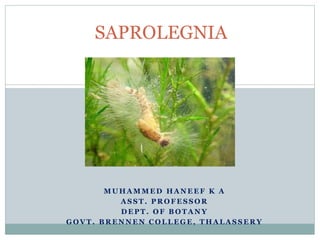
- 1. M U H A M M E D H A N E E F K A A S S T . P R O F E S S O R D E P T . O F B O T A N Y G O V T . B R E N N E N C O L L E G E , T H A L A S S E R Y SAPROLEGNIA
- 2. For M.Sc Botany, Kannur University Systematic Position Kingdom - Mycota Division - Eumycota Sub-division - Mastigomycotina Class - Oomycetes Order - Saprolegniales Family - Saprolegniaceae
- 3. For M.Sc Botany, Kannur University Introduction The genus Saprolegnia is represented by about 30 species. Most of the species are aquatic saprophytes, found on dead decaying organic material or on the bodies of floating dead insects. Some species like S. parasitica and S. ferax are parasite on fishes or on their eggs and they cause salmon disease. In India, the genus is represented by seven species; of these, S. monoeca, S. ferax, S. litoralis and S. parasitica are more common. Saprolegnia can be grown easily in the laboratory by keeping a dead fly in pond water in a test tube for few days.
- 4. For M.Sc Botany, Kannur University Vegetative Structure The mycelium of Saprolegnia is profusely branched and coenocytic. Two types of hyphae can be recognised during vegetative phase. [1] Intra-material or rhizoidal hyphae These hyphae are short and are capable to absorb food materials from the substratum or the host. [II] Extra-material or external hyphae These hyphae grow on the surface of the host and spread in all directions in the water. They form visible part of the fungus and develop sex organs during the reproductive phase. They are profusely branched, coenocytic and aseptate, but septa are formed at the bases of sex organs.
- 5. For M.Sc Botany, Kannur University The cell wall is made up of cellulose and glucans. There are many nuclei in the vacuolated cytoplasm. The reserve food material is in the form of oil globules and glycogen. The thallus is eucarpic, i.e., only a part of the thallus is utilised in the formation of fruiting bodies.
- 6. For M.Sc Botany, Kannur University Vegetative Reproduction Takes place by fragmentation and chlamydospore formation Fragmentation. Vegetative hyphae Id up into small segments and each segment capable to grow into a new mycelium Chlamydospore (Gemmae) Chlamydospores are thick-walled, ovoid, ghb or irregular bodies. They develop singly or in chains and may be terminal or internal in position On maturation, chlamydom are detached from the hyphae, they germinate A suitable substratum and produce new mycelia
- 7. For M.Sc Botany, Kannur University Asexual Reproduction Asexual reproduction takes place by zoospore which are produced in zoosporangia The zoosporangia is an elongated cylindrical structure, formed at the tip of the hyphae. Intercalary zoosporangia are also found in S. formosa, Zoosporangia are formed only on extra material hyphae. The tip of the hypha become swollen, accumulates numerous nuclei a cytoplasm becomes dense This structure is now separated by a basal septum The mature sporangium club shaped. The multinucleate protoplast sporangium is cleaved into uninucleate protoplasts Each uninucleate portion differentiates into biflagellate pear-shaped primary zoospore.
- 8. For M.Sc Botany, Kannur University
- 9. For M.Sc Botany, Kannur University One of the flagellum of the zoospore is of whiplash type and the other of tinsel type. Zoospores are released through an apical pore in the zoosporangium. They swim freely for some time and then come to rest, withdraw their flagella and each secretes a thick wall. These resting zoospores are now known as primary cysts. During favourable conditions, the cysts germinate and produce biflagellate kidney-shaped secondary zoospores. The secondary zoospores have lateral flagella; They swim for sometime and then germinate, producing new mycelia. The phenomenon of the production of two types of zoospores (primary and secondary) is known as diplanetism and these zoospores are called diplanetic.
- 10. For M.Sc Botany, Kannur University One of the characteristic features of Saprolegnia is the proliferation of zoosporangium. Following the discharge of zoospores from the zoosporangium, growth is renewed from the septum at the base of the sporangium and a new apex is formed inside the old zoosporangium. This apex develops into a new zoosporangium inside the old one. The zoospores of this new zoosporangium are discharged through the pore in the old zoosporangium. In some species of Saprolegnia this process is repeated several times and as such one can find several empty zoosporangia one inside the other.
- 11. For M.Sc Botany, Kannur University Sexual Reproduction Sexual reproduction usually takes place during unfavourable conditions and is of typically oogamous type. Male and female sex organs, known as antheridia and oogonia respectively, usually develop terminally on the lateral branches of the mycelium. But in S. litoralis sex organs are intercalary in position. Most s of Saprolegnia are homothallic or monoecious but a few are heterothallic or dioecious species Oogonia are usually formed singly or in chains the tips of the main hypha or its lateral branches. The tip of the hyphal branch that develops into an oogonium swells considerably and eventually becomes spherical or globose.
- 12. For M.Sc Botany, Kannur University
- 13. For M.Sc Botany, Kannur University Many nuclei along with cytoplasmic contents migrate into the swollen portion and then it is separated from the rest of the mycelium by a septum. As the oogonium increases in size, a large vacuole develops in its centre and the multinucleate protoplast forms a thin peripheral layer. The nuclei undergo some mitotic divisions and the protoplast is cleaved into as many segments as the number of nuclei. The mature oogonium is a thick-walled globose structure with usually 4-10 naked uninucleate spherical eggs. Occasionally, there are up to 32 eggs in an oogonium.
- 14. For M.Sc Botany, Kannur University Antheridium Antheridia are also formed at the tips of the main hyphae or their lateral branches. The tips which develop into antheridia are relatively broad than the supporting hyphae and they contain dense elops in cytoplasm and many nuclei. These swollen tips are cut off from the hyphae by septa. The hyphae which bear antheridia are known as sporangiophores. The mature antheridium is a club shaped structure with dense cytoplasmic contents and many nuclei.
- 15. For M.Sc Botany, Kannur University Fertilization As antheridia mature, sporangiophores elongate and extend towards oogonia and finally one or more antheridia become closely attached to an oogonium. Thereafter, each antheridium gives off many delicate protuberances, known as fertilization tubes, which penetrate the oogonium. In some species of Saprolegnia fertilization tubes are branched. The male nuclei pass into the oogonium through the fertilization tubes and come in contact with the eggs. Only one male nucleus fuses with an egg. The fertilized egg secretes a thick wall and this diploid structure is known as oospore.
- 16. For M.Sc Botany, Kannur University
- 17. For M.Sc Botany, Kannur University Germination of oospore Oospores undergo a prolonged resting period. In favourable conditions, the oospore germinates by producing an unbranched germ tube. The diploid nucleus of the oospore undergoes a meiotic division, followed by several mitotic divisions. The germ tube develops into a new mycelium.
- 18. For M.Sc Botany, Kannur University Sometimes the tip of the germ tube develops into a zoosporangium with many biflagellate zoospores (equal to the number of the haploid nuclei). The zoospores are released by the rupture of the wall of the sporangium. They swim for sometimes and then come to rest. They withdraw their flagella and each zoospore secretes a thick wall around it self. The cyst, thus formed, germinates by producing a germ tube. In S. ferax, antheridia do not develop close to oogonia and as such fertilization of the e not possible. Thus the eggs act as parthenospores and germinate directly.
- 19. For M.Sc Botany, Kannur University Life cycle