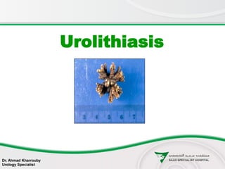
Urolithiasis (urinary stones disease) presentation
•Download as PPTX, PDF•
276 likes•54,580 views

Urinary stones disease, Urolithiasis, Urology.

Recommended
More Related Content
What's hot
What's hot (20)
Viewers also liked
Viewers also liked (20)
Similar to Urolithiasis (urinary stones disease) presentation
Similar to Urolithiasis (urinary stones disease) presentation (20)
More from Ahmad Kharrouby
More from Ahmad Kharrouby (8)
Recently uploaded
Recently uploaded (20)
Urolithiasis (urinary stones disease) presentation
- 1. Urolithiasis Dr. Ahmad Kharrouby Urology Specialist
- 2. Urolithiasis (from Greek oûron-urine and lithos-stone) is the condition where urinary stones are formed or located anywhere in the urinary system. Urolithiasis
- 3. Background
- 4. Kidney stones Ureteral stones Bladder stones Urethral stones Urolithiasis
- 5. Urolithiasis is a common disease that is estimated to produce medical costs of $2.1 billion per year in the United States alone. Urolithiasis has been a part of the human condition for millennia and have even been found in Egyptian mummies. Background
- 6. Renal colic affects approximately 1.2 million people each year in USA and accounts for approximately 1% of all hospital admissions. Most active emergency departments (EDs) manage patients with acute renal colic every day Background
- 7. Epidemiology
- 8. Urolithiasis occurs in all parts of the world A lifetime risk: 2-5% for Asia 8-15% for the West 20% for the Kingdom of Saudi Arabia Hot Climate Dietary habits Hereditary factors Epidemiology
- 9. The lower the economic status, the lower the likelihood of renal stones Most at 20-49 years Peak incidence at 35-45 years Male-to-female ratio of 3:1 Epidemiology
- 10. Chemical types and etiology
- 11. Four main chemical types: Calcium stones Struvite (magnesium ammonium phosphate) stones Uric acid stones Cystine stones Chemical Types
- 12. Calcium stones account for 75% of Urolithiasis. Radio-opaque Multiple factors and etiologies Mostly incidental Calcium stones
- 13. Incidental Hyperparathyroidism Increased gut absorption of calcium Renal calcium leak Renal phosphate leak Hperuricosuria Hperoxaluria Hypocitraturia Hypomagnesuria Calcium Stone Known etiologies
- 14. Calcium Stone
- 15. Account for 15% of renal calculi Infectous stones Gram-negative rods capable of splitting urea into ammonium, which combines with phosphate and magnesium More common in females Urine pH is typically greater than 7 Struvite (magnesium ammonium phosphate) stones
- 16. Stag horn stones are non obstructive thus painless Slowly growing Discovered incidentally Struvite (magnesium ammonium phosphate) stones
- 17. Account for 6% of renal calculi Urine pH less than 5.5 High purine intake eg. organ meats legumes malignancy 25% of patients have gout Uric acid stones
- 18. Uric Acid Stones
- 19. Uric Acid Stones
- 20. 2% of renal calculi Autosomal recessive trait Intrinsic metabolic defect resulting in failure of renal tubular reabsorption of: Cystine Ornithine Lysine Arginine Urine becomes supersaturated with cystine, with resultant crystal deposition Cystine stones
- 22. Prognosis
- 23. 80 % pass spontaneously 20% require hospital admission or intervention because of: unrelenting pain inability to retain enteral fluids proximal UTI inability to pass the stone renal failure Prognosis
- 24. Prognosis Recurrence rates after an initial episode of ureterolithiasis: 14% at 1 year 35% at 5 years 52% at 10 years
- 25. History
- 26. History The presentation is variable. Patients with urinary calculi may report Pain Infection Hematuria Asymptomatic
- 27. Silent Kidney stones Small nonobstructing stones in the kidneys only occasionally cause symptoms. If present, symptoms are usually moderate and easily controlled.
- 28. The passage of stones into the ureter is associated with classic renal colic because of: subsequent acute obstruction proximal urinary tract dilation ureteral spasm Acute renal colic is probably the most excruciatingly painful event a person can endure Obstructive ureteral stone
- 29. Classic Renal Colic Acute onset of severe flank pain radiating to the groin Gross or microscopic hematuria Nausea, and vomiting not associated with an acute abdomen in 50%
- 30. Staghorn calculi are often relatively asymptomatic. Branched kidney stone occupying the renal pelvis and at least one calyceal system. Manifest as infection and hematuria. Staghorn stone
- 31. Acute renal failure Asymptomatic bilateral obstruction Solitary Kidney with obstructive stone
- 32. Location and characteristics of pain from ureteral stones Depends on the level of obstruction and its degree: ureteropelvic junction pelvic brim ureterovesical junction
- 33. UPJ Stone Stones obstructing the ureteropelvic junction may present with mild-to-severe deep flank pain without radiation to the groin
- 34. Ureteral Stone Cause abrupt, severe, colicky pain in the flank and ipsilateral lower abdomen with radiation to the testicles or the vulvar area. Intense nausea, with or without vomiting, usually is present.
- 35. Upper ureter Tends to radiate to the flank and lumbar areas
- 36. Mid Ureter Cause pain that radiates anteriorly and caudally. Can easily mimic appendicitis on the right or acute diverticulitis on the left.
- 37. Distal Ureter and UVJ stones Cause pain that tends to radiate into the groin or testicle in the male or labia majora in the female At the ureterovesical junction also may cause irritative voiding symptoms mimicking cystitis, such as: urinary frequency dysuria
- 39. Bladder Stones Usually asymptomatic and are passed relatively easily during urination. Rarely, a patient reports positional urinary retention (obstruction precipitated by standing, relieved by recumbency).
- 40. Phases of an attack
- 41. Phases of an attack The entire process typical lasts 3-18 hours Acute phase: peak in most patients within 2 hours of onset (30 min to 6 hours) Constant Phase 1- 4 hours maximum 12 hours Relief phase 1.5-3 hours
- 42. Physical exam
- 43. Dramatic costovertebral angle tenderness unremarkable abdominal evaluation painful testicles but normal-appearing constant body positional movements (eg, writhing, pacing) Tachycardia Hypertension Microscopic hematuria Physical exam
- 44. Diagnosis
- 45. The diagnosis of nephrolithiasis is often made on the basis of clinical symptoms alone, although confirmatory tests are usually performed. Diagnosis
- 46. Laboratory tests
- 47. The recommended based on EUA recommendations: Urinary sediment/dipstick test: To demonstrate blood cells Serum creatinine level: To measure renal function Labarotary Testing
- 48. May be helpful: CBC in febrile patients Serum electrolyte assessment in vomiting patients 24-Hour urine profile on outpatient basis Additional Lab Tests
- 49. Imaging studies
- 50. Noncontrast abdominopelvic CT scan: The imaging modality of choice for assessment of urinary tract disease, especially acute renal colic. IV contrast and delayed images might be required in selected cases Imaging studies
- 51. Renal ultrasonography: Renal stone Hydronephrosis or ureteral dilation Misses 30 % of stones Plain abdominal radiograph (flat plate or KUB) misses 40 % of stones Imaging studies
- 52. Imaging studies IVP (urography) historically, the criterion standard In rare select situations: Plain renal tomography Retrograde pyelography Nuclear renal scanning
- 53. Management
- 54. Emergency Renal Colic IV access to allow : Fluid Analgesics: Paracetamol NSAID Opiod Antiemetic In case of infection: Urine culture Blood culture accordingly e.g. febrile Antibiotics
- 55. Approach Considerations In emergency settings what should be kept in mind is the small percentage suffering renal damage or sepsis. These include: Evident infection with obstruction A solitary functional kidney Bilateral ureteral obstruction Renal failure
- 56. Important The most morbid and potentially dangerous aspect of stone disease is the combination of urinary tract obstruction and upper urinary tract infection. Pyelonephritis Pyonephrosis Urosepsis Early recognition and immediate surgical drainage are necessary in these situations
- 57. Approach Considerations The size of the stone is an important predictor of spontaneous passage. A stone less than 4 mm in diameter has an 80% chance of spontaneous passage; this falls to 20% for stones larger than 8 mm in diameter
- 58. Approach Considerations Hospital admission is clearly necessary when any of the following is present: Oral analgesics are insufficient to manage the pain. Intractable vommiting Ureteral obstruction from a stone occurs in a solitary or transplanted kidney. Bilateral ureteral obstruction Ureteral obstruction from a stone occurs in the presence of a urinary tract infection (UTI) Fever Sepsis Pyonephrosis
- 59. Approach Considerations Relative indications to consider for a possible admission include comorbid conditions diabetes dehydration renal failure immunocompromised state perinephric urine extravasation pregnancy
- 60. Approach Considerations Most patients with acute renal colic can be treated on an ambulatory basis.
- 61. Approach Considerations Aggressive medical therapy has shown promise in increasing the spontaneous stone passage rate and relieving discomfort while minimizing narcotic usage
- 62. Clinic Follow up Patients who do not meet admission criteria to be discharged on medical expulsive therapy from the ED in anticipation that the stone will pass spontaneously at home. Arrangements should be made for follow-up with a urologist in 2-3 days.
- 63. Active medical expulsive therapy Paracetamol PRN for pain with or without Codeine NSAID PRN for pain Oral opiod analogue for severe pain Alpha blockers Antiemetic PRN for nausea and/or vommiting Prednisone 20 mg twice daily for 6 days With MET, stones 5-8 mm in size often pass, especially if located in the distal ureter.
- 64. Approach Considerations An important aspect of medical and preventive therapy is maintaining a good fluid intake and subsequent high urinary volume.
- 65. Emergency Advice Patients should be told to return for : fever uncontrolled pain uncontrolled vomiting Patients should be discharged with a urine strainer and encouraged to submit any recovered calculi to a urologist for chemical analysis
- 66. General recommendation not to wait longer than 4 weeks for a stone to pass spontaneously before considering intervention. Approach Considerations
- 67. Approach Considerations Larger stones (ie, ≥ 7 mm) that are unlikely to pass spontaneously require some type of surgical procedure. Such patients require mandatory urology follow up
- 68. Approach Considerations About 15-20% of patients require invasive intervention eventually as emergency or electively due to: stone size continued obstruction Infection intractable pain
- 69. Indications for Surgery The primary indications for surgical treatment include: Pain Infection Obstruction Indications for urgent intervention: Obstruction complicated by evident infection Obstruction complicated by acute renal failure Solitary kidney Bilateral obstruction
- 70. Surgical options Obstruction relief: Ureteral stent insertion Percutaneous nephrostomy Definitive surgical treatment: ESWL Ureteroscopy PCNL Open, laparoscopic and robotic pyelo-lithotomy, ureterolithotomy, cystolithotomy Open anatrophic nephrolithotomy
- 71. Surgical options For an obstructed and infected collecting system secondary to stone disease Emergency surgical relief is required with no contraindications: percutaneous nephrostomy for critical patients ureteral stent placement for stable patients
- 72. Surgical options The vast majority of symptomatic urinary tract calculi are now treated with noninvasive or minimally invasive techniques Open surgical excision of a stone from the urinary tract is now limited to isolated atypical cases
- 73. Surgical options ESWL and ureteroscopy are internationaly recognized as first-line treatments for ureteral stones. The 2005 American Urological Association (AUA) staghorn calculus guidelines recommend percutaneous nephrostolithotomy as the cornerstone for management
- 74. Ureteral Stent Guarantees drainage of urine from the kidney into the bladder and bypass any obstruction. Relieves renal colic pain even if the actual stone remains. Dilate the ureter, making ureteroscopy and other endoscopic surgical procedures easier to perform later.
- 75. Percutaneous nephrostomy Indicated if stent placement is inadvisable or impossible. In particular patients with pyonephrosis who have a UTI or urosepsis exacerbated by an obstructing calculus
- 76. Extracorporeal shockwave lithotripsy ESWL, the least invasive of the surgical methods of stone removal Utilizes an underwater energy wave focused on the stone to shatter it into passable fragments It is especially suitable for stones that are smaller than 2 cm and lodged in the upper or middle calyx the upper ureter
- 77. Extracorporeal shockwave lithotripsy The patient, under varying degrees of anesthesia The shock head delivers shockwaves developed from an Electrohydraulic Electromagnetic piezoelectric source
- 78. Ureteroscopy Ureteroscopic manipulation of a stone is a commonly applied method of stone removal A small endoscope, which may be Rigid Semirigid Flexible is passed into the bladder and up the ureter to directly visualize the stone
- 79. Ureteroscopy Flexible ureteroscopy allows tackling of even lower calyceal stones Stones are fragmented using Swiss lithoclast Laser Ultrasonic lithotripter Stones are retrieved using a stone basket
- 80. Percutaneous nephrostolithotomy Percutaneous procedures are generally reserved for large and/or complex renal stones and failures from the other 2 modalities Percutaneous nephrostolithotomy is especially useful for stones larger than 2 cm in diameter
- 81. Percutaneous nephrostolithotomy In some cases, a combination of SWL and a percutaneous technique is necessary to completely remove all stone material from a kidney.
- 82. Open Surgery Open surgery has been used less and less often since the development of the previously mentioned techniques It now constitutes less than 1% of all interventions. Disadvantages include longer hospitalization longer convalescence increased requirements for blood transfusion.
- 83. Approach Considerations Metabolic evaluation and treatment at clinic are indicated for patients at greater risk for recurrence, including: multiple stones personal or family history of previous stone formation stones at a younger age residual stones after treatment
- 84. Medical Therapy for Stone Disease Urinary calculi composed predominantly of calcium cannot be dissolved medical therapy is important in the long-term chemoprophylaxis of further calculus growth or formation
- 85. Medical Therapy for Stone Disease Uric acid and cystine calculi can be dissolved with medical therapy. Suitable option in patients with uric acid stones who do not require urgent surgical intervention Is based on alkalization of the urine.
- 86. Medical Therapy for Stone Disease Sodium bicarbonate can be used as the alkalizing agent But potassium citrate is usually preferred because of the availability of slow-release tablets and the avoidance of a high sodium load
- 87. Medical Therapy for Stone Disease The dosage of the alkalizing agent should be adjusted to maintain the urinary pH between 6.5 and 7.0.
- 88. Chemoprophylaxis Prophylactic therapy might include: most importantly, augmentation of fluid intake. limitation of dietary components addition of stone-formation inhibitors or intestinal calcium binders avoid excessive salt and protein intake
- 89. Chemoprophylaxis Better to base medical therapy for long-term chemoprophylaxis of urinary calculi on the results of a 24-hour urinalysis for chemical constituents
- 90. Long-Term Monitoring Metabolic evaluation is done by a typical 24-hour urine determination of: urinary volume pH specific gravity Calcium Citrate Magnesium Oxalate Phosphate uric acid.
- 91. Long-Term Monitoring Most common findings are Hypercalciuria Hyperuricosuria Hyperoxaluria Hypocitraturia low urinary volume
- 92. Chemoprophylaxis Chemoprophylaxis of uric acid and cystine calculi consists primarily of long-term alkalinization of urine.
- 93. Chemoprophylaxis If hyperuricosuria or hyperuricemia is documented in patients with pure uric acid stones, allopurinol (300 mg qd) is recommended
- 94. Chemoprophylaxis Pharmaceuticals that can bind free cystine in the urine: D-penicillamine 2-alpha-mercaptopropionyl-glycine Help reduce stone formation in cystinuria. Captopril has been shown to be effective in some trials
- 95. Dietary Measures In almost all patients in whom stones form, an increase in fluid intake and, therefore, an increase in urine output is recommended. This is likely the single most important aspect of stone prophylaxis The goal is a total urine volume in 24 hours in excess of 2 liters.
- 96. Dietary Measures The only other general dietary guidelines are to avoid excessive salt and protein intake. Moderation of calcium and oxalate intake is also reasonable Beware to advice moderation not avoid calcium intake as it will result in calcium deficiency disorders, most importantly osteoperosis.
- 97. Thank you
- 98. References • Main references: • Medscape article nephrolithiasis by J Stuart Wolf Jr, MD, FACS updated feb 11, 2013 • Campbell-Walsh Urology 10th edition • Smith and Tanagho's General Urology, Eighteenth Edition • Images used in this presentation are from different web based resources • N.B. The presentation is directed to general medical audience in the hospital mainly nurses and physicians with special focus on the acute management.