
Recommended
More Related Content
What's hot
What's hot (20)
Similar to Mitochondria , peroxisome and lysosome
Similar to Mitochondria , peroxisome and lysosome (20)
Recently uploaded
Recently uploaded (20)
Mitochondria , peroxisome and lysosome
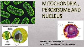
- 1. PRESENTER : J. AISHWARYA M.Sc. 3RD YEAR MEDICAL BIOCHEMISTRY 18-May-21 1
- 3. MITOCHONDRION - HISTORY • A double membrane bound organelle found in cytosol of eukaryotic cells • Mito – thread, chondrion – granule like • First observed - Richard Altman ( 1894) • Term mitochondria was coined by Carl Benda (1898) MAIN FUNCTION: • They produce enzymes for the metabolic conversion of food to energy. 18-May-21 3
- 4. ORIGIN OF MITOCHONDRIA • Derived from bacteria – process by Endosymbiosis • Mitochondria arose - 2 billion years ago when a bacterium fused with an archael cell / established a symbiotic relationship with a primitive eukaryotic cell • The closest extant relatives of Bacteria that gave rise to mitochondria - Rickettsia. • The first person to recognize mitochondria as descendents of endosymbiotic bacteria - Ivan 18-May-21 4
- 6. EVIDENCES TO SUPPORT ENDOSYMBIOTIC THEORY • Mitochondria – self replicating bodies like bacteria, divide by binary fission • Two membranes , inner membrane composition similar to bacteria • Mitochondrial DNA – similar to bacterial DNA • mi ribosomes, enzymes & transport proteins – similar to bacteria • similar size • Antibiotics – inhibits mitochondrial protein synthesis 18-May-21 6
- 7. MORPHOLOGY • Size 0.05 – 1.0 µm in diameter • Length 1 – 10 µm long • Shape Bean shaped , in fibroblast it is elongated and thread like. • Number Depends on type, size and functional state of cell. Eg : an average liver cell contain around 1500 mitochondria. • Location Cells with high energy requirement 18-May-21 7
- 8. GENERAL PROPERTIES • Mitochondria occupy - 20% of the cytoplasmic volume of a eukaryotic cell. • Often depicted as short, bacterium-like bodies - diameter of 0.5–1 μm • Remarkably dynamic and plastic, moving about the cell, constantly changing shape, dividing, and fusing • Mitochondria are often associated with the microtubular cytoskeleton - which determines their orientation and distribution in different cell types. 18-May-21 8
- 9. • In highly polarized cells - neurons, mitochondria can move long distances (up to a meter or more ) being propelled along the tracks of the microtubular cytoskeleton. • In other cells, mitochondria remain fixed at points of high energy demand- for example, in skeletal or cardiac muscle cells, they pack between myofibrils, and in sperm cells they wrap tightly around the flagellum 18-May-21 9
- 10. SHAPE, DISTRIBUTION • In higher plants : • Rod shape with hemispheroidal ends, Some are cup or filamentous shape. • Vary from globular to threadlike or branched • In Animals • Long filaments , not spatial in arrangement 18-May-21 10
- 11. NUMBERS • Depends on what the cell needs to do • Flagellated protozoa or sperm, they are found around the base of the flagellum. • Cardiac muscle, they surround the contractile parts. • Hummingbird flight muscle is the richest sources of mitochondria. When energy is not enough, more mitochondria are created ,they grow, move, and combine with other mitochondria 18-May-21 11
- 12. INTERACTION WITH OTHER MEMBRANE • Contacts between mitochondria and ER - facilitate the exchange of lipids between the two membrane systems. • Facilitate fission and fusion - involved in the distribution and partitioning of mitochondria within cells 18-May-21 12
- 14. • Double membrane • creates 3 compartments – • OUTER MEMBRANE • INTERMEMBRANE SPACE • INNER MEMBRANE • CRISTAE • MATRIX • The outer and inner membrane is composed of phospholipid bilayers and proteins. • The two membranes have different properties. 18-May-21 14
- 15. OUTER MEMBRANE • Simple phospholipid bilayer. • Contain large number of integral protein structures called porins, which allows molecules to freely diffuse from one side of the membrane to the other. • Porins pass molecules less then 5000 D - a special class of β-barrel-type membrane protein that creates aqueous pores across the membrane • Ions, nutrient molecules, ATP, ADP etc can pass through the outer membrane with ease. • The outer mitochondrial membrane is composed - 50% phospholipids by weight and contains a variety of enzymes 18-May-21 15
- 16. INNER MEMBRANE SPACE • It is also known as Perimitochondrial space. • It has high proton concentration. • The space is approximately 70 A. • Because the outer membrane is freely permeable to small molecules- concentration of small molecules such as ions and sugars in the intermembrane space is same as that of the cytosol. • Proteins present, participate in ATP synthesis 18-May-21 16
- 17. INNER MEMBRANE • Is freely permeable only to oxygen, CO₂ , H₂O. • The inner mitochondrial membrane contains proteins that perform redox reactions in oxidative phosphorylation, ATP synthase, transport proteins, protein import machinery, mitochondria fusion and fission protein. • Several antiport systems exist , allowing exchange of anions between the cytosol and the mitochondrial 18-May-21 17
- 18. CRISTAE • Are folds of inner mitochondrial membrane, which expand its surface area , enhancing its ability to produce ATP. • Stalked particles or inner membrane spheres : cristae is covered with this inner membrane spheres called stalked particles or knobs or heads. 18-May-21 18
- 19. MATRIX • It is the space enclosed by the inner membrane. • Gel like consistency ,Dense , homogenous. • Contains 2/3 rd of total protein of mitochondria. • Matrix have enzymes, DNA genome, ribosomes, tRNA, granules, fibrils and tubules. • The matrix is important in the production of ATP with the aid of the ATP synthase contained in the inner membane. • Major enzymes include enzymes involved in - Synthesis of nucleic acid and proteins, Fatty acid oxidation. 18-May-21 19
- 20. MITOCHINDRIAL DNA (mt DNA) • Small, Double stranded ,covalently closed ,circular molecule. • Occurs in multiple copies • It has 16569 bp , 37 genes • Most usually remains attached to inner mitochondrial membrane. • Stores biological info required for growth and multiplication of mitochondria. • Encode RNA s and proteins - essential for mitochondrial function. • It codes 2rRNAs , 22 tRNAs and 13 mitochondrial membrane proteins. • Can undergo replication and duplication 18-May-21 20
- 21. 18-May-21 21
- 22. BIOGENESIS • They grow - importing most of their proteins from the cytoplasm and by internal synthesis of some proteins and replication of the genome. • Similar to cells, mitochondria divide and fuse with other mitochondria maintaining their number of cells. • The balance between fusion and fission - major determinant of mitochondrial number, length, and degree of interconnection • Fusion > fission, the mitochondria tend to become more elongated and interconnected • Fission > fusion ,- more numerous and distinct mitochondria. 18-May-21 22
- 23. TARGETING OF MITOCHONDRIAL PROTEINS 18-May-21 23
- 24. • Mitochondrial proteins synthesized – 80S cytosolic & 70S matrix ribosomes • 99 % of mit proteins – synthesized as precursors in cytosol • translocated post-translationally • Mit protein import – membrane receptos & translocons • initiated – binding of mit targeting sequence to import receptor in outer membrane • TOM (translocase of outer membrane) & TIM (translocase of inner membrane) • Cooperative mechanism TARGETING OF MITOCHONDRIAL PROTEINS 18-May-21 24
- 25. 18-May-21 25
- 27. Mitochondrial targeting signal sequences • Signal sequences – 2 classes • N-terminal cleavable matrix targeting sequence • Non- cleavable internal targeting sequence • Presequences – rich in basic & hydroxyl AA, 10-30 AA residues 18-May-21 27
- 28. 18-May-21 28
- 29. 1. Targeting mit proteins to matrix • Matrix proteins contain presequences • synthesized by 80S cytosolic ribosome • maintained in unfolded state – cytosolic chaperone Hsp70 18-May-21 29
- 30. 18-May-21 30
- 31. 2. Targeting mit proteins to IMM • similar to targeting proteins to matrix side – except there is an additional stop transfer signal – located after presequence • arrests translocation • OXA complex – insert proteins synthesized by 70S ribosome 18-May-21 31
- 32. 3. Targeting to outer membrane 18-May-21 32
- 34. 18-May-21 34
- 35. DIAGNOSIS 18-May-21 35 • Unfortunately, mitochondrial genetic disorders can be difficult to diagnose, and many affected people may never receive a specific diagnosis. • In some cases, the pattern of symptoms may be suggestive of a specific mitochondrial condition. If the disease-causing gene(s) associated with the particular condition is known, the diagnosis can then be confirmed with genetic testing. • In these cases, a physician may evaluate the levels of certain substances in a sample of blood or cerebrospinal fluid. • Exercise testing •Magnetic resonance spectroscopy (detects abnormalities in the brain's chemical makeup) •Imaging studies of the brain such as MRI or CT scan •Electroencephalography (EEG) •Tests that evaluate the heart including electrocardiography and echocardiography •Muscle biopsy
- 36. 18-May-21 36
- 37. PEROXISOME History of Peroxisomes • First observed by electron microscopy in animal cells (1950s), then in plant cells (1960s) • Christian deDuve (1965) - Isolated from liver cells by centrifugation • Called them peroxisomes because they generate and destroy H2O2 18-May-21 37
- 38. ORIGIN • Peroxisomes are a vestige of an ancient organelle that performed all the oxygen metabolism in the primitive ancestors of eukaryotic cells. • When the oxygen produced by photosynthetic bacteria first accumulated in the atmosphere, it would have been highly toxic to most cells. • Peroxisomes might have lowered the intracellular concentration of oxygen. 18-May-21 38
- 39. • Single membrane • Roughly spherical • 0.2 - 1.7µm • Composition varies • Peroxisomes are also called Microbodies • They also resemble lysosomes in being filled with enzymes • They are self replicating • contain enzymes to oxidize organic substances like fats • Hydrogen peroxide is broken down right away by the enzyme catalase into oxygen and water. • Peroxisomes are abundant in the liver where they produce bile salts and cholesterol and break down fats PROPERTIES 18-May-21 39
- 40. • do not contain DNA or ribosomes • all of their proteins are encoded in the nucleus • They contain oxidative enzymes, such as catalase and urate oxidase, at such high concentrations that the peroxisomes stand out in electron micrographs because of the presence of a crystalloid protein core 18-May-21 40
- 41. 18-May-21 41
- 42. 18-May-21 42
- 43. Peroxisomes Use Molecular Oxygen and Hydrogen Peroxide to Perform Oxidation Reactions • RH2 + O2 → R + H2O2 • removes H2 atoms • H2O2 + R′H2 → R′ + 2H2O. • This type of oxidation reaction important in liver and kidney cells, where the peroxisomes detoxify various harmful molecules that enter the bloodstream. • In addition, when excess H2O2 accumulates in the cell, catalase converts it to H2O through the reaction 2H2O2 → 2H2O + O2 18-May-21 43
- 44. FUNCTIONS • Fatty acid oxidation • Bile acid synthesis • lipid biosynthesis – cholesterol & dolichol • plants – two pathways 18-May-21 44
- 46. PROTEIN TARGETING IN PEROXISOME • A Short Signal Sequence Directs the Import of Proteins into Peroxisomes • A specific sequence of three amino acids (Ser–Lys–Leu) located at the C-terminus of peroxisomal proteins functions as - import signal • transport from cytosol to peroxisome – post translationally • proteins synthesized in membrane free ribosomes • Peroxins – 23 types • ATP dependent process • Sequence- Ser – Lys - Leu 18-May-21 46
- 47. 18-May-21 47
- 48. 18-May-21 48
- 49. 18-May-21 49
- 51. NUCLEUS • light microscope - nucleus is the largest visible compartment • The presence of a nucleus distinguishes eukaryotic cells from prokaryotic cells • Houses all of the eukaryotic cell’s genome and acts as a center for controlling cellular activities 18-May-21 51
- 52. STRUCTURE • A double membrane called nuclear envelope encloses the nucleus. • The lumen separates the two membranes and is continuous with the Endoplasmic Reticulum. • Macromolecules pass between the nucleus and cytoplasm through the Nuclear Pore complexes (NPCs) - channels spanning the envelope. • Nucleolus is the most clearly visible structure. • Regions other than the nucleolus are referred to as the nucleoplasm. • Other sub-compartments include speckles, cajal bodies and PML bodies etc. • Inside the nucleus the DNA can be found in the compacted and highly stained form, heterochromatin or in the less densely compacted form the euchromatin. 18-May-21 52
- 53. NUCLEAR MEMBRANE • Nuclear Membrane is a fence between nucleus and cytoplasm to stave off free transmission of molecules. • Provides nucleus an identity of separate biochemical compounds. • The nuclear membrane consists of: • 1. Outer nuclear membrane • 2. Inner nuclear membrane • 3. Perinuclear space • 4. Nuclear pores • 5. Nuclear lamina 18-May-21 53
- 54. OUTER NUCLEAR MEMBRANE • The outer nuclear membrane is continuous with endoplasmic reticulum, therefore the lumen of nuclear membrane is directly connected with lumen of ER. • The outer nuclear membrane is functionally homologous to ER membrane. • The cytoplasmic surface of outer nuclear membrane has ribosomes that are different in composition of protein and these ribosomes are enriched in membrane proteins (for cytoskeleton binding) 18-May-21 54
- 55. PERINUCLEAR SPACE • Space is present between ONM and INM and is called Perinuclear space or lumen of envelope. • The thickness of each nuclear membrane is 7-8nm thick while perinuclear space is 20-40nm thick. 18-May-21 55
- 56. INNER NUCLEAR MEMBRANE • Proteins that are specific to nucleus are present in INM such as those that bind the nuclear lamina. • Including Lamin B receptor (LBR), lamin aassociated polypeptide (LAP) 1, LAP2, emerin, MAN1 and nurim. • Most of these proteins interact with lamins and chromatin. • Mutations in emerin and nuclear lamins have been associated with muscular dystrophies and lipodystrophy. • Integral proteins of the inner nuclear membrane are synthesized on the rough ER and reach the inner nuclear membrane by lateral diffusion in the connected ER and nuclear envelope membranes. 18-May-21 56
- 57. WHAT ARE NUCLEAR PORES? • Small polar molecules, ions and macro-molecules can only move in between the nucleus and cytoplasm through channels. • The large circles with diameter of 120nm and molecular mass of ̴125 million Dalton and 30X the size of ribosomes are pores that are collectively called as Nuclear Pore complex. • They are composed of several proteins of 30 different types. • Those specialized proteins are named as Nucleoporins • The complex consists of an assembly of eight spokes attached to rings on the cytoplasmic and nuclear sides of the nuclear envelope. • The spoke-ring assembly surrounds a central channel containing the central transporter. • Cytoplasmic filaments extend from the cytoplasmic ring, and filaments forming the nuclear basket extend from the nuclear ring. 18-May-21 57
- 58. NUCLEAR PORE CHANNEL (NPC) • The Phospholipid bilayer is only permeable for non-polar micromolecules. • The only channel through which transmission of polar micromolecules and macromolecules occurs is through Nuclear Pore Complex. • NPCS are the points where lNM and ONM are continuous 18-May-21 58
- 59. NUCLEAR LAMINA • In multicellular eukaryotes, a fibrous mesh work supports the inner nuclear membrane called Nuclear Lamina • The nuclear lamina is present inside the nuclear envelope. • Lamins are 60-80 kilo Dalton fibrous proteins that makeup the nuclear lamina • Some associated proteins are also present. • Lamins belong to a class of intermediate filament proteins. • Nuclear Lamina disease: • 1. Emery-Dreifuss muscular dystrophy • 2. Hutchinson-Gilford progeria syndrome 18-May-21 59
- 60. TRANSPORT THROUGH NUCLEAR PORE COMPLEX • Proteins < 50kDa – passively pass • Proteins > 50kDa – actively transported • proteins – imported & exported – signal sequence - Nucleus Localisation signal (NES / NIS ) • Nuclear import receptor – Importin • Nuclear export receptor – Exportin • transported post-translationally & in fully folded confirmation • NES - Leu rich • NIS – Lys rich • Karyopherins – homologous sequences • Interact with FG (phe, gly) repeats of nucleophorins 18-May-21 60
- 61. 18-May-21 61
- 62. DISORDERS Cancer associated morphological changes: • The most commonly used quantitative nuclear morphometric parameter is the measurement of nuclei size. • The nuclear-cytoplasmic ratio (also known as the “karyoplasmic ratio”) 18-May-21 62
- 63. LAMINOPATHIES • group of diseases caused by alterations in nuclear morphology are laminopathies • genetic disorders arising from mutations in the genes encoding lamins or lamin- interacting proteins, such as NE integral membrane proteins, and components of the LINC complex 18-May-21 63
- 64. 18-May-21 64
- 65. 18-May-21 65
- 66. 18-May-21 66
- 67. 18-May-21 67
- 68. 18-May-21 68
- 69. 18-May-21 69
Editor's Notes
- 15 AUTOSOMAL , I X LINKED Dysmorphism (large fontanelle, high forehead, abn ears, micrognathia, low/broad nose, redundant skin folds) - ZELLWEGER