BY:ADLIN FARIHA BINTI AZHARAHMAD FAZWAN BIN JUNAIDISITI AISYAH BINTI RAMLICASE PRESENTATION
Demographic detailsName: Rose KhadeejaTihanieDate of birth: 24th December 2007Age: 3 years oldGender: femaleEthnic group:MalayDate of admission: 15th December 2010Date of transfer: 26th December 2010Ward of admission: 8CInformant: MotherAddress:  Rawang
Presenting complaints
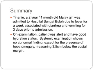
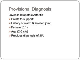
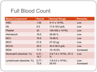
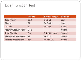
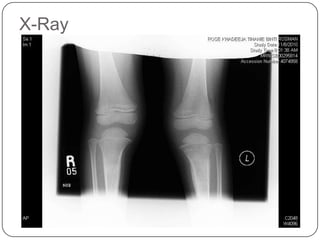
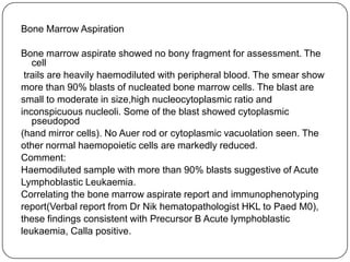
Rose KhadeejaTihanie, a 2 year 11 month old Malay girl was admitted to Hospital Sungai Buloh on 15th December 2010 (2 weeks ago) due to fever for 1 week associated with diarrhea and vomiting for 3 days prior to admission.
History of presenting complaintsTihani was previously well until she started to develop pain in her left knee and fever on 4th September in early morning (3.00 a.m.). She cannot sleep that night because of the pain.The next morning, mother claims that Tihani cannot walk because of the pain. However, there was no history of trauma noted.Her parents then brought her to a paediatric clinic in Kuching. However, no diagnosis was established.She was then brought to Sarawak general hospital and was diagnose to have muscle pain. She was given medication to relief her pain.After 3 days taking the medication, the pain resolves and she was able to walk.
On 22th September, she was again develop fever and joint pain. The joints that involves were both her knee but more on her left side, left ankle, back of her shoulder on left side and also her right elbow.She was then brought to private clinic but she was referred to Hospital Kuala Kubu.At Hospital Kuala Kubu, blood test perform shows normal results. She was given medication to relief her symptoms.After 1 week on medication, she was able to walk again.
On 1st November she was again develop fever and left knee pain and swelling and was brought to PUSRAWI. Then, she was referred to Hospital Sungai Buloh. The staffs in Hospital Sungai Buloh treated her for juvanile idiopathic arthritis.After staying in the hospital for a week, her mother asked permission to discharge earlier because she needs to go to work.On the day of discharge, Tihanie was still unable to walk but the pain and swelling subsided.
Tihanie started to have fever for one week and diarrhea and vomiting for 3 days prior to admission.The fever was on and off and associated with chills and rigor. Her mother said that she was warm to touch and documented temperature at home was 38°C.Tihanie will vomit after meal since 3 days prior to admission. Her mother described the vomitus as watery and non projectile. There was no blood or bile stain in the vomitus.For her diarrhea, she passes loose and yellowish colored stool for about 5 time a day. There was no blood in the stool.Tihanie was still unable to walk and 3 days prior to admisssion, her mother notice that she was weaker and looking pale.
Systemic review
Past medical and surgical historyThere was no significant past medical and surgical history.Drug historyShe was not on any  long term medication.
Allergy She has no known allergy.Birth historyShe was delivered in Pahang Hospital at term via spontaneous vertex delivery weight 3kg without complication.Antenatal and postnatal history was uneventful.
Neonatal historyHer neonatal history was uneventful.Feeding historyShe was exclusively breastfed for 3 months.After 3months old, she was introduced to formula milk.At 6 months old, she was given semisolid foodAbout 1 year of age, she started to have family diet.She had loss of appetite since she was ill.
Immunization history  Her immunization was up to her age.Developmental historyGross motor:	She was able to walk up and down stairs and can ride a  bicycle.Fine motor and vision:	She can draw “O” and “+”.Speech and language:	She can count numbers and can name more than 3 colors.Personal and social:	She can play with others.
Family historyHer parents and other siblings were healthy.There was no family history of asthma, diabetes, hypertension and blood disorder.The was no family members that was having the same symptoms.Social historyShe lives with parents and two other siblings in a terrace house at Rawang.Their family total income for a month was about RM 6000.
Effects of the illnessHer mother has to take a long leave from work to take care of her in hospital.
Physical Examination
General ExaminationPatient was lying comfortably in supine position, supported by one pillow. She was conscious, alert and responsive to people. There were 2 branullasinserted on the dorsum of both feet.  Vital SignsPulse Rate: 146 bpmBlood Pressure: 98/55 mmHgRespiratory Rate: 30 bpmTemperature: 37 °C
ArthropometryWeight	           : 11 kgHeight	           : 95 cm Impression	:  The height is on the 50th centile while the weight is between 3rd and 10th centile.Hydrational statustongue and mucous membranes in the oral cavity were moistNormal skin turgor.Capillary refill time was less than 2 secondsImpression: Her hydration status was good.
HeadThere were no conjunctival pallor and no jaundice.No angular stomatitis, no central cyanosis.Lymph NodesNo palpable cervical, axillary,  or inguinal lymph nodesUpper LimbsPalms are pink and warm to touch.No finger clubbing, koilonychia , good capillary filling time.No swelling of the elbows or the wrists.Lower LimbsNo ankle edema, no swelling in the knee joints or ankles.
Systemic Examination
Cardiovascular SystemInspection: chest moves symmetrically with respiration. There was no chest wall deformity, no scar, no dilated veins, no precordial bulge, no sign of respiratory distress and no visible pulsation noted.Palpation: apex beat was felt at 4th intercostals space, mid-clavicular line. There was no left parasternal heaves and no thrills at left sternal edge, pulmonary area and aortic area.Auscultation: normal 1st and 2nd heart sound was heard. There was no additional heart sound or murmur.Impression: No abnormal findings
Respiratory SystemInspection: the chest moves symmetrically with respiration on bothsides. There was no suprasternal, intercostals and subcostal recession. There was no chest deformity and no scar seen.Palpation: the trachea is centrally located and chest expansion was symmetrical on both sides. The apex beat was located at 4th intercostals space, mid-clavicular line. Normal vocal fremitus was notedPercussion:both sides of lungs were resonance. There was normal liver and cardiac  dullness.Auscultation: the air entry was adequate on both sides of the lung. Normal  vesicular breath sound was heard. There were no added sounds .Impression: No abnormal findings
Gastrointestinal SystemInspection: abdomen was symmetrically distended and moves with respiration. The umbilicus was centrally located and inverted. There was no scar, no dilated vein, no visible pulsation and peristalsis noted. Palpation: abdomen was soft and non tender. On deeppalpation, the liver was palpable up to 3.5 cm below costal margin, the spleen was not palpable. Both kidneys were not ballotable Percussion: no dullness was notedAuscultation: normal bowel sound present with no renal bruit.Impression: There is hepatomegaly measuring 3.5cm below the costal margin.
Central Nervous SystemHigher function: Patient was conscious, alert and can communicate with others.Cranial nerves: cranial nerves were intact.Motor function: Muscle bulk and muscle tone was normal. Muscle power for all extremities grading 5/5.  Biceps, triceps, supinator, knee, and ankle reflexes were present. Sensory functions: Normal sensation to touch, temperature and joint position sense.Impression: No abnormal findings
SummaryTihanie, a 2 year 11 month old Malay girl was admitted to Hospital Sungai Buloh due to fever for a week associated with diarrhea and vomiting for 3 days prior to admisssion.On examination, patient was alert and have good hydration status.  Systemic examination shows no abnormal finding, except for the presence of hepatomegaly, measuring 3.5cm below the costal margin.
Provisional DiagnosisJuvenile Idiopathic ArthritisPoints to support:History of warm & swollen joint
Female (6:1)
Age (2-6 y/o)
Previous diagnosis of JIADifferential DiagnosisSeptic ArthritisDengue Fever
Investigation
Full Blood Count
Liver Function Test
X-Ray
Impression: there is pancytopenia, indicating bone marrow suppression or failure.Differential Diagnosis:LeukaemiaAplastic AnemiaFifth Disease (parvovirus B19)
Microbiology
Peripheral Blood FilmHB:Moderateanaemia.RBC:Microcytosis with anisopoikilocytosis. No nrbc seen.WBC:Markedly reduced. Occasional lymphocytes, atypical lymphocytes and band neutrophils. An occasional suspicious looking mononuclear seen.PLT:Reduced. No platelet clumping.Impression: Severe pancytopenia. Acute leukaemia cannot be rule out from this smear. Advise for BMA TRO haematological malignancy.
Bone Marrow AspirationBone marrow aspirate showed no bony fragment for assessment. The cell trails are heavily haemodiluted with peripheral blood. The smear showmore than 90% blasts of nucleated bone marrow cells. The blast aresmall to moderate in size,highnucleocytoplasmic ratio andinconspicuous nucleoli. Some of the blast showed cytoplasmicpseudopod(hand mirror cells). No Auer rod or cytoplasmicvacuolation seen. Theother normal haemopoietic cells are markedly reduced.Comment:Haemodiluted sample with more than 90% blasts suggestive of AcuteLymphoblastic Leukaemia.Correlating the bone marrow aspirate report and immunophenotypingreport(Verbal report from Dr Nikhematopathologist HKL to Paed M0),these findings consistent with Precursor B Acute lymphoblasticleukaemia, Calla positive.
Final DiagnosisJuvenile arthritis secondary to acute lymphoblastic leukaemia.
Discussion
Definition Leukaemia is a malignant disorders of heamatopoietic stem cells characteristically associated with increase number of white cells in bone marrow or/and peripheral blood.                                 (Davidson’s Principles and Practice of Medicine, 21st edition)
Types of leukemiaAcute Chronic Proliferation of primitive stem cells leading to an accumulation of blasts, predominantly in the bone marrow  which will lead to bone marrow failure.Acute lymphoblastic leukaemia and acute myeloid leukaemiaThe malignant clone is able to differentiate, resulting in accumulation of more mature cells.Chronic lymphoblastic leukaemia and chronic myeloid leukaemia.
Epidemiology Acute lymphoblastic leukaemia shows a peak incidence in children age 1-5 years oldAcute myeloid leukaemia has the lowest incidence in young adult life and there is striking rise over the age of 50Chronic lymphoblastic leukaemia and chronic myeloid leukaemia mainly in middle and old age.
Risk factorsEnvironmental factors-family incomefather with higher social contact number of elder siblingsfather who smokesthe distance of the house from a power line(Based on A CASE-CONTROL STUDY ON THE ASSOCIATION BETWEEN ENVIRONMENTAL FACTORS AND THE OCCURRENCE OF ACUTE LEUKEMIA AMONG CHILDREN IN KLANG VALLEY, MALAYSIA by Abdul Rahman HI, Shah SA, Alias H, Ibrahim HM, 2008)
Genetic factorsGenetic diseases such as Down syndrome.People who have an identical twin who develops leukemia are more likely to develop it themselves.
Clinical menifestationPersistent infections and fever (76%) – infections due to low healthy white blood cellsMalaise- lack of red blood cellsPallor (71%)- lack of red blood cellsAbnormal bruising (34%)-low plateletsVomiting  (3%)– acute leukaemia affects brain and spinal cords which part or central nervous system.Pain in joint and bones (37%)-bone marrow filling with leukaemia cells.Abdominal discomfort, loss of appetite and weight loss-when leukaemia cells accumulate in spleen, liver and kidney, the organs become swollen, and leads to abdominal pain and patient feels full after eating small quantities of food.
Signs Hepatomegaly (84%)Spleenomegaly (76%)Lymphadenopathy(84%)   Due to reticulo-endothelial system infiltration which the increase of worn out blood cells result in phagocytosis of macrophage and monocytes to the blast cells            (Percentage based on Acute leukaemia in malaysia children, sinniah et.al, 1971)
Investigations Full blood count-often raised white countBlood films- to detect types of leukaemia that presence in peripheral bloodRenal profile- due to tumourlysis syndrome, need to monitor hyperkalemia, hyperuricaemia, hyperphosphotemia and hypocalcemiaBone marrow aspiration-hypercellular replacement of normal cells by blast cells which more than 20%.Chromosome analysis- AML with t(8;21), CML with t(9;22)
Managements Aims- to destroy the leukemia clone cells without destroying the residual normal stem cells.Remission induction-the tumor is destroyed by chemotherapy. The patient goes through a period of severe hypoplasia.
2. Remission consolidation-if remission is achieved, residual disease is attacked by therapy during consolidation phase. This consists of a number of courses chemotherapy resulting bone marrow hypoplasia. Central nervous system prophylaxis stop the cancer from spreading to brain and nervous system.It kills cancer cells that may be in the brain and spinal cord, even though no cancer has been detected.3. Remission maintenance – with chemotherapy drug to prevent disease recurrence. It involves lower drug doses and continue up to three years.
Supportive therapyPeriod of bone marrow failure:Anaemia- treated with red cell transfuseBleeding- platelets transfusion. Prophylactic platelet transfusion should be given to maintain the platelet count >10x10⁹/LInfection – parental broad-spectrum antibiotics therapy. Empirical therapy is given by combination an aminoglycosides and broad spectrum penicillin by at least 3 days after fever resolved.Patient with ALL are susceptible to Pneumocycticjirovecii. Prophylaxis co-trimoxazole is giving during chemotherapy
2. Metabolic monitoringFrequent monitoring of fluid balance and renal, hepatic and haemostatic function is necessary.Renal toxicity may occur with some antibiotics (eg: aminoglycosides)Cellular breakdown during induction therapy (tumor lysis syndrome ) releases intracellular ions and nucleic acid breakdown products, causing hyperkalaemia, hyperuricaemia, hyperphosphataemia and hypoclacaemia. This may cause renal failureAllopurinol and intravenous hydration are given to prevent this. In patient in high risk of tumourlysis syndrome prophylactic rasburicase (a recombinant urateoxidase enzyme) can be used.

8. Acute Lymphoblastic Leukemia

  • 1.
    BY:ADLIN FARIHA BINTIAZHARAHMAD FAZWAN BIN JUNAIDISITI AISYAH BINTI RAMLICASE PRESENTATION
  • 2.
    Demographic detailsName: RoseKhadeejaTihanieDate of birth: 24th December 2007Age: 3 years oldGender: femaleEthnic group:MalayDate of admission: 15th December 2010Date of transfer: 26th December 2010Ward of admission: 8CInformant: MotherAddress: Rawang
  • 3.
    Presenting complaintsRose KhadeejaTihanie,a 2 year 11 month old Malay girl was admitted to Hospital Sungai Buloh on 15th December 2010 (2 weeks ago) due to fever for 1 week associated with diarrhea and vomiting for 3 days prior to admission.
  • 4.
    History of presentingcomplaintsTihani was previously well until she started to develop pain in her left knee and fever on 4th September in early morning (3.00 a.m.). She cannot sleep that night because of the pain.The next morning, mother claims that Tihani cannot walk because of the pain. However, there was no history of trauma noted.Her parents then brought her to a paediatric clinic in Kuching. However, no diagnosis was established.She was then brought to Sarawak general hospital and was diagnose to have muscle pain. She was given medication to relief her pain.After 3 days taking the medication, the pain resolves and she was able to walk.
  • 5.
    On 22th September,she was again develop fever and joint pain. The joints that involves were both her knee but more on her left side, left ankle, back of her shoulder on left side and also her right elbow.She was then brought to private clinic but she was referred to Hospital Kuala Kubu.At Hospital Kuala Kubu, blood test perform shows normal results. She was given medication to relief her symptoms.After 1 week on medication, she was able to walk again.
  • 6.
    On 1st Novembershe was again develop fever and left knee pain and swelling and was brought to PUSRAWI. Then, she was referred to Hospital Sungai Buloh. The staffs in Hospital Sungai Buloh treated her for juvanile idiopathic arthritis.After staying in the hospital for a week, her mother asked permission to discharge earlier because she needs to go to work.On the day of discharge, Tihanie was still unable to walk but the pain and swelling subsided.
  • 7.
    Tihanie started tohave fever for one week and diarrhea and vomiting for 3 days prior to admission.The fever was on and off and associated with chills and rigor. Her mother said that she was warm to touch and documented temperature at home was 38°C.Tihanie will vomit after meal since 3 days prior to admission. Her mother described the vomitus as watery and non projectile. There was no blood or bile stain in the vomitus.For her diarrhea, she passes loose and yellowish colored stool for about 5 time a day. There was no blood in the stool.Tihanie was still unable to walk and 3 days prior to admisssion, her mother notice that she was weaker and looking pale.
  • 8.
  • 9.
    Past medical andsurgical historyThere was no significant past medical and surgical history.Drug historyShe was not on any long term medication.
  • 10.
    Allergy She hasno known allergy.Birth historyShe was delivered in Pahang Hospital at term via spontaneous vertex delivery weight 3kg without complication.Antenatal and postnatal history was uneventful.
  • 11.
    Neonatal historyHer neonatalhistory was uneventful.Feeding historyShe was exclusively breastfed for 3 months.After 3months old, she was introduced to formula milk.At 6 months old, she was given semisolid foodAbout 1 year of age, she started to have family diet.She had loss of appetite since she was ill.
  • 12.
    Immunization history Her immunization was up to her age.Developmental historyGross motor: She was able to walk up and down stairs and can ride a bicycle.Fine motor and vision: She can draw “O” and “+”.Speech and language: She can count numbers and can name more than 3 colors.Personal and social: She can play with others.
  • 13.
    Family historyHer parentsand other siblings were healthy.There was no family history of asthma, diabetes, hypertension and blood disorder.The was no family members that was having the same symptoms.Social historyShe lives with parents and two other siblings in a terrace house at Rawang.Their family total income for a month was about RM 6000.
  • 14.
    Effects of theillnessHer mother has to take a long leave from work to take care of her in hospital.
  • 15.
  • 16.
    General ExaminationPatient waslying comfortably in supine position, supported by one pillow. She was conscious, alert and responsive to people. There were 2 branullasinserted on the dorsum of both feet. Vital SignsPulse Rate: 146 bpmBlood Pressure: 98/55 mmHgRespiratory Rate: 30 bpmTemperature: 37 °C
  • 17.
    ArthropometryWeight : 11 kgHeight : 95 cm Impression : The height is on the 50th centile while the weight is between 3rd and 10th centile.Hydrational statustongue and mucous membranes in the oral cavity were moistNormal skin turgor.Capillary refill time was less than 2 secondsImpression: Her hydration status was good.
  • 18.
    HeadThere were noconjunctival pallor and no jaundice.No angular stomatitis, no central cyanosis.Lymph NodesNo palpable cervical, axillary, or inguinal lymph nodesUpper LimbsPalms are pink and warm to touch.No finger clubbing, koilonychia , good capillary filling time.No swelling of the elbows or the wrists.Lower LimbsNo ankle edema, no swelling in the knee joints or ankles.
  • 19.
  • 20.
    Cardiovascular SystemInspection: chestmoves symmetrically with respiration. There was no chest wall deformity, no scar, no dilated veins, no precordial bulge, no sign of respiratory distress and no visible pulsation noted.Palpation: apex beat was felt at 4th intercostals space, mid-clavicular line. There was no left parasternal heaves and no thrills at left sternal edge, pulmonary area and aortic area.Auscultation: normal 1st and 2nd heart sound was heard. There was no additional heart sound or murmur.Impression: No abnormal findings
  • 21.
    Respiratory SystemInspection: thechest moves symmetrically with respiration on bothsides. There was no suprasternal, intercostals and subcostal recession. There was no chest deformity and no scar seen.Palpation: the trachea is centrally located and chest expansion was symmetrical on both sides. The apex beat was located at 4th intercostals space, mid-clavicular line. Normal vocal fremitus was notedPercussion:both sides of lungs were resonance. There was normal liver and cardiac dullness.Auscultation: the air entry was adequate on both sides of the lung. Normal vesicular breath sound was heard. There were no added sounds .Impression: No abnormal findings
  • 22.
    Gastrointestinal SystemInspection: abdomenwas symmetrically distended and moves with respiration. The umbilicus was centrally located and inverted. There was no scar, no dilated vein, no visible pulsation and peristalsis noted. Palpation: abdomen was soft and non tender. On deeppalpation, the liver was palpable up to 3.5 cm below costal margin, the spleen was not palpable. Both kidneys were not ballotable Percussion: no dullness was notedAuscultation: normal bowel sound present with no renal bruit.Impression: There is hepatomegaly measuring 3.5cm below the costal margin.
  • 23.
    Central Nervous SystemHigherfunction: Patient was conscious, alert and can communicate with others.Cranial nerves: cranial nerves were intact.Motor function: Muscle bulk and muscle tone was normal. Muscle power for all extremities grading 5/5. Biceps, triceps, supinator, knee, and ankle reflexes were present. Sensory functions: Normal sensation to touch, temperature and joint position sense.Impression: No abnormal findings
  • 24.
    SummaryTihanie, a 2year 11 month old Malay girl was admitted to Hospital Sungai Buloh due to fever for a week associated with diarrhea and vomiting for 3 days prior to admisssion.On examination, patient was alert and have good hydration status. Systemic examination shows no abnormal finding, except for the presence of hepatomegaly, measuring 3.5cm below the costal margin.
  • 25.
    Provisional DiagnosisJuvenile IdiopathicArthritisPoints to support:History of warm & swollen joint
  • 26.
  • 27.
  • 28.
    Previous diagnosis ofJIADifferential DiagnosisSeptic ArthritisDengue Fever
  • 29.
  • 30.
  • 31.
  • 32.
  • 33.
    Impression: there ispancytopenia, indicating bone marrow suppression or failure.Differential Diagnosis:LeukaemiaAplastic AnemiaFifth Disease (parvovirus B19)
  • 34.
  • 35.
    Peripheral Blood FilmHB:Moderateanaemia.RBC:Microcytosiswith anisopoikilocytosis. No nrbc seen.WBC:Markedly reduced. Occasional lymphocytes, atypical lymphocytes and band neutrophils. An occasional suspicious looking mononuclear seen.PLT:Reduced. No platelet clumping.Impression: Severe pancytopenia. Acute leukaemia cannot be rule out from this smear. Advise for BMA TRO haematological malignancy.
  • 36.
    Bone Marrow AspirationBonemarrow aspirate showed no bony fragment for assessment. The cell trails are heavily haemodiluted with peripheral blood. The smear showmore than 90% blasts of nucleated bone marrow cells. The blast aresmall to moderate in size,highnucleocytoplasmic ratio andinconspicuous nucleoli. Some of the blast showed cytoplasmicpseudopod(hand mirror cells). No Auer rod or cytoplasmicvacuolation seen. Theother normal haemopoietic cells are markedly reduced.Comment:Haemodiluted sample with more than 90% blasts suggestive of AcuteLymphoblastic Leukaemia.Correlating the bone marrow aspirate report and immunophenotypingreport(Verbal report from Dr Nikhematopathologist HKL to Paed M0),these findings consistent with Precursor B Acute lymphoblasticleukaemia, Calla positive.
  • 37.
    Final DiagnosisJuvenile arthritissecondary to acute lymphoblastic leukaemia.
  • 38.
  • 39.
    Definition Leukaemia isa malignant disorders of heamatopoietic stem cells characteristically associated with increase number of white cells in bone marrow or/and peripheral blood. (Davidson’s Principles and Practice of Medicine, 21st edition)
  • 40.
    Types of leukemiaAcuteChronic Proliferation of primitive stem cells leading to an accumulation of blasts, predominantly in the bone marrow which will lead to bone marrow failure.Acute lymphoblastic leukaemia and acute myeloid leukaemiaThe malignant clone is able to differentiate, resulting in accumulation of more mature cells.Chronic lymphoblastic leukaemia and chronic myeloid leukaemia.
  • 41.
    Epidemiology Acute lymphoblasticleukaemia shows a peak incidence in children age 1-5 years oldAcute myeloid leukaemia has the lowest incidence in young adult life and there is striking rise over the age of 50Chronic lymphoblastic leukaemia and chronic myeloid leukaemia mainly in middle and old age.
  • 42.
    Risk factorsEnvironmental factors-familyincomefather with higher social contact number of elder siblingsfather who smokesthe distance of the house from a power line(Based on A CASE-CONTROL STUDY ON THE ASSOCIATION BETWEEN ENVIRONMENTAL FACTORS AND THE OCCURRENCE OF ACUTE LEUKEMIA AMONG CHILDREN IN KLANG VALLEY, MALAYSIA by Abdul Rahman HI, Shah SA, Alias H, Ibrahim HM, 2008)
  • 43.
    Genetic factorsGenetic diseasessuch as Down syndrome.People who have an identical twin who develops leukemia are more likely to develop it themselves.
  • 44.
    Clinical menifestationPersistent infectionsand fever (76%) – infections due to low healthy white blood cellsMalaise- lack of red blood cellsPallor (71%)- lack of red blood cellsAbnormal bruising (34%)-low plateletsVomiting (3%)– acute leukaemia affects brain and spinal cords which part or central nervous system.Pain in joint and bones (37%)-bone marrow filling with leukaemia cells.Abdominal discomfort, loss of appetite and weight loss-when leukaemia cells accumulate in spleen, liver and kidney, the organs become swollen, and leads to abdominal pain and patient feels full after eating small quantities of food.
  • 45.
    Signs Hepatomegaly (84%)Spleenomegaly(76%)Lymphadenopathy(84%) Due to reticulo-endothelial system infiltration which the increase of worn out blood cells result in phagocytosis of macrophage and monocytes to the blast cells (Percentage based on Acute leukaemia in malaysia children, sinniah et.al, 1971)
  • 46.
    Investigations Full bloodcount-often raised white countBlood films- to detect types of leukaemia that presence in peripheral bloodRenal profile- due to tumourlysis syndrome, need to monitor hyperkalemia, hyperuricaemia, hyperphosphotemia and hypocalcemiaBone marrow aspiration-hypercellular replacement of normal cells by blast cells which more than 20%.Chromosome analysis- AML with t(8;21), CML with t(9;22)
  • 47.
    Managements Aims- todestroy the leukemia clone cells without destroying the residual normal stem cells.Remission induction-the tumor is destroyed by chemotherapy. The patient goes through a period of severe hypoplasia.
  • 48.
    2. Remission consolidation-ifremission is achieved, residual disease is attacked by therapy during consolidation phase. This consists of a number of courses chemotherapy resulting bone marrow hypoplasia. Central nervous system prophylaxis stop the cancer from spreading to brain and nervous system.It kills cancer cells that may be in the brain and spinal cord, even though no cancer has been detected.3. Remission maintenance – with chemotherapy drug to prevent disease recurrence. It involves lower drug doses and continue up to three years.
  • 49.
    Supportive therapyPeriod ofbone marrow failure:Anaemia- treated with red cell transfuseBleeding- platelets transfusion. Prophylactic platelet transfusion should be given to maintain the platelet count >10x10⁹/LInfection – parental broad-spectrum antibiotics therapy. Empirical therapy is given by combination an aminoglycosides and broad spectrum penicillin by at least 3 days after fever resolved.Patient with ALL are susceptible to Pneumocycticjirovecii. Prophylaxis co-trimoxazole is giving during chemotherapy
  • 50.
    2. Metabolic monitoringFrequentmonitoring of fluid balance and renal, hepatic and haemostatic function is necessary.Renal toxicity may occur with some antibiotics (eg: aminoglycosides)Cellular breakdown during induction therapy (tumor lysis syndrome ) releases intracellular ions and nucleic acid breakdown products, causing hyperkalaemia, hyperuricaemia, hyperphosphataemia and hypoclacaemia. This may cause renal failureAllopurinol and intravenous hydration are given to prevent this. In patient in high risk of tumourlysis syndrome prophylactic rasburicase (a recombinant urateoxidase enzyme) can be used.