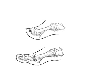
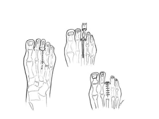
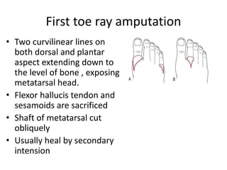
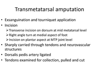
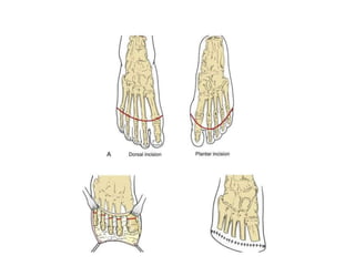
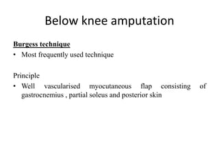
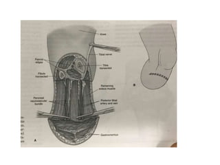
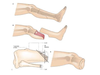
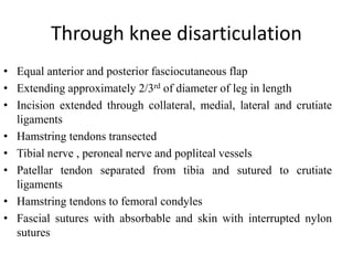
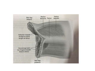
3. Types
• Guillotine / open amputation
Skin not closed over the amputation stump
Non healthy wounds
Followed by second surgery
Secondary closure
Plastic surgery
Revision of stump and reconstruction
Reamputation
• Closed amputation
Skin closed primarily
Elective amputations
4. • Minor amputations
Transphalangeal toe amputation
Transmetatarsal (Ray) toe amputation
Transmetatarsal foot amputatrion
• Major amputations
Below knee
Above knee
Through knee disarticulation
5. Minor amputations
Goals
• Remove anatomic pressure points and prevent
recurrent ulceration
• Control osteomyelitis and infection
• Preserve and restore the foot to optimum
ambulatory function
• Control and releive pain
6. Indications
• Simple toe amputation
Irreversible tissue loss / osteomyelitis of one toe distal to
proximal interphalangeal joint
• Ray amputation
Disease extends to web space/ MTP joint / Metatarsal
head
• Transmetatarsal amputation
Disease extending beyond single ray amputation
Need for multiple ray amputation
Failed previous ray amputation
Forefoot deformity causing severe disability and
intractable pain
8. Transphalangeal toe amputation
• Incision
Mid proximal phalanx to the level of bone
Circumferencial incision
Plantar flap in 1st toe
• Stripping of periosteum
• Transection of bone with oscillating saw
1cm proximal to skin
• Tendons pulled and sharply cut to retract
• Skin is closed in anteroposterior orientation
9.
10. Ray amputation
• Incision
Racquet shape incision on dorsum
Extending on each side of toe in semielliptical
fashion
Ratio of incision length to width of 3:1 – aids in
primary closure
• Deepened till bone
• Toe disarticulated at MTP joint
11. • Periosteum of bone elevated proximally
• Metatarsal head is excised at proximal
diaphyseal end using oscillating saw/ bone
nibbler
• Cut ends smoothened with bone file
• Wound irrigation
• Primary closure / left open for daily dressing
12.
13. First toe ray amputation
• Two curvilinear lines on
both dorsal and plantar
aspect extending down to
the level of bone , exposing
metatarsal head.
• Flexor hallucis tendon and
sesamoids are sacrificed
• Shaft of metatarsal cut
obliquely
• Usually heal by secondary
intension
14. Transmetatarsal amputation
• Exsanguination and tourniquet application
• Incision
Transverse incision on dorsum at mid metatarsal level
Right angle turn at medial aspect of foot
Incision on plantar aspect at MTP joint level
• Sharply carried through tendons and neurovascular
structures
• Dorsalis pedis artery ligated
• Tendons examined for collection, pulled and cut
15. • Periosteum elevated on metatarsal till intended
resection
• Retract skin flaps and cut the bone with oscillating gigli
saw 2cm proximal to skin incision
• Insertion of peroneus longus, brevis and and tibialis
brevis preserved
• First and fifth metatarsal transected in oblique fashion
• 2nd to 4th metatarsal cut in parabolic fashion with long
dorsal and bevelled plantar
16.
17. • Flexor tendons are cut
• Tourniquet released
• Irrigation and hemostasis
• Drain depending on bleeding
• Skin closed with mattress
• Dressing uptil midleg
• Limb splinted in neutral position to prevent equinus
contracture
18. Postoperative management
Simple toe / ray amputation
• Early mobilisation and evaluation by prosthetist for
shoe fitting
Transmetatarsal amputation
• Non weight bearing mobilisation until wound heals
completely
• Drain removal in 24-48hours
• Suture till 2-4weeks
• Once mobilisation – monitoring for transfer ulcers
19. Major lower limb amputations
Indications
• Acute limb ischemia
• Chronic critical limb ischemia
• Major infection in diabetics
20. Guillotine amputation
• Question regarding uncontrolled pedal sepsis
• Two staged amputation is planned
• Advantages
Remove septic focus quickly
Direct examination of calf muscle compartments
for septic extension
BKA can be planned once infection resolves
21.
22. • Limb prepped till knee
• Circular incision above the malleolus down to
tibia and fibula
• Bones cut with gigli saw
• Vascular bundles are ligated
• Irrigation and hemostasis
• Bulky dressings apllied
23. Below knee amputation
Burgess technique
• Most frequently used technique
Principle
• Well vascularised myocutaneous flap consisting of
gastrocnemius , partial soleus and posterior skin
24. • Supine position
• Limb is prepped to groin level
Incision
• Anterior skin incision 12cm below tibial tuberosity
• Continued transversely for a distance approximately
one third of calf circumference
• Incision extended along vertical axis length
approximately one and half times length of transverse
incision
• Posterior flap then completed transversely
25.
26. • Skin incision is deepened through fascia with ligation of GSV
Anterior compartment dissection
• Muscles divided at same level
• Tibialis anterior, extensor hallucis longus and extensor
digitorum longus,fibularis longus
• Central in anterior compartment
• Anterior tibial vessels and peroneal nerve
27.
28. Transection of bones
• Tibia cleared of lateral and posterior attachments
• Interroseus membrane incised
• Division of tibia at skin incision level with anterior bevelling
• Lateral compatment muscles (fibularis longus and brevis )
divided
• Fibula is transected 2cm proximal to level of tibial transection
29. Posterior compartment
• Muscles divided obliquely with removal of enough soleus
• Posterior tibial and peroneal vessels ligated
• Bone edges are smoothened
• Fascial edges are closed with interruppted absorbable sutures
with or without drain
• Skin closed with mattress suture
30.
31.
32. Through knee disarticulation
• Equal anterior and posterior fasciocutaneous flap
• Extending approximately 2/3rd of diameter of leg in length
• Incision extended through collateral, medial, lateral and crutiate
ligaments
• Hamstring tendons transected
• Tibial nerve , peroneal nerve and popliteal vessels
• Patellar tendon separated from tibia and sutured to crutiate
ligaments
• Hamstring tendons to femoral condyles
• Fascial sutures with absorbable and skin with interrupted nylon
sutures
33. Above knee amputation
Indication
• Amputation with a fixed-knee contracture
• A nonfunctional limb, or
• Insufficient circulation to heal a BKA
34.
35. • Transverse fish-mouth incision in the lower thigh. Deepened through
the subcutaneous tissue to allow the edges to separate
• The greater saphenous vein needs to be identified and ligated.
• The dissection is carried down to the femur, which is cleared using a
periosteal elevator to a level 2 to 3 cm proximal to the skin incision.
• The superficial femoral artery is dissected free and suture ligated.
• The bone is transected with a gigli saw, and the posterior muscle flap
is divided.
36. • The sciatic nerve is identified and pulled ,highly ligated, and
divided.
• Irrigated and hemostasis ensured.
• The fascia is then closed with absorbable suture.
• The subcutaneous tissue is closed with absorbable suture to
minimize tension on the skin edges and the skin is closed with
monofilament suture or staples.
• Alternative incisions can be made to accommodate surgical
wounds.