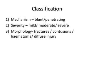
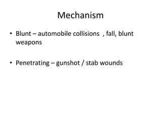
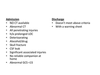
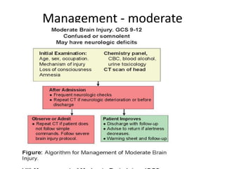
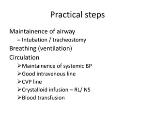
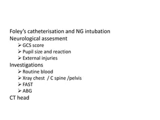
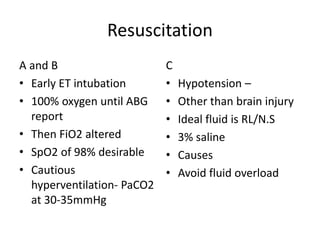
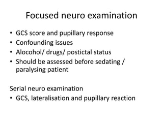
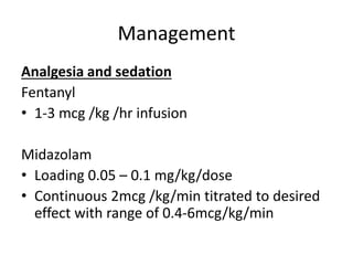
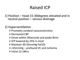
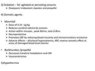
Head injury
Dr Suhas U
Incidence
Anatomy and
physiology
Classification
Management
Incidence
• Leading cause of morbidity , mortality ,
disability and socioeconomic losses
• 1.5 – 2 million injured per year
• 1million succumb to death every year
• Causes
RTA- 60%
Falls – 20-25%
Violence 10%
• Alcohol / drug – 15-20%
Scalp
Skull
• Cranial vault
• Base
• Calvaria – thin at temporal region (temporalis)
Meninges
Duramater
Arachnoidmater
Piamater
• Epidural space – meningeal arteries, dural
sinuses
• Subdural space – bridging veins
• Subarachnoid space - CSF
Ventricles
• From choroid
plexus of lateral
and 3 rd ventricle
• CSF production at
20ml/hr
Tentorium cerebelli
• Separates supra from infratentorial
compartment
• Connected through incisura
• Uncal herniation – I/L 3rd nerve + C/L
hemiparesis
Physiology
Intracranial pressure
• Normal ICP 10mmHg
• ICP >20mmHg – risk for transtentorial
herniation
MONRO KELLIE DOCTRINE
• Total volume of intracranial content remains
constant
Classification
1) Mechanism – blunt/penetrating
2) Severity – mild/ moderate/ severe
3) Morphology- fractures / contusions /
haematoma/ diffuse injury
Mechanism
• Blunt – automobile collisions , fall, blunt
weapons
• Penetrating – gunshot / stab wounds
Severity – GCS
• Total score – 15
• Mild head injury – 13-15
• Moderate – 9-12
• Coma / severe head injury - <8
• In a hemiplegia patient while assessing GCS ,
best motor response to be used for scoring
Morphology
Skull fractures - Never to be underestimated
• Calvaria / base
• Linear / stellate
• Open / closed
Intracranial injuries- Diffuse
Concussion
• Transient nonfocal
neurological
disturbance
• Mainly LOC
• Ischemia/ hypoxia
• CT being normal
DAI
• High velocity
decelaration injuries
• Multiple punctate
hemorrhages at GW
matter junction
• Poor outcome
EDH
• 0.5 – 3%
• 9% when GCS <8
• 10-40years
• M:F :: 4:1
• Temporal(80%) >frontal
• Meningeal arteries
>dural veins
• CT – biconvex or
lenticular
SDH
• 30% of severe brain
injuries
• M:F::3-6:1
• Older age
• Sheering of bridging
veins
• CT – concavoconvex
Contusions / ICH
• 20-30%
• Frontal and temporal lobes
• Mutiple contusions Coalese to form IC
haematoma / mass effect – 20%
• Repeat CT after 24hours
Management – minor (13-15)
• 80% head injury patients
• History of disorientation , amnesia , transient
LOC in a patient who is conscious and talking
• Most patients – uneventful recovery
• 3% - deteriorate
• Primary survey – Pupil, GCS , paresis
• Investigations – CT – indications
MTBI - CT
Admission
• NO CT available
• Abnormal CT
• All penetrating injuries
• h/o prolonged LOC
• Deterioarating
• Alocohol/drug
• Skull fracture
• CSF leak
• Significant associated injuries
• No reliable companion at
home
• Abnormal GCS <15
Discharge
• Doesn’t meet above criteria
• With a warning sheet
Management - moderate
Management - Severe
• Unable to follow even after cardiopulmonary
resusitation
• Significant morbidity and mortality
• Initial management focuses at resuscitation
Practical steps
Maintainence of airway
– Intubation / tracheostomy
Breathing (ventilation)
Circulation
Maintainence of systemic BP
Good intravenous line
CVP line
Crystalloid infusion – RL/ NS
Blood transfusion
Foley’s catheterisation and NG intubation
Neurological assesment
GCS score
Pupil size and reaction
External injuries
Investigations
Routine blood
Xray chest / C spine /pelvis
FAST
ABG
CT head
Resuscitation
A and B
• Early ET intubation
• 100% oxygen until ABG
report
• Then FiO2 altered
• SpO2 of 98% desirable
• Cautious
hyperventilation- PaCO2
at 30-35mmHg
C
• Hypotension –
• Other than brain injury
• Ideal fluid is RL/N.S
• 3% saline
• Causes
• Avoid fluid overload
Focused neuro examination
• GCS score and pupillary response
• Confounding issues
• Alocohol/ drugs/ postictal status
• Should be assessed before sedating /
paralysing patient
Serial neuro examination
• GCS, lateralisation and pupillary reaction
Management
Analgesia and sedation
Fentanyl
• 1-3 mcg /kg /hr infusion
Midazolam
• Loading 0.05 – 0.1 mg/kg/dose
• Continuous 2mcg /kg/min titrated to desired
effect with range of 0.4-6mcg/kg/min
Raised ICP
1) Position – head 15-30degrees elevated and in
neutral position – venous drainage
2) Hyperventilation
Promotes cerebral vasoconstriction
Decreased CBF
Onset within 30seconds and peaks 8min
ICP lowered by 25% in most
Maintain 30-35mmHg PaCO2
<25mmHg – profound VC and ischemia
Initial 12-18hrs
3) Sedation – for agitated or persisting seizures
 Diazepam/ midazolam /opiates and propofol
4) Osmotic agents
• Mannitol
 Dose of 0.25- 1g/kg
 Reduces cerebral edema by osmosis
 Action within minutes , peak 60min, lasts 6-8hrs
 Neuroprotective
 Promotes CBF by reducing blood viscosity and microcirculatory resistance
 Adverse effects – dilutional hyponatremia, ARF, reverse osmotic effect at
areas of damaged blood brain barrier
• Barbiturates /propofol
 Decreases Cerebral metabolism and CBF
 Vasoconstriction
5)Hypothermia
Post Traumatic Seizures
• 9% have early post traumatic seizures(blunt)
• 42% (penetrating)
• Seizures predisposes to secondary brain insult
Hypoxia
Hypercarbia
Increased excitatory neurotransmitter
Increased ICP
Post traumatic
seizures
Immediate
At time of injury
Instant depolarisation of cortex to injury
Generally doesn’t recur
Early
After impact but <24hrs
manifestation of intracranial
injury
late
>1week
Scarring and
mechanical irritation
of brain
PTS management
First line – Fosphenytoin
Loading dose – 15-20mg/kg
Maintainence dose – 4-6mg/kg/24hr divided in
2doses
Second line – Phenobarbitol
Loading dose - 15-20mg/kg
Maintainence – 3-5mg/kg/24hrs in 2 doses
Role of Surgery
• Head injury doesn’t mean haematoma nor surgery
doesn’t mean cure
• Aimed to reduce ICP and not complete evacuation
• At surgery , adequate decrease in ICP judged by a lax
and pulsatile brain
• Indications of surgery / aims
Removable of a large haematoma producing mass effect
Operate on open compound injury to prevent CSF leak and
meningitis
Indications of surgery in acute head
injury
• Large intracranial haematoma producing mass
effect and raised ICP
Patient is not improving/ deteriorating
• Compound head inury
Scalp laceration with CSF leak
Surgery for foreign body
Profuse CSF rhinorrhoea/ otorrhoea for >3weeks
Surgery in chronic head injury
• Chronic SDH
• Subdural effusion
• Post traumatic hydrocephalus
• CSF fistulae
• Osteomylitis / brain abscess
Conservative management of IC
haematoma
• Patient with good GCS(mostly children and
oldage )
Showing progressive improvement
Static with normal coma score
• Multiple small contusion/ haematoma
None producing mass effect
• Patient with haematoma in brain death /
impending brain death
Indication for reoperation
• For same haematoma
Recollection
Inadequate evacuation and patient not showing
improvement
ICP remaining high postop
• Following removal of one haematoma
New haematoma – large
Small haematoma increased in size
Postop abscess
Surgery for compound head injury
1) open – blunt / penetrating
< 24hrs – removal of bone / foreign bodies
>24hrs – onset of meningitis is high
• So antibiotics to target and then observed
2) Closed – CSF leak >3 weeks
Xray skull
• Linear / depressed fracture
• Midline position of pineal gland(if calcified)
• Air fluid levels in sinus
• Pneumocephalus
• Facial fractures
• Foreign bodies
Conclusion
1. Aim of treating a head inury patient is to
treat the primary brain damage and prevent
secondary brain damage
2. Surgery in head injury is required in small
percentage of patients
3. Aim of surgery is to remove ICH to reduce ICP
and to operate on open compound injury to
prevent CSF leak and meningitis

Head injury

  • 1.
    Head injury Dr SuhasU Incidence Anatomy and physiology Classification Management
  • 2.
    Incidence • Leading causeof morbidity , mortality , disability and socioeconomic losses • 1.5 – 2 million injured per year • 1million succumb to death every year • Causes RTA- 60% Falls – 20-25% Violence 10% • Alcohol / drug – 15-20%
  • 3.
  • 4.
    Skull • Cranial vault •Base • Calvaria – thin at temporal region (temporalis)
  • 5.
    Meninges Duramater Arachnoidmater Piamater • Epidural space– meningeal arteries, dural sinuses • Subdural space – bridging veins • Subarachnoid space - CSF
  • 6.
    Ventricles • From choroid plexusof lateral and 3 rd ventricle • CSF production at 20ml/hr
  • 7.
    Tentorium cerebelli • Separatessupra from infratentorial compartment • Connected through incisura • Uncal herniation – I/L 3rd nerve + C/L hemiparesis
  • 8.
    Physiology Intracranial pressure • NormalICP 10mmHg • ICP >20mmHg – risk for transtentorial herniation
  • 9.
    MONRO KELLIE DOCTRINE •Total volume of intracranial content remains constant
  • 10.
    Classification 1) Mechanism –blunt/penetrating 2) Severity – mild/ moderate/ severe 3) Morphology- fractures / contusions / haematoma/ diffuse injury
  • 11.
    Mechanism • Blunt –automobile collisions , fall, blunt weapons • Penetrating – gunshot / stab wounds
  • 12.
  • 14.
    • Total score– 15 • Mild head injury – 13-15 • Moderate – 9-12 • Coma / severe head injury - <8 • In a hemiplegia patient while assessing GCS , best motor response to be used for scoring
  • 15.
    Morphology Skull fractures -Never to be underestimated • Calvaria / base • Linear / stellate • Open / closed
  • 16.
    Intracranial injuries- Diffuse Concussion •Transient nonfocal neurological disturbance • Mainly LOC • Ischemia/ hypoxia • CT being normal DAI • High velocity decelaration injuries • Multiple punctate hemorrhages at GW matter junction • Poor outcome
  • 17.
    EDH • 0.5 –3% • 9% when GCS <8 • 10-40years • M:F :: 4:1 • Temporal(80%) >frontal • Meningeal arteries >dural veins • CT – biconvex or lenticular
  • 18.
    SDH • 30% ofsevere brain injuries • M:F::3-6:1 • Older age • Sheering of bridging veins • CT – concavoconvex
  • 19.
    Contusions / ICH •20-30% • Frontal and temporal lobes • Mutiple contusions Coalese to form IC haematoma / mass effect – 20% • Repeat CT after 24hours
  • 20.
    Management – minor(13-15) • 80% head injury patients • History of disorientation , amnesia , transient LOC in a patient who is conscious and talking • Most patients – uneventful recovery • 3% - deteriorate • Primary survey – Pupil, GCS , paresis • Investigations – CT – indications
  • 21.
  • 22.
    Admission • NO CTavailable • Abnormal CT • All penetrating injuries • h/o prolonged LOC • Deterioarating • Alocohol/drug • Skull fracture • CSF leak • Significant associated injuries • No reliable companion at home • Abnormal GCS <15 Discharge • Doesn’t meet above criteria • With a warning sheet
  • 23.
  • 24.
    Management - Severe •Unable to follow even after cardiopulmonary resusitation • Significant morbidity and mortality • Initial management focuses at resuscitation
  • 25.
    Practical steps Maintainence ofairway – Intubation / tracheostomy Breathing (ventilation) Circulation Maintainence of systemic BP Good intravenous line CVP line Crystalloid infusion – RL/ NS Blood transfusion
  • 26.
    Foley’s catheterisation andNG intubation Neurological assesment GCS score Pupil size and reaction External injuries Investigations Routine blood Xray chest / C spine /pelvis FAST ABG CT head
  • 27.
    Resuscitation A and B •Early ET intubation • 100% oxygen until ABG report • Then FiO2 altered • SpO2 of 98% desirable • Cautious hyperventilation- PaCO2 at 30-35mmHg C • Hypotension – • Other than brain injury • Ideal fluid is RL/N.S • 3% saline • Causes • Avoid fluid overload
  • 28.
    Focused neuro examination •GCS score and pupillary response • Confounding issues • Alocohol/ drugs/ postictal status • Should be assessed before sedating / paralysing patient Serial neuro examination • GCS, lateralisation and pupillary reaction
  • 29.
    Management Analgesia and sedation Fentanyl •1-3 mcg /kg /hr infusion Midazolam • Loading 0.05 – 0.1 mg/kg/dose • Continuous 2mcg /kg/min titrated to desired effect with range of 0.4-6mcg/kg/min
  • 30.
    Raised ICP 1) Position– head 15-30degrees elevated and in neutral position – venous drainage 2) Hyperventilation Promotes cerebral vasoconstriction Decreased CBF Onset within 30seconds and peaks 8min ICP lowered by 25% in most Maintain 30-35mmHg PaCO2 <25mmHg – profound VC and ischemia Initial 12-18hrs
  • 31.
    3) Sedation –for agitated or persisting seizures  Diazepam/ midazolam /opiates and propofol 4) Osmotic agents • Mannitol  Dose of 0.25- 1g/kg  Reduces cerebral edema by osmosis  Action within minutes , peak 60min, lasts 6-8hrs  Neuroprotective  Promotes CBF by reducing blood viscosity and microcirculatory resistance  Adverse effects – dilutional hyponatremia, ARF, reverse osmotic effect at areas of damaged blood brain barrier • Barbiturates /propofol  Decreases Cerebral metabolism and CBF  Vasoconstriction 5)Hypothermia
  • 32.
    Post Traumatic Seizures •9% have early post traumatic seizures(blunt) • 42% (penetrating) • Seizures predisposes to secondary brain insult Hypoxia Hypercarbia Increased excitatory neurotransmitter Increased ICP
  • 33.
    Post traumatic seizures Immediate At timeof injury Instant depolarisation of cortex to injury Generally doesn’t recur Early After impact but <24hrs manifestation of intracranial injury late >1week Scarring and mechanical irritation of brain
  • 34.
    PTS management First line– Fosphenytoin Loading dose – 15-20mg/kg Maintainence dose – 4-6mg/kg/24hr divided in 2doses Second line – Phenobarbitol Loading dose - 15-20mg/kg Maintainence – 3-5mg/kg/24hrs in 2 doses
  • 35.
    Role of Surgery •Head injury doesn’t mean haematoma nor surgery doesn’t mean cure • Aimed to reduce ICP and not complete evacuation • At surgery , adequate decrease in ICP judged by a lax and pulsatile brain • Indications of surgery / aims Removable of a large haematoma producing mass effect Operate on open compound injury to prevent CSF leak and meningitis
  • 36.
    Indications of surgeryin acute head injury • Large intracranial haematoma producing mass effect and raised ICP Patient is not improving/ deteriorating • Compound head inury Scalp laceration with CSF leak Surgery for foreign body Profuse CSF rhinorrhoea/ otorrhoea for >3weeks
  • 37.
    Surgery in chronichead injury • Chronic SDH • Subdural effusion • Post traumatic hydrocephalus • CSF fistulae • Osteomylitis / brain abscess
  • 38.
    Conservative management ofIC haematoma • Patient with good GCS(mostly children and oldage ) Showing progressive improvement Static with normal coma score • Multiple small contusion/ haematoma None producing mass effect • Patient with haematoma in brain death / impending brain death
  • 39.
    Indication for reoperation •For same haematoma Recollection Inadequate evacuation and patient not showing improvement ICP remaining high postop • Following removal of one haematoma New haematoma – large Small haematoma increased in size Postop abscess
  • 40.
    Surgery for compoundhead injury 1) open – blunt / penetrating < 24hrs – removal of bone / foreign bodies >24hrs – onset of meningitis is high • So antibiotics to target and then observed 2) Closed – CSF leak >3 weeks
  • 41.
    Xray skull • Linear/ depressed fracture • Midline position of pineal gland(if calcified) • Air fluid levels in sinus • Pneumocephalus • Facial fractures • Foreign bodies
  • 42.
    Conclusion 1. Aim oftreating a head inury patient is to treat the primary brain damage and prevent secondary brain damage 2. Surgery in head injury is required in small percentage of patients 3. Aim of surgery is to remove ICH to reduce ICP and to operate on open compound injury to prevent CSF leak and meningitis