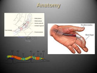
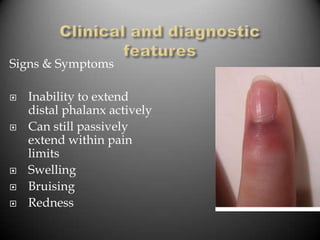
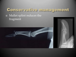
Dan Purtell
Hand / Occupational
      Therapist
Trigger   finger / thumb

De   Quervain’s

Mallet   Finger

PIPJ   Dislocations

Dupuytren’s     disease

Simple    #’s
   Not clearly /
    consistently defined
   Triggering of the digital
    flexor tendons at the
    fibrooseous tunnel
    formed by the
    metacarpal neck and
    A1 pulley
   In thumb the sesamoid
    bones may also be site
    of constriction
   Generally affects FDS
    rather than FDP
    because it lies directly
    under A1 pulley
   Primary trigger
    finger most
    commonly found in
    middle aged
    women, 2–6 X more
    than men
   Most commonly
    affects thumb (30-
    50%)
   Then ring, long,
    index, & little
   Stenosing tendovaginitis
   Digital flexors susceptible to compression
    and shear at level of wrist and MP joints
    where they enter fibrooseous tunnels
   Blunt trauma or sustained tool use causing
    direct compression at A1 pulley
   Secondary TF in individuals with connective
    tissue disorders
   Discrepancy between
    the size of the A1 finger
    pulley lumen and
    tendon volume
   Hypertrophy of the
    pulley
   Poor tendon
    vascularity between A1
    and A2 pulley makes
    tendons more
    susceptible to
    degenerative changes
   Tendons develop
    nodules from
    tenosynovitis
   Pain over site of tendon disorder aggravated
    by movement
   Symptoms vary from stiffness, to uneven
    movement, catching, blocking, or complete
    locking of tendon
   Pain can also be referred to distal joint or
    proximally up forearm
   Local swelling and thickening creating a
    palpable nodule over distal palmar crease
    (A1 pulley area)
   Local tenderness over A1
   Patient may present with acute, subacute or
    chronic disorder
Patients often reluctant to
have cortico-steroid injection,
even more reluctant to have
surgery

Conservative measures should
be trialled for 4-6 weeks

   Thermoplastic hand based
    splint to limit MCP flexion
    (stops triggering through A1
    pulley)
   Splinting works better for
    fingers than for thumbs
   Trigger Thumb -
    Thermoplastic barrel
    splint, IP joint at 10
    degrees flexion

   Passive tendon gliding
    exercises
   Soft tissue massage of
    nodule and tendon.
   +/- Ultrasound
   +/- NSAID gel
   Cortisone injection
    for trigger fingers
    and thumbs relieves
    symptoms in 47% to
    94% of affected
    digits.
   Most reported success rates are
    above 90%.
   Decompression of pulley to
    allow flexor tendon to glide

   Turowski, 1997: n=59, 97%
    complete resolution. No post-op
    nerve or tendon damage.
   Eastwood, 1992: Percutaneous
    release, n=35, 94% complete
    resolution. No complications.
    Not for thumb
   Thorpe, 1988: n = 53, 60%
    complete resolution, success
    correlated with surgeon skill.
   Oedema and wound management
   Scar management once wound healed
   Active tendon gliding exercises
   Stretching long finger flexors
   Strengthening only if necessary

Conclusion
 Surgery remains the most successful treatment
  option
 Splinting and exercise program good alternative for
  those patients reluctant to consider a CSI or surgery
   de Quervain’s
    tenosynovitis is
    the entrapment
    tendonitis/tenosy
    novitis of the
    abductor pollicis
    longus and
    extensor pollicis
    brevis tendons at
    the styloid
    process of the
    radius
   Most cases it is a
    tendinopathy like
    trigger finger,
    tennis elbow etc
    that leads to
    tenosynovitis.
   Finklesteins test and clinical Hx confirm
    diagnosis.
   10 x more Common in Women than men.
   Common in pre-post natal and menopause.
   Often caused by repetitive strain or
    sustained posture of the wrist = strain on
    EPB and APL.
   Can occur post direct trauma to the area
    (rare)
   Treatment:
       Steroid (won’t fix
        tendinopathy)

       Splint and rest

       Kinesio tape

       Surgery (last resort)
   Surgery
   Any injury that causes
    a mallet deformity of
    the distal phalangeal
    joint
   Disruption of the terminal extensor tendon as it
    inserts into distal phalanx +/- fracture
MECHANISM OF
 INJURY

   Flexion force on
    extended DIP joint
   Direct crush
   Ball to tip of finger
   Often occurs in
    ball sports.
Signs & Symptoms

   Inability to extend
    distal phalanx actively
   Can still passively
    extend within pain
    limits
   Swelling
   Bruising
   Redness
Volar plate laxity = swan
   neck deformity
   depends on size of fragment and position of
    joint
     If complete tendon rupture without
      fracture
     If # is less than 30% joint surface
    → Conservative management -
      splinting

   If # greater than 30% joint surface or joint is
    significantly displaced needs surgical
    intervention
   Mallet splint reduces the
    fragment
   Splint DIP in hyperextension 6 – 8/52
   Splint strictly 24/24
   Clear instructions and demonstration re
    changing and wearing routine
   Advice re skin care
   PIP flexion exercises
   May return to sport with
    splinting
   Off the shelf stack
    splint often fit
    poorly

   Patient may end up
    with a lag at DIP
    joint
   Wean splint slowly after
    6-8 weeks
   Keep on at night and for
    work a further 2 weeks
   Initially active flexion
    exercises to gain full
    flexion
   Start off 30 degrees flexion
    first week and increase
    slowly ie 20 degrees per
    week
   May take 4 – 6 weeks to
    regain full flexion
   If lag reoccurs →
    RESPLINT
   Passive flexion only
    added if needed
   Large Fragment
   Fixed with K-wire or 2
- Conservative approach
only if small fragment with
no joint subluxation.
 - Splint for 6/52 in barrel
splint
DIPJ can be free.
- Larger fragments with joint
subluxation can be ORIF’d.
= Boutonniere Deformity
    = FFD of PIPJ
   = Very hard to fix
   Mechanism of Injury – hyperextension of the
    PIP joint
   with or without dislocation
   often initial injury seems trivial
 Dislocation of the
  PIPJ
 Avulsion # of
  middle phalanx
 Disruption of volar
  plate over the PIP
  joint
Signs & Symptoms

• Swelling
• Bruising
• Pain volar aspect of
  PIP joint
• Instability or pain
  on stress of volar
  plate
• Decreased range of
  motion particularly
  flexion
 If dislocation without #
       OR
 If # fragment less than 30% joint
 surface

→ reduce then manage
 conservatively in dorsal
 blocking splint (DBS)
Dorsal blocking splint

•   PIPJ in 30 degrees
    flexion

•volar structures
off
stretch

•slowly increase
out
  to neutral
  splint 4 - 6/52
  weekly adjustments PIPJ from 30
   flexion → full extension as stability
   increases
• Coban for swelling
• Flexion exercises within splint
• Isolated FDP & FDS flexion important to
  prevent adherance to volar plate
• Early mobilisation also assists oedema
• If collaterals involved need to buddy
   strap when out of splint
• Tape/splint for work for 6 -8/52
• Watch for FFC PIPJ in late stages → may
   need to include extension splinting
   If volar plate repaired post surgical
    management same but progress
    slower
   Occasionally flexion exercises are
    delayed if stability is a concern
   5th Metacarpal
       Assess ROM and digit
        Rotation
       Usually managed
        conservatively
   4th Metacarpal Spiral #
       Assess ROM and digit
        Rotation
       Tendency to rotate
       Impacted #’s result in
        extension lag = poor
        function.
Treat conservatively in splint
unless joint subluxation
occurring
Treat conservatively in splint
unless joint subluxation or
stenners lesion is present.

- Usually characterised by
lots of oedema, nil end point
of stability.
- Very difficult to assess with
certainty
- U/s scan to confirm

Common hand injuries

  • 1.
    Dan Purtell Hand /Occupational Therapist
  • 2.
    Trigger finger / thumb De Quervain’s Mallet Finger PIPJ Dislocations Dupuytren’s disease Simple #’s
  • 3.
    Not clearly / consistently defined  Triggering of the digital flexor tendons at the fibrooseous tunnel formed by the metacarpal neck and A1 pulley  In thumb the sesamoid bones may also be site of constriction  Generally affects FDS rather than FDP because it lies directly under A1 pulley
  • 5.
    Primary trigger finger most commonly found in middle aged women, 2–6 X more than men  Most commonly affects thumb (30- 50%)  Then ring, long, index, & little
  • 6.
    Stenosing tendovaginitis  Digital flexors susceptible to compression and shear at level of wrist and MP joints where they enter fibrooseous tunnels  Blunt trauma or sustained tool use causing direct compression at A1 pulley  Secondary TF in individuals with connective tissue disorders
  • 7.
    Discrepancy between the size of the A1 finger pulley lumen and tendon volume  Hypertrophy of the pulley  Poor tendon vascularity between A1 and A2 pulley makes tendons more susceptible to degenerative changes  Tendons develop nodules from tenosynovitis
  • 8.
    Pain over site of tendon disorder aggravated by movement  Symptoms vary from stiffness, to uneven movement, catching, blocking, or complete locking of tendon  Pain can also be referred to distal joint or proximally up forearm  Local swelling and thickening creating a palpable nodule over distal palmar crease (A1 pulley area)  Local tenderness over A1  Patient may present with acute, subacute or chronic disorder
  • 9.
    Patients often reluctantto have cortico-steroid injection, even more reluctant to have surgery Conservative measures should be trialled for 4-6 weeks  Thermoplastic hand based splint to limit MCP flexion (stops triggering through A1 pulley)  Splinting works better for fingers than for thumbs
  • 10.
    Trigger Thumb - Thermoplastic barrel splint, IP joint at 10 degrees flexion  Passive tendon gliding exercises  Soft tissue massage of nodule and tendon.  +/- Ultrasound  +/- NSAID gel
  • 11.
    Cortisone injection for trigger fingers and thumbs relieves symptoms in 47% to 94% of affected digits.
  • 12.
    Most reported success rates are above 90%.  Decompression of pulley to allow flexor tendon to glide  Turowski, 1997: n=59, 97% complete resolution. No post-op nerve or tendon damage.  Eastwood, 1992: Percutaneous release, n=35, 94% complete resolution. No complications. Not for thumb  Thorpe, 1988: n = 53, 60% complete resolution, success correlated with surgeon skill.
  • 13.
    Oedema and wound management  Scar management once wound healed  Active tendon gliding exercises  Stretching long finger flexors  Strengthening only if necessary Conclusion  Surgery remains the most successful treatment option  Splinting and exercise program good alternative for those patients reluctant to consider a CSI or surgery
  • 14.
    de Quervain’s tenosynovitis is the entrapment tendonitis/tenosy novitis of the abductor pollicis longus and extensor pollicis brevis tendons at the styloid process of the radius
  • 15.
    Most cases it is a tendinopathy like trigger finger, tennis elbow etc that leads to tenosynovitis.
  • 16.
    Finklesteins test and clinical Hx confirm diagnosis.
  • 17.
    10 x more Common in Women than men.  Common in pre-post natal and menopause.  Often caused by repetitive strain or sustained posture of the wrist = strain on EPB and APL.  Can occur post direct trauma to the area (rare)
  • 18.
    Treatment:  Steroid (won’t fix tendinopathy)  Splint and rest  Kinesio tape  Surgery (last resort)
  • 19.
    Surgery
  • 20.
    Any injury that causes a mallet deformity of the distal phalangeal joint
  • 21.
    Disruption of the terminal extensor tendon as it inserts into distal phalanx +/- fracture
  • 23.
    MECHANISM OF INJURY  Flexion force on extended DIP joint  Direct crush  Ball to tip of finger  Often occurs in ball sports.
  • 24.
    Signs & Symptoms  Inability to extend distal phalanx actively  Can still passively extend within pain limits  Swelling  Bruising  Redness
  • 25.
    Volar plate laxity= swan neck deformity
  • 26.
    depends on size of fragment and position of joint  If complete tendon rupture without fracture  If # is less than 30% joint surface → Conservative management - splinting  If # greater than 30% joint surface or joint is significantly displaced needs surgical intervention
  • 27.
    Mallet splint reduces the fragment
  • 28.
    Splint DIP in hyperextension 6 – 8/52  Splint strictly 24/24  Clear instructions and demonstration re changing and wearing routine  Advice re skin care  PIP flexion exercises  May return to sport with splinting
  • 29.
    Off the shelf stack splint often fit poorly  Patient may end up with a lag at DIP joint
  • 30.
    Wean splint slowly after 6-8 weeks  Keep on at night and for work a further 2 weeks  Initially active flexion exercises to gain full flexion  Start off 30 degrees flexion first week and increase slowly ie 20 degrees per week  May take 4 – 6 weeks to regain full flexion  If lag reoccurs → RESPLINT  Passive flexion only added if needed
  • 31.
    Large Fragment  Fixed with K-wire or 2
  • 33.
    - Conservative approach onlyif small fragment with no joint subluxation. - Splint for 6/52 in barrel splint DIPJ can be free. - Larger fragments with joint subluxation can be ORIF’d.
  • 34.
    = Boutonniere Deformity = FFD of PIPJ = Very hard to fix
  • 36.
    Mechanism of Injury – hyperextension of the PIP joint  with or without dislocation  often initial injury seems trivial
  • 37.
     Dislocation ofthe PIPJ  Avulsion # of middle phalanx  Disruption of volar plate over the PIP joint
  • 38.
    Signs & Symptoms •Swelling • Bruising • Pain volar aspect of PIP joint • Instability or pain on stress of volar plate • Decreased range of motion particularly flexion
  • 39.
     If dislocationwithout # OR If # fragment less than 30% joint surface → reduce then manage conservatively in dorsal blocking splint (DBS)
  • 40.
    Dorsal blocking splint • PIPJ in 30 degrees flexion •volar structures off stretch •slowly increase out to neutral
  • 43.
     splint4 - 6/52  weekly adjustments PIPJ from 30 flexion → full extension as stability increases • Coban for swelling • Flexion exercises within splint • Isolated FDP & FDS flexion important to prevent adherance to volar plate • Early mobilisation also assists oedema
  • 44.
    • If collateralsinvolved need to buddy strap when out of splint • Tape/splint for work for 6 -8/52 • Watch for FFC PIPJ in late stages → may need to include extension splinting
  • 45.
    If volar plate repaired post surgical management same but progress slower  Occasionally flexion exercises are delayed if stability is a concern
  • 46.
    5th Metacarpal  Assess ROM and digit Rotation  Usually managed conservatively
  • 47.
    4th Metacarpal Spiral #  Assess ROM and digit Rotation  Tendency to rotate  Impacted #’s result in extension lag = poor function.
  • 48.
    Treat conservatively insplint unless joint subluxation occurring
  • 49.
    Treat conservatively insplint unless joint subluxation or stenners lesion is present. - Usually characterised by lots of oedema, nil end point of stability. - Very difficult to assess with certainty - U/s scan to confirm