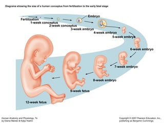
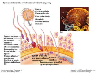
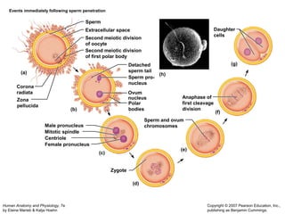
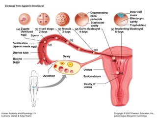
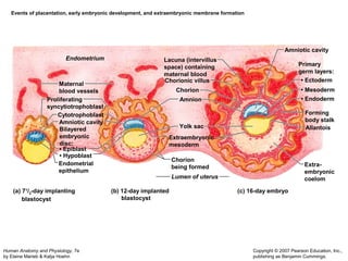
1) The document describes the key stages of human embryonic development from fertilization through the formation of the basic body plan and extraembryonic membranes over the first 8 weeks.
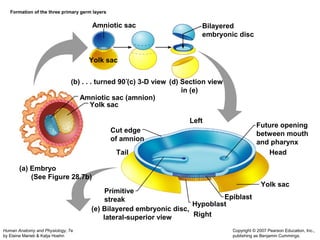
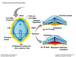
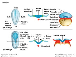
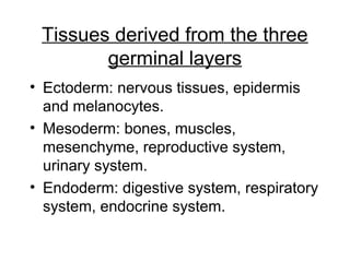
2) It explains how the three primary germ layers (ectoderm, mesoderm and endoderm) form and give rise to the major tissues and organ systems.
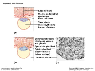
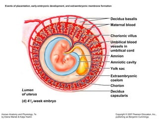
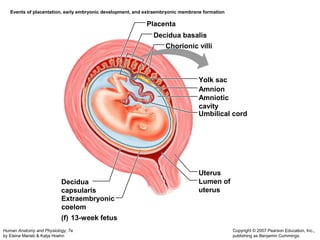
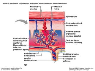
3) The role and formation of the embryonic membranes - amnion, yolk sac and allantois - as well as the placenta for nutrient exchange are covered.