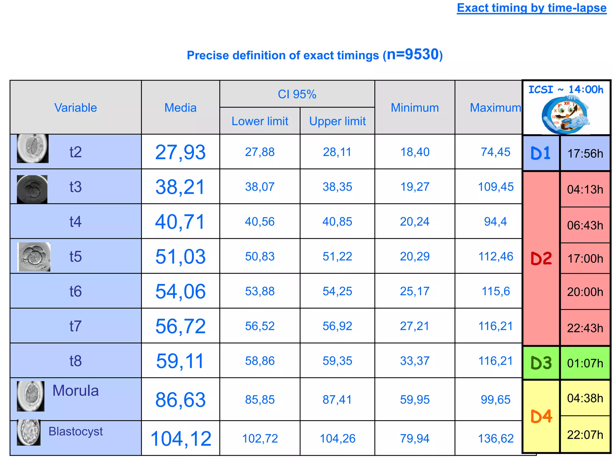
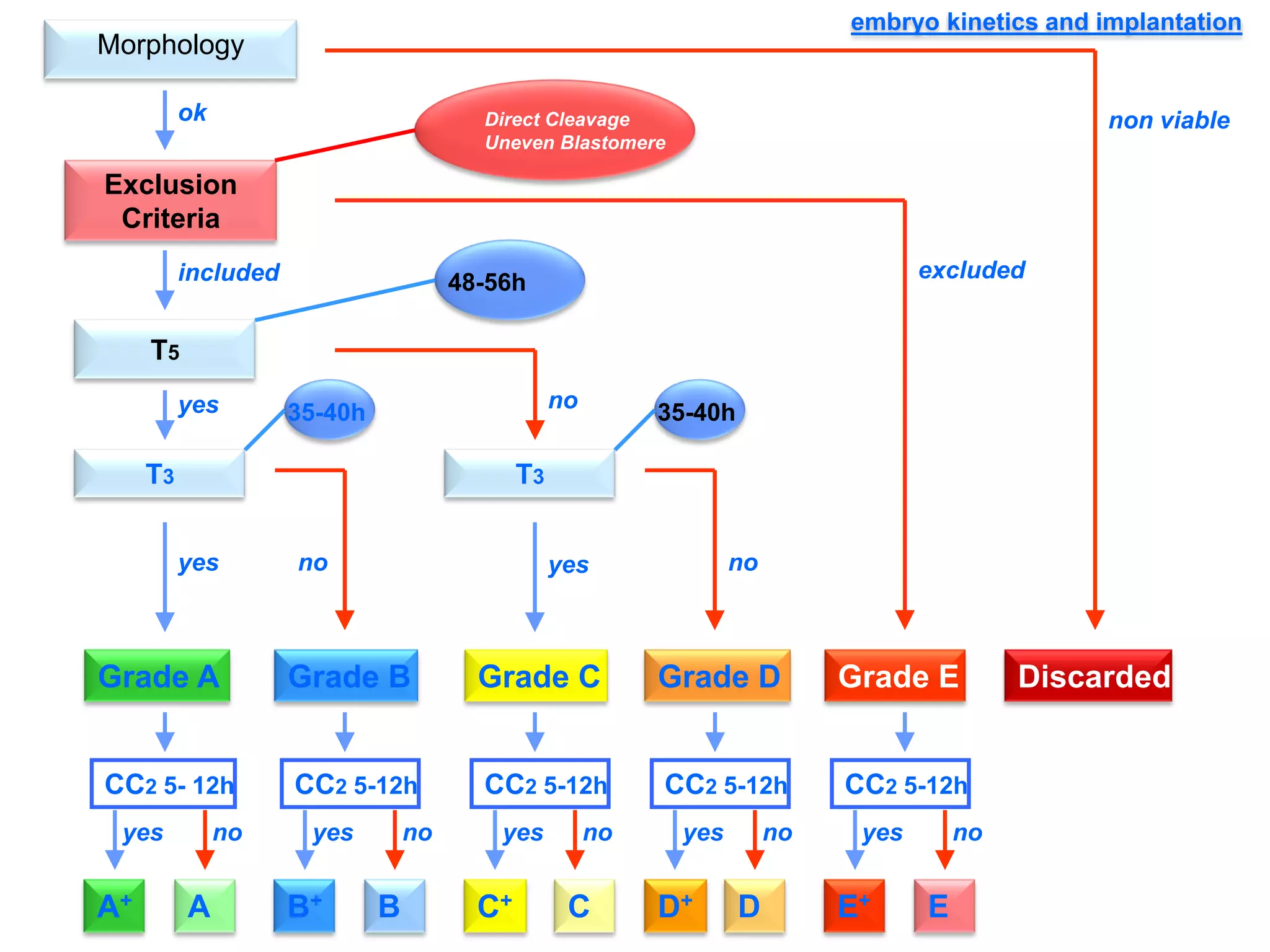
Time-Lapse
-Cleavage-stage development is a dynamic process in which embryo
morphology may change significantly over a time span (h).
-Conventional grading practices may not detect subtle differences between
individual embryos, such as the time to progress from one cleavage division
to the next.
-The use of an automated instrument with programmable time-lapse image
acquisition, allows data to be collected for individual embryos during
development to quantify the exact timing of each cell division.
5
15
25
35
45
Day 3Embryo CategoriesASEBIR
%Inside
%Outside
%Inside 33,2 17,1 20,4 29,4
%Outside 35,6 15,7 20,6 28,1
A B C D
p=0,941
Exact timing by time-lapse
Precise definition of exact timings (n=9530)
Variable Media
CI 95%
Minimum Maximum
Lower limit Upper limit
t2 27,93 27,88 28,11 18,40 74,45
t3 38,21 38,07 38,35 19,27 109,45
t4 40,71 40,56 40,85 20,24 94,4
t5 51,03 50,83 51,22 20,29 112,46
t6 54,06 53,88 54,25 25,17 115,6
t7 56,72 56,52 56,92 27,21 116,21
t8 59,11 58,86 59,35 33,37 116,21
Morula
86,63 85,85 87,41 59,95 99,65
Blastocyst
104,12 102,72 104,26 79,94 136,62
ICSI ~ 14:00h
D1 17:56h
D2
04:13h
06:43h
17:00h
20:00h
22:43h
D3 01:07h
D4
04:38h
22:07h
Implantation Success; ESDMorphology
included
ok
Grade A Grade B Grade C Grade D Grade E Discarded
non viable
excluded
yes no
yes nono yes
A+ A E+ EB+ B C+ C D+ D
CC2 5- 12h CC2 5-12h CC2 5-12h CC2 5-12h CC2 5-12h
yes no yes no yes no yes no yes no
Exclusion
Criteria
Direct Cleavage
Uneven Blastomere
T5
48-56h
T3
35-40h
T3
35-40h
embryo kinetics and implantation
Time-lapse + respiration
Respiration Peak = Citokinesis
Time-hours post ICSI
Embryo respiration and Implantation
5
5,2
5,4
5,6
5,8
6
6,2
6,4
6,6
6,8
7
0 1 2 3 4 5 6 7 8 9 10
fmol/sec
Cleavage O2 consumption pattern
Not Implanted
Implanted
Cumulus cells — Transcriptomics at an early stage
- To identify reliable genomic biomarkers
expressed in cumulus cells that accurately
and non-invasively can predict the oocyte
developmental competence and reinforce
the already used morphological criteria.
- Gene expression assessed using microarray
platforms
- The oocyte triggering stimulates the
expression of genes involved in the
granulosa luteinization process and
ovulation finality − a crucial step during
follicular development to produce a fully
healthy oocyte
- Several genes are also differentially
associated to successful pregnancy and
implantation
Investigating spent culture medium
Pyruvate
Glucose
Amino acids
Sugars
Uptake
Hormones
Lactate
Ammonia
Enzymes
Production
Other factors
- A noninvasive metabolomic profile of the culture medium surrounding the
embryo with an analysis of the components produced or depleted by
them can be achieved by different technologies.
- Multivariable analysis could be applied and models based on viability
index may be used in the embryo selection process.
Proteomics
• Multiple protein analysis of
human embryo secretions
offers the chance to expand our
perspective of a noninvasive
quantification of human
embryonic viability and even
chromosomal normality
avoiding any type of
manipulation.

L6 key slides meseguer (2)

  • 1.
    Time-Lapse -Cleavage-stage development isa dynamic process in which embryo morphology may change significantly over a time span (h). -Conventional grading practices may not detect subtle differences between individual embryos, such as the time to progress from one cleavage division to the next. -The use of an automated instrument with programmable time-lapse image acquisition, allows data to be collected for individual embryos during development to quantify the exact timing of each cell division.
  • 2.
    5 15 25 35 45 Day 3Embryo CategoriesASEBIR %Inside %Outside %Inside33,2 17,1 20,4 29,4 %Outside 35,6 15,7 20,6 28,1 A B C D p=0,941
  • 3.
    Exact timing bytime-lapse Precise definition of exact timings (n=9530) Variable Media CI 95% Minimum Maximum Lower limit Upper limit t2 27,93 27,88 28,11 18,40 74,45 t3 38,21 38,07 38,35 19,27 109,45 t4 40,71 40,56 40,85 20,24 94,4 t5 51,03 50,83 51,22 20,29 112,46 t6 54,06 53,88 54,25 25,17 115,6 t7 56,72 56,52 56,92 27,21 116,21 t8 59,11 58,86 59,35 33,37 116,21 Morula 86,63 85,85 87,41 59,95 99,65 Blastocyst 104,12 102,72 104,26 79,94 136,62 ICSI ~ 14:00h D1 17:56h D2 04:13h 06:43h 17:00h 20:00h 22:43h D3 01:07h D4 04:38h 22:07h
  • 5.
    Implantation Success; ESDMorphology included ok GradeA Grade B Grade C Grade D Grade E Discarded non viable excluded yes no yes nono yes A+ A E+ EB+ B C+ C D+ D CC2 5- 12h CC2 5-12h CC2 5-12h CC2 5-12h CC2 5-12h yes no yes no yes no yes no yes no Exclusion Criteria Direct Cleavage Uneven Blastomere T5 48-56h T3 35-40h T3 35-40h embryo kinetics and implantation
  • 7.
    Time-lapse + respiration RespirationPeak = Citokinesis Time-hours post ICSI Embryo respiration and Implantation 5 5,2 5,4 5,6 5,8 6 6,2 6,4 6,6 6,8 7 0 1 2 3 4 5 6 7 8 9 10 fmol/sec Cleavage O2 consumption pattern Not Implanted Implanted
  • 8.
    Cumulus cells —Transcriptomics at an early stage - To identify reliable genomic biomarkers expressed in cumulus cells that accurately and non-invasively can predict the oocyte developmental competence and reinforce the already used morphological criteria. - Gene expression assessed using microarray platforms - The oocyte triggering stimulates the expression of genes involved in the granulosa luteinization process and ovulation finality − a crucial step during follicular development to produce a fully healthy oocyte - Several genes are also differentially associated to successful pregnancy and implantation
  • 9.
    Investigating spent culturemedium Pyruvate Glucose Amino acids Sugars Uptake Hormones Lactate Ammonia Enzymes Production Other factors - A noninvasive metabolomic profile of the culture medium surrounding the embryo with an analysis of the components produced or depleted by them can be achieved by different technologies. - Multivariable analysis could be applied and models based on viability index may be used in the embryo selection process.
  • 10.
    Proteomics • Multiple proteinanalysis of human embryo secretions offers the chance to expand our perspective of a noninvasive quantification of human embryonic viability and even chromosomal normality avoiding any type of manipulation.