Resources
• Anatomy andphysiology. The unity of FORM and FUNCTION by
SALADIN 9th Edition
• First Aid 2022
• Anatomy and Physiology OpenStax
3.
Objectives
• Section 29.1
a.describe the process of sperm migration and fertilization;
b. explain how an egg prevents fertilization by more than
one sperm;
c. describe the major events that transform a fertilized egg
into an embryo; and
d. describe the implantation of the preembryo in the uterine
wall.
• Section 29.2
a. describe the formation and functions of the placenta;
b. explain how the conceptus is nourished before the
placenta takes over this function;
c. describe the embryonic membranes and their functions;
d. identify the major tissues derived from the primary germ
layers;
e. describe the major events of fetal development; and
f. describe the fetal circulatory system
• Section 29.3
a. describe how and why the circulatory system
changes at birth;
b. explain why the first breaths of air are relatively
difficult for a neonate;
c. describe the major physiological problems of a
premature infant; and
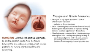
d. discuss some common causes of birth defects.
• Section 29.4
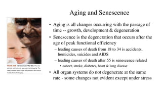
a. define senescence and distinguish it from aging;
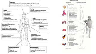
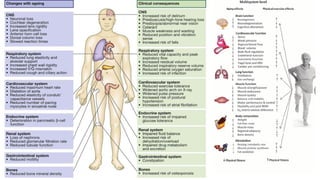
b. describe some major changes that occur with
aging in each organ system;
c. summarize some current theories of senescence;
and
d. be able to explain how exercise and other factors
can slow the rate of senescence.
•
4.
Introduction
• It ismiraculous that a one-celled, fertilized egg transforms into an independent, fully developed
individual
• Embryology—the study of prenatal development
• Developmental biology—examines changes in form and function from fertilized egg through old
age
• Embryo—term has varied meanings
• Some authorities assert that: the fertilized egg or the two-cell stage is an embryo
• Other authorities (including this textbook) assert that an individual becomes an embryo when: it
is 16 days old and consists of three primary germ layers: Ectoderm, mesoderm, and ectoderm
• Embryogenesis—events leading up to this stage
• Preembryonic stage is the first 16 days after fertilization
5.
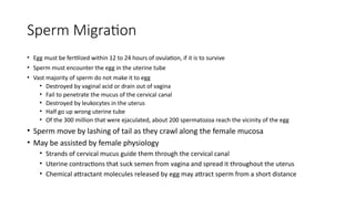
Sperm Migration
• Eggmust be fertilized within 12 to 24 hours of ovulation, if it is to survive
• Sperm must encounter the egg in the uterine tube
• Vast majority of sperm do not make it to egg
• Destroyed by vaginal acid or drain out of vagina
• Fail to penetrate the mucus of the cervical canal
• Destroyed by leukocytes in the uterus
• Half go up wrong uterine tube
• Of the 300 million that were ejaculated, about 200 spermatozoa reach the vicinity of the egg
• Sperm move by lashing of tail as they crawl along the female mucosa
• May be assisted by female physiology
• Strands of cervical mucus guide them through the cervical canal
• Uterine contractions that suck semen from vagina and spread it throughout the uterus
• Chemical attractant molecules released by egg may attract sperm from a short distance
6.
Sperm Capacitation
• Spermatozoareach distal uterine tube in half an hour or less but
cannot fertilize the egg for 10 hours
• Capacitation: process that migrating sperm must undergo to make it possible
to penetrate an egg
• Membrane of fresh sperm is toughened by cholesterol which prevents premature
release of acrosomal enzymes (which could damage sperm ducts)
• Female fluids leach cholesterol from the sperm plasma membrane and dilute inhibitory
factors in semen
• Sperm membrane becomes fragile and permeable to Ca2+
which diffuses into sperm
causing more powerful lashing of the tail
7.
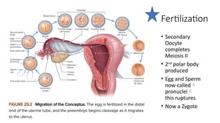
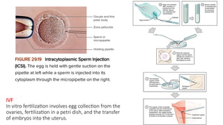
Fertilization
• Sperm areviable for up to 6 days after ejaculation
• Conception optimal if sperm are deposited a few days before ovulation to 14 hours after
• When sperm encounters an egg, it undergoes an acrosomal reaction—exocytosis of the acrosome,
releasing the enzymes needed to penetrate the egg Penetrates granulosa cells, then zona
pellucida
• Two acrosomal enzymes
• Hyaluronidase—digests the hyaluronic acid that binds granulosa cells together
• Acrosin—a protease similar to trypsin
• Sperm head and midpiece enter egg
• Egg destroys the sperm mitochondria
• Passes only maternal mitochondria on to the offspring
• Fertilization combines the haploid (n) set of sperm chromosomes with the haploid set of egg
chromosomes producing a diploid (2n) set
• Polyspermy—fertilization by two or more sperm which would produce a doomed fertilized egg
8.
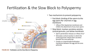
Fertilization & theSlow Block to Polyspermy
• Two mechanisms to prevent polyspermy
• Fast block: binding of the sperm to the
egg opens Na+
channels in egg
membrane
• Inflow of Na+
depolarizes membrane and
inhibits the attachment of any more sperm
• Slow block: involves secretory vesicles,
cortical granules, just below membrane
• Sperm penetration releases an inflow of Ca2+
• Stimulates cortical reaction in which the
cortical granules release their secretion
beneath the zona pellucida
• Creates an impenetrable fertilization
membrane between the egg and the zona
pellucida
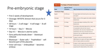
Pre-embryonic stage
• First2 weeks of development
• Cleavage: MITOTIC division that occurs for 3
days
• 30 hours 2 cell stage 4 cell stage 8 cell
stage
• 72 hours Day 3 Morula
• Day 4-5 Morula in uterine cavity
• Zona pellucida breaks down blastocyst
released
• Blastocyst Outer cells Trophoblast will
become part of the placenta
• Inner cell mass Embryoblast becomes
embryo
11.
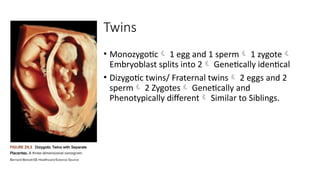
Twins
• Monozygotic 1egg and 1 sperm 1 zygote
Embryoblast splits into 2 Genetically identical
• Dizygotic twins/ Fraternal twins 2 eggs and 2
sperm 2 Zygotes Genetically and
Phenotypically different Similar to Siblings.
12.
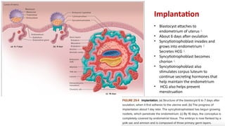
Implantation
• Blastocyst attachesto
endometrium of uterus
• About 6 days after ovulation
• Syncytiotrophoblast invades and
grows into endometrium
Secretes HCG
• Syncytiotrophoblast becomes
chorion
• Syncytiotrophoblast also
stimulates corpus luteum to
continue secreting hormones that
help maintain the endometrium
• HCG also helps prevent
menstruation
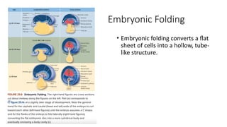
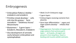
Embryogenesis
• Embryoblast flattens/disklike
endoderm and ectoderm
• Primitive streak develops cells
sink into this groove form
mesoderm Gelatinous tissue/
Mesenchyme
• Blastomere 3 germ layers
Ectoderm, Mesoderm, Endoderm
• Also development of amniotic
cavity between embryoblast and
cytotrophoblast
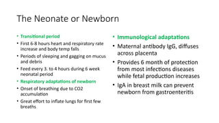
• Week 2 to 9= Embryonic stage
• 3 germ layers
• Embryo begins receiving nutrients from
placenta
• Germ layer differentiation organs and
organ systems develop initiates fetal
stage
• Organogenesis: Formation of organs and
organ systems from primary germ
layers at 8 weeks, all organs are
present in 3cm long fetus. Heart is
beating and muscles exhibit contracts.
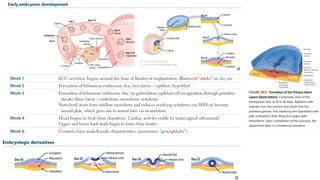
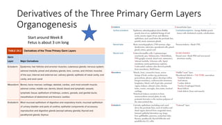
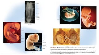
16.
Derivatives of theThree Primary Germ Layers
Organogenesis
Start around Week 8
Fetus is about 3 cm long
17.
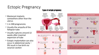
Ectopic Pregnancy
• Blastocystimplants
somewhere other than the
uterus
• 1 in 300 pregnancies
• Usually the ampulla of the
fallopian tube
• Usually ruptures around 12
weeks after maximal
expansion of tube
• Ectopic implantation can also
occur in abdominal cavity
9% result in live birth via
cesarian section
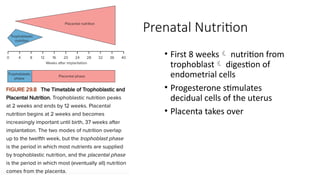
19.
Prenatal Nutrition
• First8 weeks nutrition from
trophoblast digestion of
endometrial cells
• Progesterone stimulates
decidual cells of the uterus
• Placenta takes over
20.
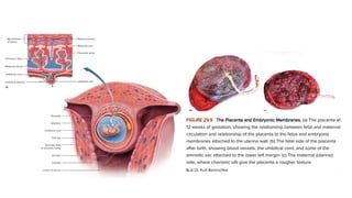
Placentation
• Placenta formationoccurs from
Day 11 through 12 weeks
• Chorionic villi
• Extensions of
syncytiotrophoblast into
endometrium by digestion and
growth of roots of tissue
• Mesenchyme extends into
chorionic villi to form embryonic
blood vessels
• Placental Sinus
• Pools of maternal blood that
merge and surround villi
• Blood stimulates rapid growth of
chorionic villi
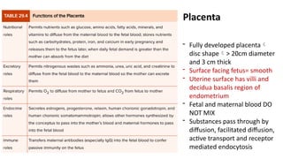
22.
- Fully developedplacenta
disc shape> 20cm diameter
and 3 cm thick
- Surface facing fetus= smooth
- Uterine surface has villi and
decidua basalis region of
endometrium
- Fetal and maternal blood DO
NOT MIX
- Substances pass through by
diffusion, facilitated diffusion,
active transport and receptor
mediated endocytosis
Placenta
23.
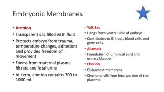
Embryonic Membranes
• Amnion
•Transparent sac filled with fluid
• Protects embryo from trauma,
temperature changes, adhesions
and provides freedom of
movement
• Forms from maternal plasma
filtrate and fetal urine
• At term, amnion contains 700 to
1000 mL
• Yolk Sac
• Hangs from ventral side of embryo
• Contributes to GI tract, blood cells and
germ cells
• Allantois
• Foundation of umbilical cord and
urinary bladder
• Chorion
• Outermost membrane
• Chorionic villi from fetal portion of the
placenta.
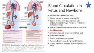
24.
Blood Circulation in
Fetusand Newborn
• Fetus= from 8 weeks until birth
• Organs mature to support external life
• Spaces in mesoderm become lined with
endothelium and merge into blood vessels and
lymphatic vessels
• Side by side endothelial tubes fuse to form heart
• Fetal Circulation
• Umbilical-placental circuit via umbilical cord
• Circulatory shunts
• Ductus venosus connects to IVC
• Foramen Ovale connects right and left atria
• Ductus arteriosus connects pulmonary trunk to
aorta
25.
The Neonate orNewborn
• Transitional period
• First 6-8 hours heart and respiratory rate
increase and body temp falls
• Periods of sleeping and gagging on mucus
and debris
• Feed every 3. to 4 hours during 6 week
neonatal period
• Respiratory adaptations of newborn
• Onset of breathing due to CO2
accumulation
• Great effort to inflate lungs for first few
breaths
• Immunological adaptations
• Maternal antibody IgG, diffuses
across placenta
• Provides 6 month of protection
from most infections diseases
while fetal production increases
• IgA in breast milk can prevent
newborn from gastroenteritis