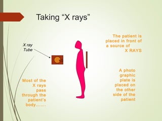
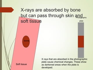
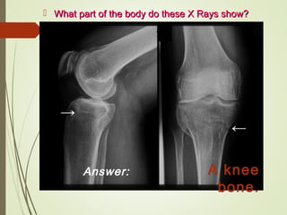
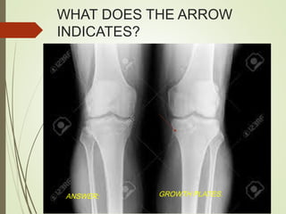
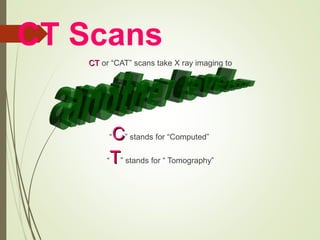
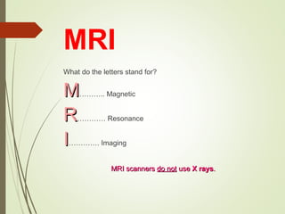
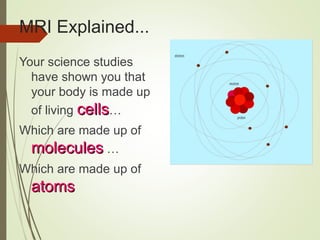
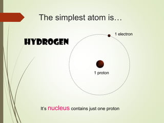
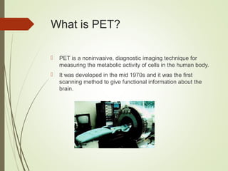
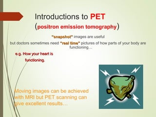
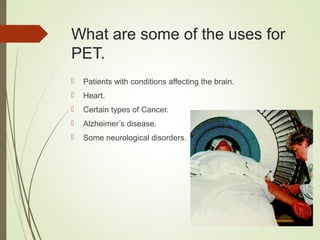
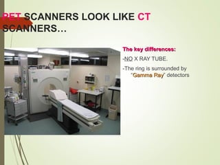
2. X-rays
Very little ordinary
light can pass
through skin. It’s
either absorbedabsorbed at
the surface or
reflectedreflected back…..
To “see” inside we
need a kind of
“light” with moremore
energyenergy…
Skin
Ordinary Light
X rays
3. Taking “X rays”
The patient is
placed in front of
a source of
X RAYS
X ray
Tube
A photo
graphic
plate is
placed on
the other
side of the
patient
Most of the
X rays
pass
through the
patient’s
body…….
4. X-rays are absorbed by bone
but can pass through skin and
soft tissue
bone
Soft tissue
Photographic
plate
X rays that are absorbed in the photographic
plate cause chemical changes. These show
as darkened areas when the plate is
developed.
5. X-rays are also partly absorbed by some tissues
in the body this creates a more subtle picture.
bone
Soft tissue
Photographic
plate
6. What part of the body do these X Rays show?What part of the body do these X Rays show?
Answer: A knee
bone.
7. WHAT DOES THE ARROW
INDICATES?
ANSWER: GROWTH PLATES.
8. CT Scans
CTCT or “CAT” scans take X ray imaging to
“CC” stands for “Computed”
“TT” stands for “ Tomography”
9. In short….
CT scanners are
complex X ray
machines
attached to
very clever
computers
using complicated
mathematics to
build up
images of our insides.
10. The patient is placed on a bed
The scanner (X ray machine) is the shape of a ring
The patient is slowly moved through the ringThe patient is slowly moved through the ring
as the scan takes place…as the scan takes place…
11. Looking end on….
X ray tube
X ray detector
Patient
X Rays are
produced in an
X ray tube, pass
through the
patient and are
detected by the
detector
The scanner
rotates the X ray
tube and
detector so the
patient is
scanned from all
angles
12. MRI
What do the letters stand for?
MM……….. Magnetic
RR………… Resonance
II…………. Imaging
MRI scannersMRI scanners do notdo not useuse X raysX rays..
13. MRI Explained...
Your science studies
have shown you that
your body is made up
of living cellscells…
Which are made up of
moleculesmolecules …
Which are made up of
atomsatoms
electron
neutron
proton
14. The simplest atom is…
Hydrogen
1 electron
1 proton
It’s nucleus contains just one proton
15. After many years of investigation
physicists found they could affect the
tiny nuclear magnets of hydrogen
atoms using very strong magnets and
radio waves…
Bring in the magnets….Bring in the magnets….
S N
Note: The nuclear magnets can line up in two
possible directions
A pulse of radio waves can cause some of the nuclear
magnets absorb energy and “flip”
This high energy situation cannot be sustained
for long. Many will “flip” back….
When this happens energy is released as a tiny pulse of
radio waves !!!
…….watch how the magnetic field affects the tiny nuclear magnets.watch how the magnetic field affects the tiny nuclear magnets
16. The patient is placed on a bed and then moved into
a large hollow tube.
The tube contains a very powerfulThe tube contains a very powerful magnetmagnet….….
Using an MRI
Scanner…
17. Patients may be asked the
following questions:
Have you ever worked in the army or
metal working industry?
Metal fragments (especially in the eye) could becomeMetal fragments (especially in the eye) could become
dislodgeddislodged
Do you have a pacemaker?
If yes you cannot have an MRI scanIf yes you cannot have an MRI scan
Do you have any dental implants
Some could become magnetisedSome could become magnetised
Do you have any metal pins or staples in
your body?
Some could become magnetised and need to be checked thatSome could become magnetised and need to be checked that
they will hold in place during the scanthey will hold in place during the scan
18. With the patient safety check
complete the scan can
begin…
The part of the body
to be scanned is
placed in the centre
of the primaryprimary
magnetmagnet
X
The magnet field
produced has to be
very steady and
strong
This field causes the Hydrogen nuclei in the patients body
to line up with the field
19. X
Three further coils are embedded into the
tube….GRADIENT MAGNETS… these are used
to fine tune the magnetic field so particular
body parts and tissue types can be focused
on.
The patient will know when
these magnets are switched
on…they can make a loud
banging noise.
More coils provide a
pulse of radio waves that
cause some of the “nuclear
magnets” to flip….
The machine waits and records any radio signals that
are then emitted by the patients body…..
20. What is PET?
PET is a noninvasive, diagnostic imaging technique for
measuring the metabolic activity of cells in the human body.
It was developed in the mid 1970s and it was the first
scanning method to give functional information about the
brain.
21. Introductions to PET
(positron emission tomography)
““snapshot”snapshot” images are useful
but doctors sometimes need “real time”“real time” pictures of how parts of your body are
functioning…
e.g. How your heart ise.g. How your heart is
functioning.functioning.
Moving images can be achieved
with MRI but PET scanning can
give excellent results…
22. What are some of the uses for
PET.
Patients with conditions affecting the brain.
Heart.
Certain types of Cancer.
Alzheimer’s disease.
Some neurological disorders.
23. PET SCANNERS LOOK LIKE CT
SCANNERS…
The key differences:The key differences:
-NO X RAY TUBE.
-The ring is surrounded by
“Gamma RayGamma Ray” detectors
25. You will have heard of…Electrons
Protons
Neutrons
These are the building blocks of atoms.These are the building blocks of atoms.
Physicists have discovered a whole host ofPhysicists have discovered a whole host of otherother particlesparticles
that exist !!!that exist !!!
AND ASWELL:AND ASWELL: Every particle has it’s ownEvery particle has it’s own ANTIANTI
PARTICLEPARTICLE……
Its….Its….
equivalentequivalent
26. The antiparticle of the electron
is called a…
When an electron and a positron meet they annihilate…
The energy released creates 2
gamma rays
27. Looking at the scanner: end on….
Ring of
gamma ray
detectors
Patient
The radio tracer
produces positrons
which annihilate
with electrons in the
patients body
producing pairs of
gamma rays.
The energy and
position of all the
gamma rays are
recorded and
turned into an
image by a
computer.
28. The radiotracer concentrates
itself in certain tissue types…
This glucose
type
radiotracer has
concentrated
itself in high
glucose using
cells like the
brain, kidneys
and cancer
cells.
29. PET Scans are very
expensive…
The biggest cost is in the production of the RADIOTRACERS.
The hospital needs to have access to a “CYCLOTRONCYCLOTRON” to
create them
(several million euro to buy one!)
Radiotracers have to be used straight after they are
produced….they cannot be stored.
30. POINTS TO BE NOTED.
Not all cancers have positive PET Scans
Not all positive PET scans are cancers
31. Brief Introduction to MEG
(magnetoencephalography)
MEG is different to all the other techniques…MEG is different to all the other techniques…
It doesn’t put anything into a patients
body….
No X rays/Strong Magnetic Fields/Radio
waves/Radiotracers.
It just measures somethingIt just measures something
that is already there…that is already there…
32. Your body is full of tiny electric
currents!
These tiny currents produce tiny magnetic fieldsThese tiny currents produce tiny magnetic fields
MEG is able to measure some of these.MEG is able to measure some of these.
33. MEG is mostly used to
image brain activitybrain activity.
MEG scanners measuremeasure
tiny magnetic fieldstiny magnetic fields
produced by electrical
brain activity.
These fields are so
weak (10,000 million10,000 million
times weaker that the
earths magnetic field)
that the scanner needs
to be in a specially
shielded room.